
Thalamic nuclei [2011] PAGE 372 2. Boundaries and communications of 3rd ventricle of the brain. [2010] PAGE 375 3.Blood supply of spinal cord [2017] PAGE 303 4. Speech area of brain [2015][2018 supple] PAGE 381,382 5. Motor neurons of spinal cord [2010 supple] 6. Corpus callosum [2011 supple] PAGE 383 7. Circle of Willis [2014 supple] 8. Fornix [2017 supple] POAGE 392 9. Medial medullary syndrome [2017 supple] PAGE 357 10. Third ventricle of brain [2018 supple] 11. Pia mater of spinal cord [2018 supple] PAGE 294 12. Sensory and motor speech areas of brain [2022] 13. Lateral medullary syndrome:Anatomical basis of different clinical features.[2025] 14. Draw and label the diagram of the cross section of the midbrain at the level of 28 superior colliculus .What is the Weber syndrome?[2025] 15. Klienfelter syndrome.[2025]
thalamic nuclei classification anatomy 3rd ventricle boundaries communications
Weber syndrome midbrain superior colliculus cross section anatomy motor neurons spinal cord
https://www.ncbi.nlm.nih.gov/books/NBK559158
speech areas brain Broca Wernicke anatomy fornix structure connections pia mater spinal cord
Anatomy Exam Answers - Complete Guide
1. Thalamic Nuclei [2011]
Classification of Thalamic Nuclei
| Nucleus | Main Input | Main Output | Function |
|---|---|---|---|
| Ventral Posterior Lateral (VPL) | Medial lemniscus, spinothalamic tract | Somatosensory cortex | Relays spinal somatosensory inputs |
| Ventral Posterior Medial (VPM) | Trigeminal lemniscus, taste | Somatosensory + taste cortex | Relays cranial nerve sensory + taste |
| Lateral Geniculate (LGN) | Retina | Primary visual cortex (area 17) | Vision relay - "Lateral = Light" |
| Medial Geniculate (MGN) | Inferior colliculus | Primary auditory cortex (area 41/42) | Audition relay - "Medial = Music" |
| Ventral Lateral (VL) | Globus pallidus, deep cerebellar nuclei | Motor, premotor, supplementary motor cortex | Relays basal ganglia + cerebellar outputs |
| Ventral Anterior (VA) | Substantia nigra reticulata, GPi | Frontal lobe (widespread) | Motor relay for basal ganglia |
- Pulvinar - largest thalamic nucleus; input from superior colliculus; projects to parietotemporo-occipital association cortex; behavioral orientation to relevant stimuli
- Mediodorsal (MD) - input from amygdala, olfactory cortex; projects to prefrontal cortex; limbic/affective functions
- Lateral Dorsal nucleus - functions with anterior nuclei
- Input from mammillary body (via mammillothalamic tract) and hippocampus
- Projects to cingulate gyrus
- Part of Papez circuit; emotion and memory
- Centromedian nucleus - input from globus pallidus, deep cerebellar nuclei; projects to striatum and motor cortex
- Parafascicular nucleus - pain modulation
- Function: maintain conscious alertness (ARAS relay)
Source: Neuroanatomy through Clinical Cases, 3rd Ed., Table 7.3, p. 308; Kenhub Thalamic Nuclei
2. Boundaries and Communications of the 3rd Ventricle [2010]
Boundaries
- Roof - fornix, choroid plexus of the 3rd ventricle, ependyma
- Floor - optic chiasma (anteriorly), tuber cinereum, infundibulum, mammillary bodies, posterior perforated substance, tegmentum of midbrain
- Anterior wall - lamina terminalis, anterior commissure
- Posterior wall - posterior commissure, pineal body (habenular commissure above, posterior commissure below)
- Lateral walls - medial surface of thalamus (the two thalami are joined by the interthalamic adhesion/massa intermedia in ~70% of people), hypothalamus below the hypothalamic sulcus
Communications
- Anterosuperiorly - communicates with each lateral ventricle via the interventricular foramen of Monro (one on each side)
- Posteroinferiorly - communicates with the 4th ventricle via the cerebral aqueduct (of Sylvius) through the midbrain
Recesses
- Optic recess - above optic chiasma
- Infundibular recess - extends into infundibular stalk
- Pineal recess - between the two pineal stalks
- Suprapineal recess - above pineal body
3. Blood Supply of the Spinal Cord [2017]
Arterial Supply
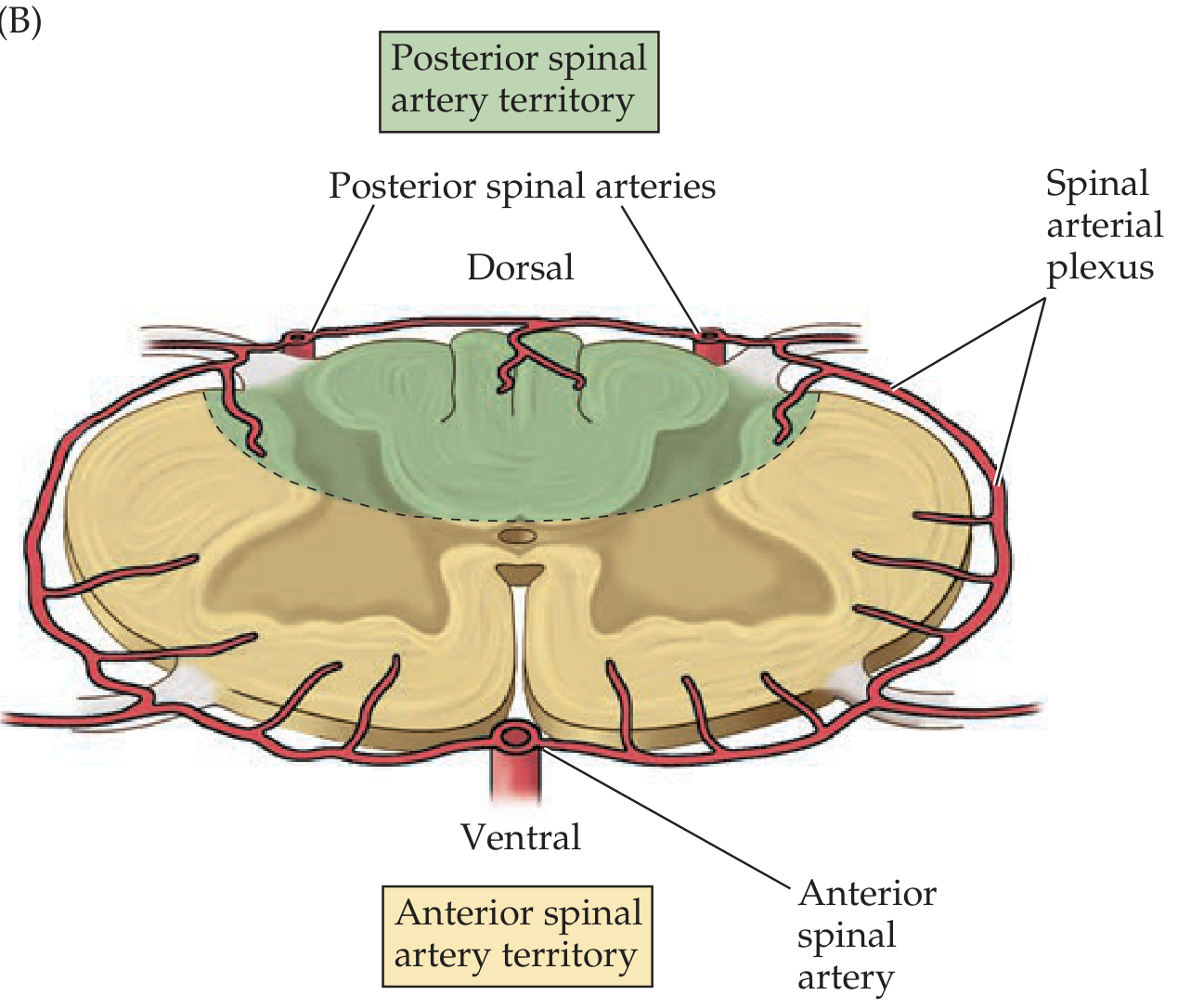
- Anterior Spinal Artery (ASA) - formed by fusion of branches from both vertebral arteries; runs in the anterior median fissure; supplies anterior 2/3 of cord (anterior horn, anterolateral funiculus, spinothalamic tracts, corticospinal tracts)
- Posterior Spinal Arteries (PSA, x2) - arise from vertebral or PICA; run along posterolateral sulci; supply posterior 1/3 (posterior columns, dorsal horn)
- 31 segmental branches enter the spinal canal, but only 6-10 reach the cord as radicular arteries
- Artery of Adamkiewicz (Arteria radicularis magna) - most important; typically arises from left intercostal/lumbar vessels at T9-T12 (sometimes L1-L2); provides major supply to lower thoracic and lumbosacral cord
- Spinal arterial plexus (vasocorona) surrounds the cord connecting anterior and posterior systems
Venous Drainage
- 6 longitudinal venous channels (anterior and posterior median veins + 4 anterolateral/posterolateral veins)
- Drain via intervertebral veins into the internal vertebral venous plexus (Batson's plexus)
Source: Neuroanatomy through Clinical Cases, 3rd Ed., p. 472-477
4. Speech Areas of the Brain [2015, 2018 supple]
Motor Speech Area (Broca's Area)
- Location: Inferior frontal gyrus (pars triangularis + pars opercularis), Brodmann areas 44 and 45, left hemisphere (dominant)
- Function: Speech production and articulation - controls the coordinated muscular activity for speaking
- Damage (Broca's aphasia): Non-fluent, effortful speech with intact comprehension; "telegraphic" speech; patient knows what they want to say but cannot say it fluently
Sensory Speech Area (Wernicke's Area)
- Location: Posterior part of superior temporal gyrus, Brodmann area 22, left hemisphere
- Function: Speech comprehension (understanding spoken and written language)
- Damage (Wernicke's aphasia): Fluent speech but with paraphasia (wrong words/neologisms), poor comprehension; patient is unaware of errors
Arcuate Fasciculus
- A bundle of white matter fibres connecting Broca's and Wernicke's areas
- Damage: Conduction aphasia - fluent speech, good comprehension, but poor repetition
Angular Gyrus (Area 39)
- At the junction of temporal-parietal-occipital lobes
- Important for reading and writing; damage causes alexia with agraphia
Supplementary Motor Area (SMA)
- Medial surface of frontal lobe (area 6)
- Initiates speech; damage causes mutism/transcortical motor aphasia
Key Teaching Points for Exams
- All speech areas are in the left (dominant) hemisphere in ~97% of right-handers
- Broca's = motor/expressive aphasia (problem with output)
- Wernicke's = sensory/receptive aphasia (problem with input/comprehension)
- Both areas connected by arcuate fasciculus
5. Motor Neurons of the Spinal Cord [2010 supple]
Alpha Motor Neurons (Lower Motor Neurons)
- Located in the anterior horn of spinal cord gray matter
- Large multipolar neurons forming Rexed lamina IX
- Send myelinated axons (alpha fibers) via ventral roots to innervate extrafusal muscle fibers
- Each motor neuron innervates multiple muscle fibers = motor unit
Gamma Motor Neurons
- Also in anterior horn, smaller than alpha neurons
- Innervate intrafusal muscle fibers (muscle spindle fibers)
- Regulate sensitivity of muscle spindle (proprioception)
- Co-activated with alpha neurons (alpha-gamma co-activation)
Upper vs. Lower Motor Neuron Distinctions
| Feature | UMN | LMN |
|---|---|---|
| Location | Cortex, corticospinal tracts | Anterior horn, ventral root |
| Tone | Increased (spasticity) | Decreased (flaccidity) |
| Reflexes | Hyperreflexia | Hyporeflexia/areflexia |
| Weakness | Less severe | More severe/wasting |
| Babinski | Present | Absent |
| Fasciculations | Absent | Present |
| Atrophy | Mild (disuse) | Severe (denervation) |
Renshaw Cells
- Inhibitory interneurons in the anterior horn
- Activated by collaterals of alpha motor neuron axons
- Provide recurrent inhibition back onto the same motor neuron (feedback control)
Topographic Organization (Somatotopy)
- Medial motor neurons innervate axial/trunk muscles
- Lateral motor neurons innervate distal limb muscles
6. Corpus Callosum [2011 supple]
Parts (Anterior to Posterior)
- Rostrum - thin anterior part, connects orbital frontal lobes
- Genu - curves anteriorly; connects prefrontal cortex (frontal forceps minor)
- Body (Trunk) - largest part; connects frontal, parietal lobes
- Splenium - posterior thickening; connects occipital, posterior temporal, parietal lobes (forceps major, tapetum)
Relations
- Superior surface - cingulate gyrus runs above it (separated by callosal sulcus); median longitudinal stria
- Inferior surface - septum pellucidum (between genu and fornix); body of fornix
- Anterior - genu curves under to form rostrum, then connects to lamina terminalis
- Posterior - splenium overhangs the pineal body and midbrain
Functions
- Interhemispheric transfer of sensory, motor, and cognitive information
- Coordinates activities between hemispheres
- Important for bimanual coordination
Corpus Callosum Agenesis
- May be partial or complete
- Can be silent or associated with intellectual disability, seizures
- Associated with Dandy-Walker malformation, lissencephaly
- Detected on MRI: absent corpus callosum, widely spaced parallel ventricles ("bat wing" appearance), colpocephaly
7. Circle of Willis [2014 supple]
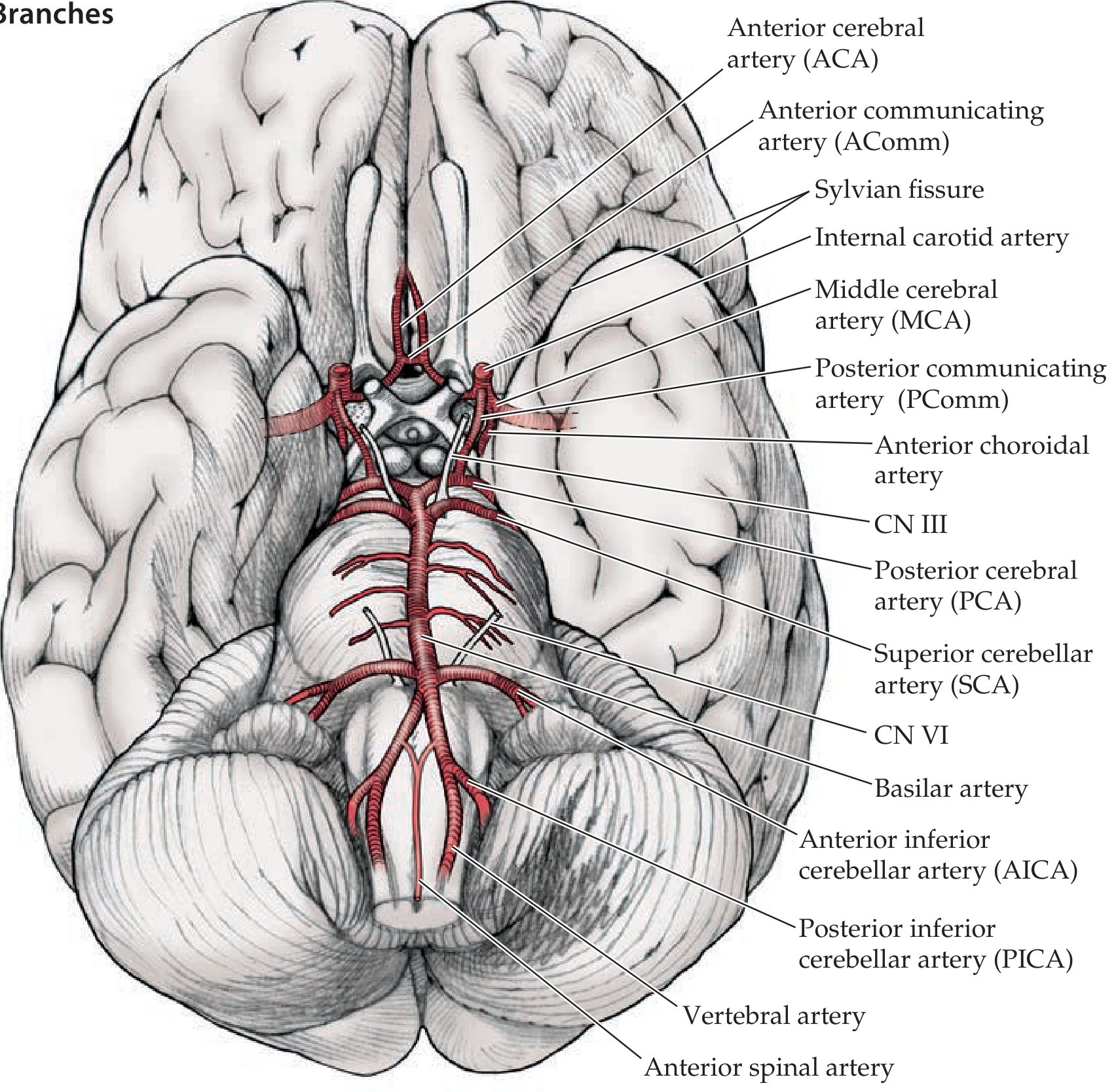
Components (from anterior, clockwise)
- Anterior communicating artery (AComm) - connects the two ACAs
- Anterior cerebral artery (ACA) x2 - terminal branch of ICA; supplies medial frontal and parietal lobes
- Internal carotid artery (ICA) x2 - enters from below
- Posterior communicating artery (PComm) x2 - connects ICA to PCA; joins anterior and posterior circulations
- Posterior cerebral artery (PCA) x2 - terminal branches of basilar artery; supplies occipital lobes, medial temporal
- Basilar artery - formed by union of both vertebral arteries; not technically in the circle but feeds it
Additional Branches Noted
- Middle cerebral artery (MCA) - largest branch of ICA (not part of circle itself)
- Anterior choroidal artery - from ICA
- Superior cerebellar artery (SCA)
- Anterior inferior cerebellar artery (AICA)
- Posterior inferior cerebellar artery (PICA)
Clinical Importance
- Allows collateral flow if one vessel is occluded
- A complete circle is present in only ~34% of individuals (variants are common)
- Common site of berry/saccular aneurysms: AComm (most common), PComm, MCA bifurcation
- PComm aneurysm classically causes CN III palsy (compresses oculomotor nerve)
Source: Neuroanatomy through Clinical Cases, 3rd Ed., p. 417
8. Fornix [2017 supple]
Structure
- A C-shaped white matter tract that arches from the hippocampus around the thalamus to the hypothalamus
- Bilateral (one from each hippocampus); the two limbs join and partially decussate at the midline forming the hippocampal commissure (psalterium)
Parts (following the fiber from posterior to anterior)
- Fimbria - begins on the medial surface of the hippocampus
- Crus of fornix (crura) - the two crura emerge from the fimbria and arch forward
- Body of fornix - two crura join below the corpus callosum (septum pellucidum lies between them and corpus callosum)
- Columns of fornix - at the anterior end of the body, the fornix divides into two columns that curve downward
- Pre-commissural fibers - pass anterior to anterior commissure to septal nuclei
- Post-commissural fibers - pass posterior to anterior commissure to mammillary bodies of hypothalamus (main destination)
Connections (Summary)
- From: Hippocampus, subiculum, entorhinal cortex
- To: Mammillary bodies (main), anterior thalamic nucleus, septal nuclei, hypothalamus
Papez Circuit
Clinical: Damage to Fornix
- Bilateral damage causes anterograde amnesia (difficulty forming new memories)
- The fornix runs in the floor of the lateral ventricle; may be damaged during surgery near the third ventricle
9. Medial Medullary Syndrome [2017 supple]
Structures Affected and Features
| Structure Damaged | Clinical Feature |
|---|---|
| Corticospinal tract (pyramid) | Contralateral hemiplegia/hemiparesis (spares face) |
| Medial lemniscus | Contralateral loss of proprioception, vibration, and discriminative touch (from body) |
| Hypoglossal nerve (CN XII) fibers | Ipsilateral LMN tongue paralysis (tongue deviates toward lesion) |
Key: "Medial = 3 M's" mnemonic
- Motor (corticospinal pyramid)
- Medial lemniscus
- Motor of tongue (CN XII)
Differences from Lateral Medullary Syndrome
- Medial: contralateral hemiplegia + contralateral dorsal column loss + ipsilateral tongue palsy
- Lateral (Wallenberg): Horner's, dysphagia, ipsilateral ataxia, alternating sensory loss (spinothalamic crossed, trigeminal ipsilateral), NO hemiplegia
10. Third Ventricle of Brain [2018 supple]
Additional Points for "Third Ventricle" Questions
- Hangs from the roof between the two thalami
- Produced by invagination of pia mater + ependyma
- Produces cerebrospinal fluid (CSF)
- Obstruction at foramen of Monro → obstructive hydrocephalus affecting one or both lateral ventricles (colloid cyst is a common cause)
- Obstruction at cerebral aqueduct → non-communicating hydrocephalus (bilateral lateral + 3rd ventricle dilation, 4th ventricle normal)
- Hypothalamic tumors (craniopharyngioma) arise near the floor of 3rd ventricle
- Pineal tumors compress the aqueduct causing Parinaud syndrome (loss of upward gaze)
11. Pia Mater of the Spinal Cord [2018 supple]
Structure
- Thin, highly vascular, transparent membrane
- Closely adherent to the surface of the spinal cord and cannot be separated from it without damage
- Composed of flattened mesothelial cells and collagen fibers
Specializations of the Spinal Pia Mater
-
Denticulate (Dentate) Ligaments
- Bilateral serrated lateral extensions of pia mater
- Run between dorsal and ventral roots, attaching laterally to the dura mater
- 21 pairs total (from C1 to just above L1)
- Function: anchor and suspend the spinal cord within the dural tube, preventing excessive movement
-
Filum Terminale (Internum)
- At the conus medullaris (L1-L2), the pia mater continues as the filum terminale internum
- Thin thread (~20 cm long) of pia + glial tissue
- Extends through the lumbar cistern to the tip of the dural sac at S2, then as the filum terminale externum (coccygeal ligament) to the coccyx
- Function: anchors the spinal cord inferiorly
-
Anterior Median Fissure - pia dips into the anterior median fissure, carrying with it the anterior spinal artery
Relation to Dentate Ligament (Exam Point)
- Denticulate ligaments lie in the coronal plane, dividing the subarachnoid space into anterior and posterior compartments
- Useful surgical landmark during spinal surgery
12. Sensory and Motor Speech Areas of the Brain [2022]
Summary Table
| Area | Location | Brodmann | Function | Lesion |
|---|---|---|---|---|
| Broca's (motor) | Inferior frontal gyrus | 44, 45 | Speech production/articulation | Non-fluent aphasia |
| Wernicke's (sensory) | Posterior superior temporal gyrus | 22 | Speech comprehension | Fluent aphasia, poor comprehension |
| Angular gyrus | TPO junction | 39 | Reading, writing, semantics | Alexia + agraphia |
| Supramarginal gyrus | Parietal | 40 | Phonological processing | Conduction aphasia (sometimes) |
| Arcuate fasciculus | White matter tract | - | Connects Broca's + Wernicke's | Conduction aphasia (poor repetition) |
| SMA | Medial frontal | 6 | Speech initiation | Mutism, transcortical motor aphasia |
13. Lateral Medullary Syndrome (Wallenberg Syndrome) - Anatomical Basis [2025]
Vascular Cause
Clinical Features with Anatomical Basis
| Clinical Feature | Structure Damaged | Location |
|---|---|---|
| Ipsilateral facial pain/temperature loss | Spinal nucleus + tract of CN V | Posterolateral medulla |
| Contralateral body pain/temperature loss | Spinothalamic tract (already crossed) | Anterolateral medulla |
| Dysphagia, dysarthria, hoarseness | Nucleus ambiguus (CN IX, X) | Lateral medulla |
| Ipsilateral Horner syndrome (miosis, ptosis, anhidrosis) | Descending sympathetic fibers (hypothalamospinal) | Posterolateral tegmentum |
| Vertigo, nausea, vomiting, nystagmus | Vestibular nuclei (inferior + medial) | Lateral medulla |
| Ipsilateral limb ataxia, falling toward lesion | Inferior cerebellar peduncle (restiform body) | Lateral medulla |
| Hiccups | Dorsolateral medulla (respiratory centers) | Rare |
| Ipsilateral soft palate paresis, loss of gag | Nucleus ambiguus | CN IX, X |
Key Examination Point: Crossed Sensory Loss
- Face ipsilateral (CN V nucleus is ipsilateral) + body contralateral (spinothalamic tract already crossed in cord)
- Motor system (pyramids) is NOT affected (pyramids are medial, not lateral)
- Posterior columns (vibration/proprioception) are NOT affected (medial location)
Diagnostic Triad
Source: Localization in Clinical Neurology, 8e, p. 5609-5670; Adams & Victor's Principles of Neurology, 12th Ed., p. 816
14. Cross Section of Midbrain at Superior Colliculus + Weber Syndrome [2025]
Cross Section of Midbrain at Level of Superior Colliculus
SUPERIOR COLLICULUS (roof/tectum)
| |
Central Gray Matter (PAG)
|
____________________
| TEGMENTUM |
| Red nucleus (large) |
| CN III nucleus |
| Medial lemniscus |
| Spinothalamic tract |
| Medial longitudinal |
| fasciculus (MLF) |
| CN III fibers (exit |
| medially through |
| tegmentum) |
|____________________|
|
SUBSTANTIA NIGRA (pars compacta + pars reticulata)
|
CEREBRAL PEDUNCLE (CRUS CEREBRI)
[lateral = fibers to cortex / corticospinal tract medial 3/5]
[Corticospinal = middle 3/5; Corticopontine = medial 1/5 + lateral 1/5]
|
INTERPEDUNCULAR FOSSA
Key Structures at This Level:
- Tectum (roof): Superior colliculi (visual reflex centers)
- Tegmentum: Red nucleus, CN III nucleus + fibers, MLF, substantia nigra, decussation of superior cerebellar peduncle (at inferior colliculus level), reticular formation, medial lemniscus, spinothalamic tract
- Cerebral peduncle (Basis pedunculi/Crus cerebri): Corticospinal, corticobulbar, and corticopontine fibers
- CN III exits between cerebral peduncles (through interpeduncular fossa), passes between PCA and SCA, travels in the wall of cavernous sinus
Weber Syndrome
| Feature | Anatomical Basis |
|---|---|
| Ipsilateral CN III palsy (ptosis, mydriasis, "down and out" eye) | CN III fibers pass through/near the cerebral peduncle |
| Contralateral hemiplegia/hemiparesis | Corticospinal fibers in the cerebral peduncle (cross in medullary pyramids) |
| No cerebellar signs | Red nucleus and cerebellum spared (compare Benedikt syndrome) |
- Benedikt = CN III palsy + contralateral tremor/ataxia (red nucleus + CN III, deeper tegmental lesion)
- Weber = CN III palsy + contralateral hemiplegia (peduncle lesion)
Source: StatPearls - Weber Syndrome (NIH); Wikipedia; Neuroanatomy
15. Klinefelter Syndrome [2025]
Definition
Genetics
- Results from non-disjunction of sex chromosomes during meiosis
- Maternal and paternal non-disjunction contribute equally
- ~15% show mosaicism (e.g., 46,XY/47,XXY) - mosaics have milder features
- Advanced maternal age is a risk factor (but less strongly than in trisomy 21)
Clinical Features
- Only consistent finding: Hypogonadism
- Body habitus: Tall, long lower limbs; eunuchoid proportions (span > height; lower segment > upper segment)
- Testes: Markedly small (2 cm); firm due to hyalinization of seminiferous tubules
- Gynecomastia - in ~50%
- Reduced facial, body, and pubic hair
- Azoospermia/oligospermia - infertility in virtually all (unless mosaic)
- Decreased serum testosterone, elevated serum FSH and LH (hypergonadotropic hypogonadism)
- Mild intellectual disability / verbal skill deficits (variable)
Histopathology
- Hyalinization and fibrosis of seminiferous tubules (ghost tubules)
- Leydig cells appear prominent/hyperplastic
- Absent/markedly reduced spermatogenesis
Associated Conditions
- Metabolic syndrome, type 2 diabetes (insulin resistance)
- Mitral valve prolapse (~50% of adults)
- 20-30x increased risk of extragonadal germ cell tumors (mediastinal teratoma)
- Increased risk of breast cancer (20x above normal males)
- Increased autoimmune diseases (SLE, RA)
- Osteoporosis (due to hypogonadism)
Diagnosis
- Karyotype (gold standard)
- Hormonal: Low testosterone, high FSH/LH
- Testicular biopsy: hyalinized tubules
Treatment
- Testosterone replacement (improves secondary sexual characteristics, bone density, mood, libido)
- Does NOT restore fertility (sperm retrieval via TESA + ICSI may help mosaics)
- Psychological support
Source: Robbins & Kumar Basic Pathology, p. 128
Quick Reference Summary Table
| Topic | Key Mnemonic / Recall Point |
|---|---|
| Thalamic nuclei | LGN = Lateral Light; MGN = Medial Music; VPL = Spine; VPM = Face |
| 3rd ventricle | Monro anteriorly, Sylvius posteriorly |
| Spinal blood supply | 1 anterior + 2 posterior spinal arteries; Artery of Adamkiewicz at T9-T12 |
| Speech | Broca's = 44/45 (motor); Wernicke's = 22 (sensory) |
| Corpus callosum | Rostrum, Genu, Body, Splenium (R-G-B-S) |
| Circle of Willis | ACA + AComm + ICA + PComm + PCA (complete ring in only 34%) |
| Fornix | Hippocampus → Mammillary body → Anterior thalamus → Cingulate = Papez circuit |
| Medial medullary | 3 M's: Motor (pyramid), Medial lemniscus, Motor of tongue (CN XII) |
| Lateral medullary | Horner + ipsilateral ataxia + contralateral hemihypalgesia; NO hemiplegia |
| Weber syndrome | Ventromedial midbrain; CN III (ipsilateral) + hemiplegia (contralateral) |
| Klinefelter | 47,XXY; small testes + gynecomastia + infertility; high FSH/LH |
| Pia mater | Denticulate ligaments (21 pairs) + filum terminale |
A tailor presents with diffuse swelling of the palm after a needle prick over the tip of the little finger
Tailor with Palm Swelling After Needle Prick on Little Finger Tip
Diagnosis: Suppurative Tenosynovitis of Little Finger → Infection of the Ulnar Bursa
Anatomical Basis - Why the Little Finger?
The Flexor Tendon Sheath System
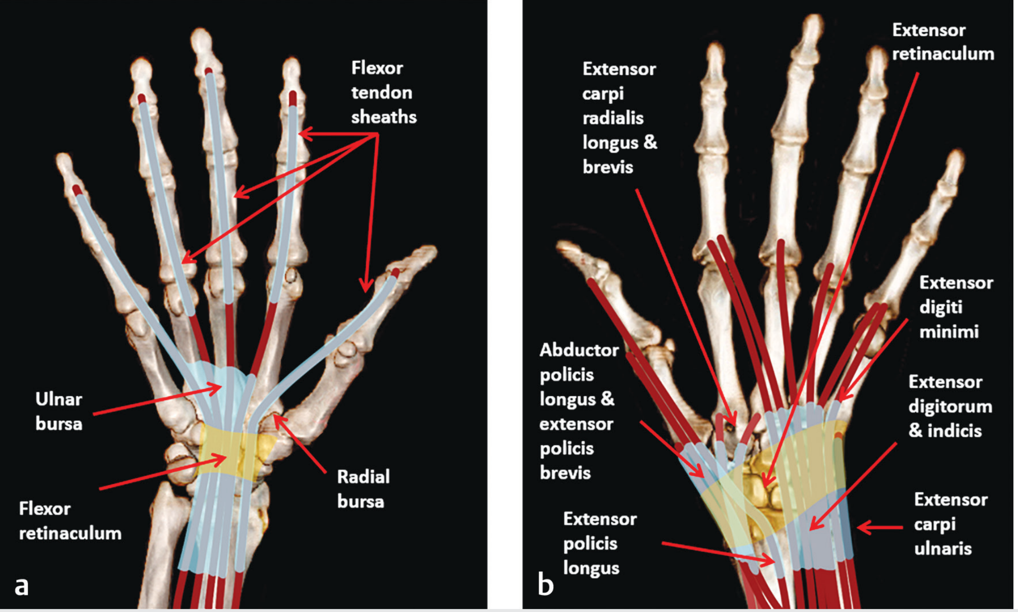
- The little finger's flexor tendon sheath runs continuously and without interruption from the fingertip all the way to (and through) the carpal tunnel, where it expands into the Ulnar Bursa (Common Flexor Sheath)
- The thumb's sheath similarly continues as the Radial Bursa
Step-by-Step Spread of Infection
Needle prick → Tip of little finger
↓
Infection enters continuous little finger flexor tendon sheath
↓
Spreads PROXIMALLY along uninterrupted sheath toward wrist
↓
Enters ULNAR BURSA in the palm
(Ulnar bursa widens in palm to cover 3rd + 4th flexor tendons too)
↓
Diffuse palmar swelling (hypothenar + palm)
↓
May spread to SPACE OF PARONA (deep to pronator quadratus, proximal to carpal tunnel)
↓
If ulnar + radial bursae communicate (occurs in 30-80% of cases) →
"HORSESHOE ABSCESS" = infection of both bursae simultaneously
Clinical Features of Ulnar Bursa Infection
Kanavel's Four Cardinal Signs of Flexor Tenosynovitis
| Sign | Description |
|---|---|
| 1. Flexed resting posture | Finger held semiflexed at rest (reduces pain by minimizing sheath tension) - earliest sign |
| 2. Fusiform (uniform) swelling | Symmetric sausage-like swelling along entire finger (not just at tip) |
| 3. Percussion tenderness | Tenderness along entire length of flexor tendon sheath |
| 4. Pain with passive extension | Exquisite pain when finger is passively straightened (stretches sheath) |
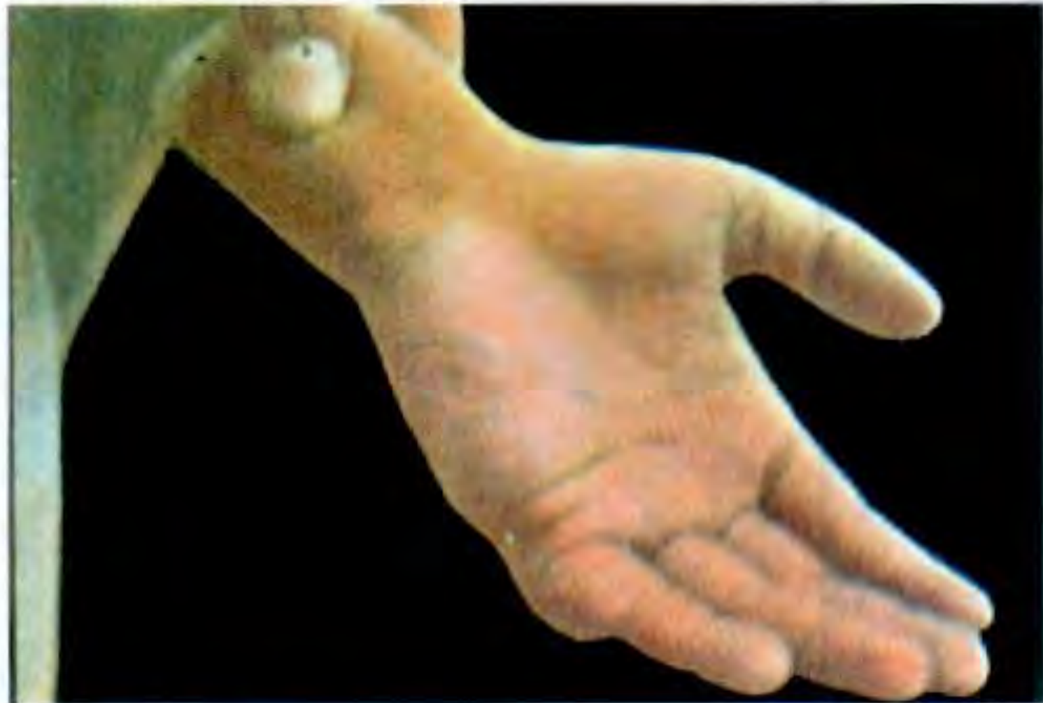
- Fullness and diffuse swelling of the palm (hypothenar eminence)
- Kanavel's point - maximum tenderness on the ulnar side of the palm, between the two palmar (transverse) creases (where the ulnar bursa bulges most)
- Oedematous swelling of the dorsum of the hand (dorsal subcutaneous tissue is lax, so pus gravitates here)
- Ring and middle fingers may also be flexed if their sheaths communicate with the ulnar bursa
Why Dorsal Swelling Despite Palmar Infection?
Microorganism
- Most common organism: Staphylococcus aureus (including MRSA)
- Also: Streptococcus spp., anaerobes (polymicrobial)
- Gram-negative organisms in immunocompromised or after open trauma
Complications (If Untreated)
- Tendon necrosis - pressure within the closed sheath cuts off blood supply (compartment syndrome of the sheath)
- Permanent flexion contracture - adhesion of tendon to sheath
- Horseshoe abscess - spread to radial bursa via ulnar-radial communication (present in 30-80%)
- Spread to Space of Parona - deep space proximal to the wrist (between FDP sheaths and pronator quadratus)
- Septicaemia - systemic spread
Treatment
- Immobilize and elevate the hand immediately
- IV antibiotics - vancomycin (covers MRSA) ± gram-negative cover if immunocompromised
- Surgical drainage - closed tendon sheath irrigation vs. open drainage
- Incision at distal end of sheath at the finger + proximal window at the palm
- Continuous irrigation with normal saline via catheter
- Post-op: Physiotherapy to prevent contracture
Sources: S Das - A Manual on Clinical Surgery, 13th Ed., p. 319; Tintinalli's Emergency Medicine, p. 1957; Imaging Anatomy, Vol 3, p. 652
Summary Box (Exam Recall)
| Point | Detail |
|---|---|
| Why little finger? | Continuous sheath from fingertip to ulnar bursa (no break) |
| Why palm swelling? | Infection tracks along sheath to ulnar bursa which widens in palm |
| Unique feature | Same continuity exists for thumb → radial bursa |
| Eponym | Kanavel's 4 signs; Kanavel's point (ulnar bursa tenderness) |
| Dangerous complication | Horseshoe abscess (ulnar + radial bursa both infected) |
| Emergency | Yes - tendon necrosis within hours if untreated |
Why is a ischio rectal abscess very painful when abscess is superficial? Right boundaries and contents of the fossa.
Ischiorectal (Ischioanal) Abscess - Complete Anatomy Answer
Why is an Ischiorectal Abscess Very Painful When Superficial?
The Key: Inferior Rectal (Haemorrhoidal) Nerve
- Somatic in innervation (not autonomic)
- Exquisitely sensitive to pain, pressure, temperature and touch
- Supplied by the same nerve fibres that carry pain from the external anal sphincter and perianal skin
- Direct pressure on the inferior rectal nerve branches running through the fat pad
- Stretching and distension of the sensitive somatic skin at the base of the fossa
- Tension in the fibrous septa of the fat (the fossa fat is divided into fibrous compartments - these septa are inextensible and do not allow easy expansion, amplifying pressure pain)
Why Deep Abscesses Are Less Painful Initially
- They are remote from the sensitive somatic skin
- Pus collects in the spacious, low-tension fatty tissue of the fossa
- Symptoms are mainly constitutional (fever, malaise, deep rectal discomfort)
- Swelling is diffuse, not localised
As Bailey & Love states: "Patients with infection in the larger fatty-filled ischiorectal space, in which tissue tension is much lower, usually present later, with less well-localised symptoms but more constitutional upset and fever."
Summary of Pain Mechanism
Superficial abscess → close to skin base
↓
Pus accumulates under somatic skin (perineum)
↓
Pressure on inferior rectal nerve (branch of pudendal nerve S2,S3,S4)
↓
Tension in fibrous fat septa (inextensible, non-compliant)
↓
Exquisite, constant, throbbing, well-localised pain
↓
Worsened by sitting, walking, defaecation
Boundaries of the Ischiorectal (Ischioanal) Fossa
Note: The modern anatomical term is ischioanal fossa, though clinically "ischiorectal" remains widely used.
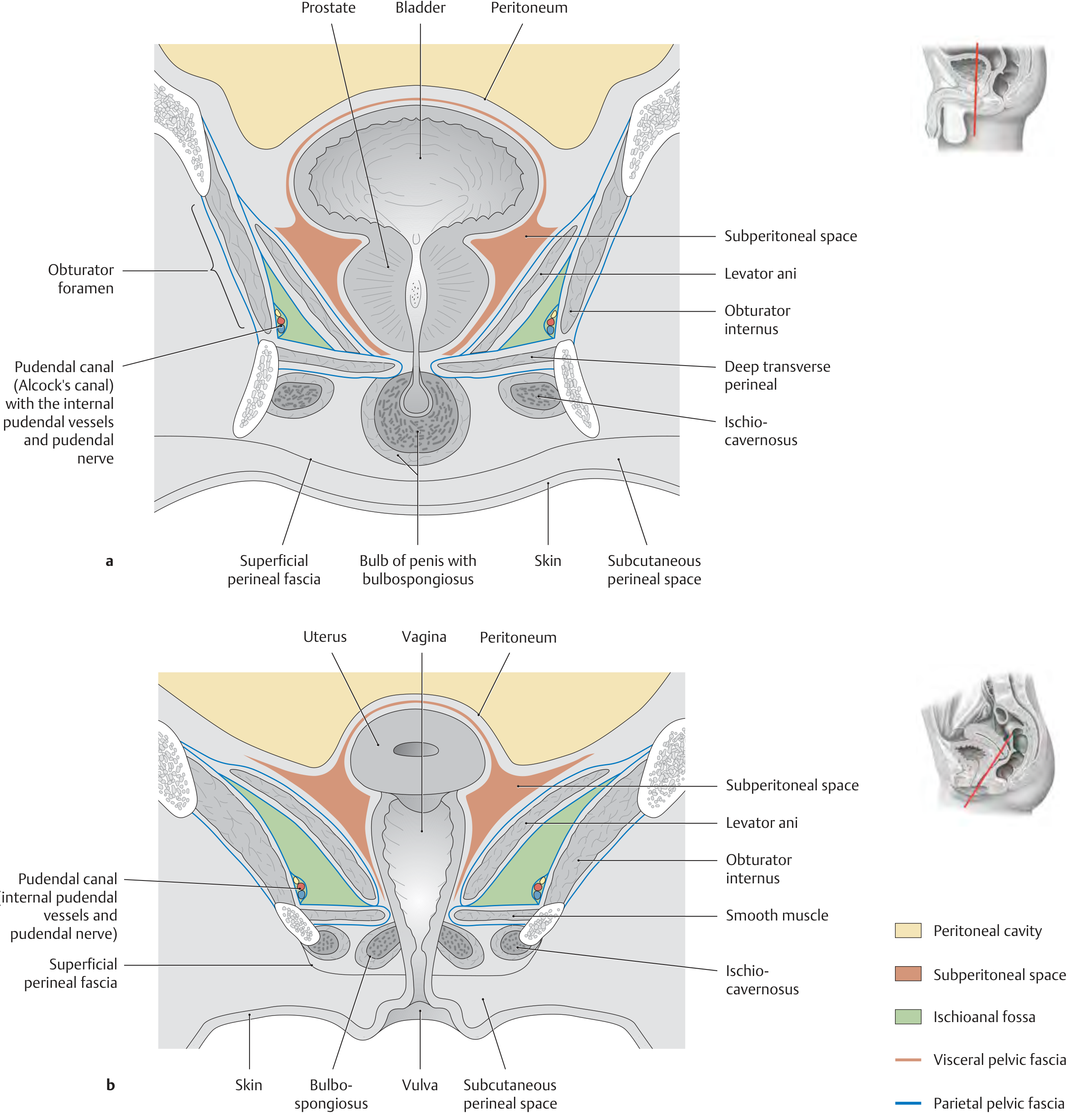
Boundaries
| Wall | Boundary |
|---|---|
| Medial | External anal sphincter + inferior fascia of the pelvic diaphragm (covering levator ani) |
| Lateral | Ischial tuberosity + obturator internus muscle covered by obturator fascia |
| Roof (apex) | Angle formed where medial and lateral walls meet - at the junction of levator ani and obturator internus (narrow apex directed superiorly toward the symphysis pubis) |
| Base (floor) | Perineal skin and superficial perineal fascia (Colles' fascia) |
| Anterior | Posterior border of the urogenital diaphragm (perineal membrane) and bodies of pubis |
| Posterior | Gluteus maximus muscle + sacrotuberous ligament |
Muscular Boundaries (Thieme Atlas)
- Superomedially: Levator ani
- Laterally: Obturator internus
- Inferiorly: Deep transverse perineal muscle
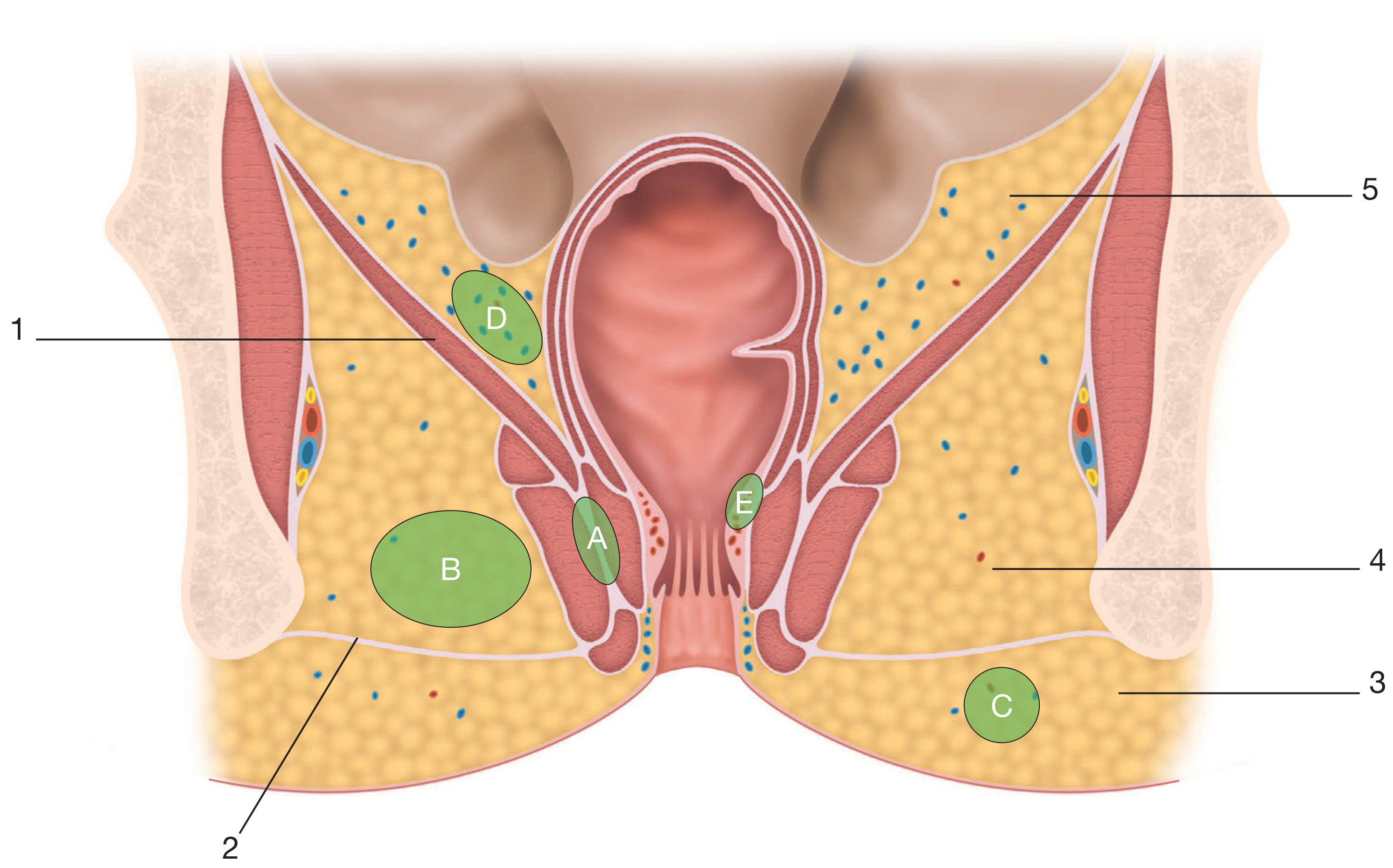
Contents of the Ischiorectal Fossa
1. Fat Body (Ischioanal Fat Pad) - Main Content
- Large mass of loose, areolar fatty tissue that fills the entire fossa
- Function: Acts as a mobile, compressible pad allowing the anal canal to expand during defaecation and the pelvic floor to descend during labour
- Divided into fibrous septa that compartmentalise the fat
- Clinically important: this fat is the space where ischiorectal abscesses develop
2. Inferior Rectal (Inferior Haemorrhoidal) Vessels and Nerve
- Inferior rectal artery - branch of the internal pudendal artery; crosses the fat pad medially to supply the external anal sphincter and perianal skin
- Inferior rectal vein - accompanies the artery
- Inferior rectal nerve - branch of the pudendal nerve (S2, S3, S4); crosses the fossa medially; supplies the external anal sphincter and perianal skin
- These structures cross the fossa from lateral to medial in its lower part
3. Pudendal Canal (Alcock's Canal) - on Lateral Wall
- A fascial tunnel formed by a split in the obturator internus fascia
- Runs along the lateral wall of the ischiorectal fossa
- Contents of Alcock's canal:
- Internal pudendal artery
- Internal pudendal vein
- Pudendal nerve (S2, S3, S4)
- Nerve to obturator internus (sometimes)
- Begins just below the ischial spine and runs to the perineal body
- The pudendal nerve gives off the inferior rectal nerve in or near this canal, which crosses the fossa
4. Perineal Branch of S4 (posterior cutaneous nerve of the perineum)
- Crosses through the posterior part of the fossa
- Supplies the skin of the posterior perineum
5. Lymphatics
- Follow the inferior rectal vessels
- Drain to the superficial inguinal nodes
The Two Fossae Communicate
Summary Table for Exam
| Feature | Detail |
|---|---|
| Shape | Pyramid/wedge - apex up, base down |
| Medial wall | External anal sphincter + levator ani fascia |
| Lateral wall | Ischial tuberosity + obturator internus + obturator fascia |
| Floor (base) | Perineal skin (somatic - inferior rectal nerve) |
| Roof (apex) | Junction of levator ani + obturator internus |
| Anterior | Urogenital diaphragm |
| Posterior | Gluteus maximus + sacrotuberous ligament |
| Main content | Ischioanal fat pad |
| Key neurovascular | Inferior rectal nerve/vessels (cross fat pad); pudendal N + internal pudendal vessels (in Alcock's canal on lateral wall) |
| Communication | Two fossae meet in deep postanal space (horseshoe abscess) |
| Why painful superficially | Somatic skin at base; inferior rectal nerve (pudendal S2-4); inextensible fat septa amplify pressure |
Sources: Bailey & Love's Short Practice of Surgery, 28th Ed., p. 1456; Color Atlas of Human Anatomy Vol. 2, p. 476-477; Thieme Atlas of Anatomy, p. 581
Make it a crisp answer for mbbs 1st year student writing 10 mark question
Ischiorectal Fossa — 10 Mark Answer
Definition
Boundaries
| Wall | Boundary |
|---|---|
| Medial | External anal sphincter + inferior fascia of levator ani |
| Lateral | Ischial tuberosity + obturator internus covered by obturator fascia |
| Roof (apex) | Junction of levator ani and obturator internus |
| Floor (base) | Perineal skin + superficial perineal fascia |
| Anterior | Posterior border of urogenital diaphragm |
| Posterior | Gluteus maximus + sacrotuberous ligament |
Apex points superiorly toward symphysis pubis; base faces downward as perineal skin.
Contents
- Ischioanal fat pad — main content; loose fatty tissue allowing anal canal expansion during defaecation
- Inferior rectal (haemorrhoidal) nerve and vessels — cross the fat pad medially; supply external anal sphincter and perianal skin
- Pudendal canal (Alcock's canal) — on the lateral wall inside the obturator fascia; contains:
- Internal pudendal artery and vein
- Pudendal nerve (S2, S3, S4)
- Perineal branch of S4 nerve — crosses the posterior fossa
- Lymphatics — drain to superficial inguinal nodes
Why is a Superficial Ischiorectal Abscess Very Painful?
The Anatomical Reason
- Directly compresses branches of the inferior rectal nerve — a somatic nerve that carries well-localised, intense pain
- Stretches the sensitive perianal skin — which is as pain-sensitive as any skin elsewhere on the body
- Builds up pressure within inextensible fibrous fat septa — the fat of the fossa is divided into tight compartments by fibrous septa; pus cannot expand freely, so pressure rises sharply → throbbing, constant pain
- Worsened by sitting, walking and defaecation — any increase in intra-abdominal pressure or movement compresses the abscess against the taut skin
Key Contrast — Deep Abscess is Less Painful
- It is remote from the somatic skin
- The spacious, low-tension fat of the upper fossa allows pus to accumulate without rapidly raising pressure
- Presents mainly with fever, malaise, and vague deep rectal discomfort
Communication Between the Two Fossae
Clinical Significance at a Glance
| Point | Fact |
|---|---|
| Most painful abscess | Superficial ischiorectal (somatic skin + nerve) |
| Nerve responsible for pain | Inferior rectal nerve (branch of pudendal nerve S2,3,4) |
| Why deep abscess is silent | Low tissue tension, away from somatic skin |
| Communication | Deep post-anal space → horseshoe abscess |
| Treatment | Surgical drainage under anaesthesia — incision over point of maximum fluctuation |
Hi, I am in MBBS, first year. WBHUS University. Cześć. I'm going to give my first professional exam. I have... Toch hier. The chapter of minerals. Potassium, iron, and electrolytes. The mineral chapter of biochemistry. And tell me the important topics to be read. About which minerals are important? Analyzing the previous year question. And also, give me the question important. For my professional exam.
WBHUS MBBS first professional exam biochemistry minerals important questions previous year
https://medical.iqcity.in/wp-content/uploads/2021/03/1st-Pro…
Minerals & Electrolytes — MBBS 1st Year Biochemistry
WBHUS 1st Professional Exam Guide
🎯 How WBHUS Asks Questions (Paper Pattern)
| Question Type | Marks | What to Expect |
|---|---|---|
| Long Essay (Group A) | 12 marks | 2-4 sub-parts; one from two choices |
| Short Essay (Group B) | 7 marks | Modified essay, 2-3 sub-parts |
| Short Notes (Group C) | 3 marks each | Four out of five |
| Explain Why (Group D) | 3 marks each | Clinical reasoning questions |
⭐ Most Important Minerals for Your Exam
TIER 1 — Highest Priority (Asked Almost Every Year)
1. 🔴 IRON (Most Important Mineral in Biochemistry)
- Dietary iron: haem iron (meat - directly absorbed) vs non-haem iron (plant - needs reduction Fe³⁺ → Fe²⁺)
- Absorbed in duodenum and upper jejunum
- Enhancers: Vitamin C (ascorbic acid), acidic pH, meat factor
- Inhibitors: Phytates, oxalates, tannins (tea), calcium, phosphates, antacids
| Protein | Function | Normal Value |
|---|---|---|
| Transferrin | Transport protein in blood; carries Fe³⁺ | TIBC = 250-370 µg/dL |
| Ferritin | Storage form (soluble, non-toxic); liver, spleen, marrow | 12-300 ng/mL |
| Haemosiderin | Insoluble storage (overflow); seen in iron overload | |
| Apotransferrin | Transferrin without iron |
- Adult male: 1 mg/day
- Menstruating female: 2 mg/day
- Pregnant woman: 3-4 mg/day
- Infant/Child: 1.5 mg/day
- Stages: Depleted stores → ↓ serum ferritin → ↓ serum iron → ↑ TIBC → microcytic hypochromic anaemia
- Why anaemia in iron deficiency? Iron is essential for haem synthesis (protoporphyrin + Fe²⁺ → haem) → without iron, haemoglobin cannot be made → RBCs are small and pale
- Excessive storage in liver, pancreas, heart, skin
- "Bronze diabetes" — diabetes + liver cirrhosis + skin pigmentation
2. 🟡 CALCIUM (Second Most Important)
- Bone and teeth mineralisation (99% of body calcium is here)
- Muscle contraction (troponin C binding)
- Blood coagulation (Factor IV)
- Nerve impulse transmission
- Enzyme activation (lipases, ATPases)
- Second messenger (calmodulin-mediated)
- Cell membrane permeability
- Total serum calcium: 9-11 mg/dL (2.2-2.7 mmol/L)
- Ionised (active) calcium: 4.5-5.6 mg/dL
- ~50% ionised (active), ~40% protein-bound (albumin), ~10% complexed
| Hormone | Source | Action on Calcium |
|---|---|---|
| PTH (Parathyroid hormone) | Parathyroid gland | ↑ blood Ca²⁺ (bone resorption, ↑ renal reabsorption, ↑ vit D activation) |
| Calcitriol (Active Vit D / 1,25-(OH)₂D₃) | Kidney (activation) | ↑ intestinal Ca²⁺ absorption, ↑ bone resorption |
| Calcitonin | Thyroid C-cells | ↓ blood Ca²⁺ (inhibits osteoclasts) |
- Cause: Vitamin D deficiency → ↓ calcium absorption → soft, unmineralised bones
- Rickets = children (open epiphyses); Osteomalacia = adults
- Features: Bow legs, rachitic rosary, craniotabes, Harrison's sulcus
3. 🟠 SODIUM & POTASSIUM (Electrolytes)
| Electrolyte | Normal Serum | Main Compartment | Function |
|---|---|---|---|
| Sodium (Na⁺) | 135-145 mEq/L | Extracellular fluid (ECF) | Osmolality, nerve impulse, fluid balance |
| Potassium (K⁺) | 3.5-5.0 mEq/L | Intracellular fluid (ICF) | Resting membrane potential, cardiac rhythm |
- Pumps 3 Na⁺ OUT and 2 K⁺ IN per cycle
- Maintains electrochemical gradient
- Electrogenic (generates slight negative charge inside)
- Inhibited by cardiac glycosides (digoxin)
| Condition | Cause | Effect |
|---|---|---|
| Hyponatraemia (<135) | Excess water, SIADH | Cerebral oedema, convulsions |
| Hypernatraemia (>145) | Dehydration, diabetes insipidus | Brain shrinkage, thirst |
| Hypokalaemia (<3.5) | Vomiting, diuretics | Muscle weakness, cardiac arrhythmia |
| Hyperkalaemia (>5.0) | Renal failure, Addison's | Peaked T waves, cardiac arrest |
4. 🟢 COPPER (Short Note — Ceruloplasmin is a repeat question!)
- Copper-carrying protein in blood (carries ~95% of serum copper)
- Functions: Ferroxidase activity (oxidises Fe²⁺ → Fe³⁺ for transferrin loading), antioxidant
- Normal: 20-40 mg/dL
- Autosomal recessive; ATP7B gene mutation
- Copper cannot be incorporated into ceruloplasmin or excreted in bile
- Result: Copper deposits in liver, brain, cornea (Kayser-Fleischer rings), kidney
- Labs: ↓ serum ceruloplasmin, ↓ serum copper, ↑ urinary copper, ↑ hepatic copper
5. 🔵 IODINE (Short Note)
- Essential for thyroid hormone synthesis (T₃ and T₄)
- Deficiency → Goitre (compensatory TSH ↑ → thyroid hypertrophy)
- Severe deficiency in pregnancy → Cretinism (irreversible mental retardation)
- RDA: 150 µg/day (200-300 µg in pregnancy)
6. 🟣 ZINC (Short Note)
- Component of >300 metalloenzymes (carbonic anhydrase, carboxypeptidase, alcohol dehydrogenase, DNA/RNA polymerase)
- Deficiency: Growth retardation, hypogonadism, delayed wound healing, loss of taste (hypogeusia), impaired immunity, night blindness
- Syndrome: Acrodermatitis enteropathica (autosomal recessive zinc malabsorption)
7. PHOSPHORUS
- 85% in bones as hydroxyapatite [Ca₁₀(PO₄)₆(OH)₂]
- Normal serum: 2.5-4.5 mg/dL
- Reciprocal relationship with calcium: ↑ Phosphate → ↓ Calcium
- Important in: ATP synthesis, 2,3-BPG (oxygen delivery from Hb)
📋 Previous Year Pattern Analysis — Most Repeated Questions
Long Essay (12 marks) — HIGH PROBABILITY
- "Describe iron metabolism — absorption, transport, storage, and regulation. What happens in iron deficiency anaemia?" (asked 2008, 2010, 2012, 2015, 2018)
- "Enumerate functions of calcium. How is blood calcium homeostasis maintained?" (repeat question every few years)
- "Describe calcium metabolism. Discuss Rickets and Osteomalacia."
Short Essay (7 marks) — HIGH PROBABILITY
- Iron absorption and factors affecting it
- Transferrin and ferritin — structure and function
- Calcium and phosphorus homeostasis
- Role of PTH in calcium regulation
Short Notes (3 marks) — VERY HIGH PROBABILITY
- Ceruloplasmin (appears in question bank explicitly)
- Ferritin
- Transferrin/TIBC
- Wilson's disease
- Haemochromatosis
- Rickets
- Zinc deficiency
- Goitre/Iodine deficiency
- Na⁺/K⁺-ATPase pump
- Haemosiderin
Explain Why (3 marks) — HIGH PROBABILITY
- "Anaemia occurs in iron deficiency" (explicitly in question bank)
- "Vitamin C enhances iron absorption"
- "Tea/Coffee inhibits iron absorption"
- "PTH increases blood calcium"
- "Hypocalcaemia causes tetany"
- "Ceruloplasmin is low in Wilson's disease"
- "Kwashiorkor patients have low serum copper"
- "Hyperkalaemia causes cardiac arrest"
📌 Quick Memory Table — All Minerals at a Glance
| Mineral | Normal Level | Key Protein | Deficiency Disease | Toxicity |
|---|---|---|---|---|
| Iron | 60-170 µg/dL | Ferritin, Transferrin, Hb | IDA, Koilonychia | Haemochromatosis |
| Calcium | 9-11 mg/dL | Calmodulin, Osteocalcin | Rickets, Tetany | Hypercalcaemia, Stones |
| Sodium | 135-145 mEq/L | — | Hyponatraemia | Hypernatraemia |
| Potassium | 3.5-5.0 mEq/L | — | Hypokalaemia | Cardiac arrest |
| Copper | 70-140 µg/dL | Ceruloplasmin | Menkes disease | Wilson's disease |
| Zinc | 70-120 µg/dL | Carbonic anhydrase | Acrodermatitis | Nausea, ↓ Cu absorption |
| Iodine | — | Thyroglobulin | Goitre, Cretinism | Thyrotoxicosis |
| Phosphorus | 2.5-4.5 mg/dL | — | Rickets (co-deficiency) | Hypocalcaemia |
✅ Your 3-Day Study Plan for Minerals
| Day | Topics |
|---|---|
| Day 1 | Iron metabolism (absorption → transport → storage → IDA → haemochromatosis) |
| Day 2 | Calcium metabolism (functions → homeostasis → PTH/Vit D/Calcitonin → Rickets) |
| Day 3 | Electrolytes (Na/K distribution, pump, disorders) + Trace minerals (Cu, Zn, I) + Short notes revision |
HIV and biochemical changes in AIDS formbbs 1st year mbbs student university exams detailed description
HIV and Biochemical Changes in AIDS
MBBS 1st Year — Detailed Study Guide
PART 1 — HIV: The Virus
Classification
- Full name: Human Immunodeficiency Virus
- Family: Retroviridae
- Genus: Lentivirus
- Type: RNA virus (single-stranded, positive-sense, diploid — two copies of RNA)
- Two types: HIV-1 (global, more virulent) and HIV-2 (mainly West Africa, less virulent)
Structure of HIV
OUTER ENVELOPE (lipid bilayer derived from host cell)
↓
┌─────────────────────────────────┐
│ gp120 (surface glycoprotein) │ ← binds CD4 receptor
│ gp41 (transmembrane protein) │ ← fusion with host cell
│ ↕ │
│ MATRIX PROTEIN (p17) │
│ ↕ │
│ CAPSID PROTEIN (p24) │ ← most important antigen for diagnosis
│ ↕ │
│ CONTENTS: │
│ • 2 copies of ssRNA │
│ • Reverse transcriptase (p51) │ ← key enzyme
│ • Integrase (p32) │ ← integrates DNA into host genome
│ • Protease (p10) │ ← cleaves viral precursor proteins
└─────────────────────────────────┘
HIV Genome (9 genes)
| Gene | Product | Function |
|---|---|---|
| gag | p17, p24, p9, p6 (capsid proteins) | Core structural proteins |
| pol | Reverse transcriptase, Integrase, Protease | Replication enzymes |
| env | gp120, gp41 | Envelope glycoproteins (entry into cells) |
| tat | Tat protein | Trans-activator — boosts viral transcription |
| rev | Rev protein | Regulates mRNA transport |
| nef | Nef protein | Downregulates CD4 and MHC Class I |
| vif, vpr, vpu | Regulatory proteins | Viral infectivity, budding |
PART 2 — Mechanism of HIV Entry and Replication
Step-by-Step: How HIV Infects a CD4+ T Cell
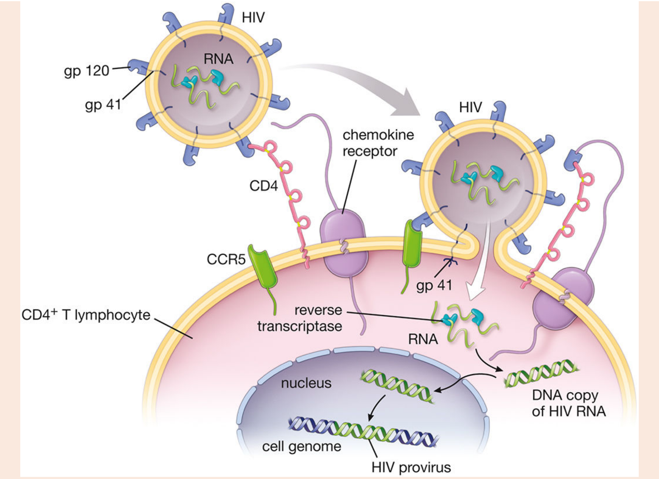
- HIV surface protein gp120 binds to the CD4 receptor on helper T lymphocytes
- This binding exposes gp120 to a second binding site - the co-receptor:
- CCR5 (C-C chemokine receptor 5) — used by macrophage-tropic strains (M-tropic)
- CXCR4 (C-X-C chemokine receptor 4) — used by T-cell-tropic strains
- Note: Individuals with a CCR5-Δ32 mutation (homozygous) are naturally resistant to HIV infection
- After gp120-CD4-CCR5 complex forms, gp41 is exposed
- gp41 anchors into the T-cell membrane and causes fusion of the viral envelope with the host cell membrane
- The viral contents (RNA + enzymes) are injected into the cytoplasm
- The enzyme reverse transcriptase converts:
- Viral single-stranded RNA → single-stranded DNA (RNA:DNA hybrid)
- Then degrades the RNA (RNase H activity of reverse transcriptase)
- Then synthesises the complementary strand → double-stranded DNA (dsDNA)
- This is the reverse of the central dogma (normally DNA → RNA)
- This is why HIV is called a retrovirus (retro = backward)
- The dsDNA is transported into the T-cell nucleus
- The enzyme integrase inserts the viral dsDNA into the host chromosome
- At this point it is called a PROVIRUS — it remains latent, hidden from immune surveillance
- This is why HIV cannot be cured — provirus persists for life
- When the T-cell is activated, RNA polymerase transcribes the provirus
- Viral RNA is produced and translated into viral proteins (structural + enzymatic)
- The enzyme protease cleaves viral polyprotein precursors into functional proteins
- New HIV particles are assembled and bud off from the T-cell, acquiring host cell membrane as their envelope
- These new virions infect other CD4+ cells
- The infected T-cell is destroyed (by cytotoxic CD8+ T cells or by viral-induced lysis)
PART 3 — Cells Targeted by HIV
| Cell Type | Receptor | Consequence |
|---|---|---|
| CD4+ Helper T lymphocytes | CD4 + CCR5/CXCR4 | Most important — loss leads to immunodeficiency |
| Macrophages/Monocytes | CD4 + CCR5 | Act as viral reservoir; not killed (HIV replicates inside them) |
| Dendritic cells | CD4 + CCR5 | Transport HIV from mucosa to lymph nodes |
| Microglia (brain) | CD4 | Causes HIV encephalopathy/dementia |
PART 4 — Natural History and Stages of HIV Infection
Stage 1 — Acute HIV Infection (Seroconversion illness, 2-4 weeks after exposure)
- Massive viral replication → high viral load → CD4 count drops temporarily
- Clinical: Fever, sore throat, lymphadenopathy, rash, myalgia — resembles infectious mononucleosis (glandular fever)
- Biochemical: Virus detectable by PCR; p24 antigen positive; antibodies NOT yet positive (window period)
- Immune response controls virus → CD4 count partially recovers
Stage 2 — Clinical Latency (Asymptomatic, can last 2-10 years)
- Virus replicates slowly; CD4 count gradually falls (~50 cells/µL per year)
- Patient feels well but is infectious
- CD4+ count: 500-200 cells/mm³
- Antibodies (anti-HIV) are now detectable — ELISA becomes positive
Stage 3 — Symptomatic HIV / AIDS (CD4 < 200 cells/mm³)
- AIDS is defined as: CD4+ count < 200 cells/mm³ (normal: 500-1500) OR presence of an AIDS-defining illness
- Overwhelming opportunistic infections and cancers develop
PART 5 — Biochemical Changes in AIDS
A. Immunological/Haematological Biochemical Changes
| Parameter | Change | Reason |
|---|---|---|
| CD4+ T cell count | ↓↓ (< 200/mm³ in AIDS) | HIV destroys helper T cells |
| CD4:CD8 ratio | ↓ (Normal: 2:1; AIDS: <1:1) | CD4 cells depleted, CD8 cells raised initially |
| Lymphocyte count | ↓ (Lymphopenia) | Loss of CD4+ cells |
| Neutrophil count | ↓ (Neutropenia) | Bone marrow suppression |
| Haemoglobin | ↓ (Anaemia) | Chronic disease, bone marrow infiltration, opportunistic infections |
| Platelet count | ↓ (Thrombocytopenia) | Immune-mediated destruction, bone marrow suppression |
B. Serum Protein Changes
| Parameter | Change | Reason |
|---|---|---|
| Serum albumin | ↓ (Hypoalbuminaemia) | Malnutrition, wasting syndrome, liver disease, protein-losing enteropathy |
| Serum globulins | ↑ (Hyperglobulinaemia) | Non-specific B-cell activation → polyclonal hypergammaglobulinaemia |
| IgA, IgG, IgM | ↑ (all raised) | Polyclonal B cell activation — yet antibodies are non-functional |
| Serum protein electrophoresis | Broad diffuse band in gamma region (polyclonal) | Non-specific immune stimulation |
| Acute phase proteins (CRP, fibrinogen) | ↑ | Chronic inflammation/infection |
| β2-microglobulin | ↑ | Released from dying CD4 cells; used as a marker of HIV progression |
C. Lipid and Metabolic Changes (HIV/AIDS Wasting Syndrome)
| Parameter | Change | Reason |
|---|---|---|
| Serum triglycerides | ↑ (Hypertriglyceridaemia) | ↑ hepatic VLDL synthesis, ↓ lipoprotein lipase activity due to high TNF-α and IL-6 |
| HDL cholesterol | ↓ | Reduced synthesis, chronic inflammation |
| LDL cholesterol | ↓ | Malnutrition, wasting |
| Serum cholesterol | ↓ (in untreated AIDS) | Malabsorption, wasting |
| Blood glucose | Variable | Opportunistic infections → stress hyperglycaemia; later: hypoglycaemia from malnutrition |
| BMI/Body weight | ↓ (Wasting) | Reduced food intake + malabsorption + cytokine-induced catabolism |
D. Liver Function Changes
| Parameter | Change | Reason |
|---|---|---|
| ALT, AST | ↑ | Opportunistic infections (CMV hepatitis, TB), co-infection with Hepatitis B or C, drug toxicity |
| Bilirubin | ↑ | Haemolysis, hepatitis, drug toxicity |
| ALP | ↑ | Hepatic infiltration by opportunistic organisms (MAC, CMV), biliary disease |
| GGT | ↑ | Liver involvement |
E. Kidney Function Changes
| Parameter | Change | Reason |
|---|---|---|
| Serum creatinine | ↑ | HIV-Associated Nephropathy (HIVAN) — direct HIV infection of podocytes |
| Proteinuria | Present | Focal segmental glomerulosclerosis (FSGS) in HIVAN |
| Electrolytes | Hyponatraemia, hypokalaemia | Vomiting, diarrhoea, adrenal insufficiency (due to opportunistic infections) |
F. Endocrine/Hormonal Changes
| Parameter | Change | Reason |
|---|---|---|
| Cortisol | ↑ initially, then ↓ | Stress response → adrenal exhaustion; CMV adrenalitis |
| Testosterone | ↓ (Hypogonadism in men) | Hypothalamic-pituitary dysfunction, malnutrition |
| TSH/T3/T4 | Euthyroid sick syndrome (↓ T3, normal TSH) | Chronic illness effect |
| Growth hormone | ↑ (but IGF-1 ↓) | GH resistance — contributes to wasting |
G. Summary — Lab Changes at a Glance
| Lab Test | AIDS Result | Significance |
|---|---|---|
| CD4+ count | < 200 cells/mm³ | AIDS-defining |
| CD4:CD8 ratio | < 1 | Inverted |
| Viral load (RNA PCR) | High (> 100,000 copies/mL) | Disease activity, treatment monitoring |
| β2-microglobulin | ↑ | HIV progression marker |
| Hb | ↓ | Anaemia |
| WBC | ↓ (lymphopenia) | Immunosuppression |
| Serum albumin | ↓ | Malnutrition, wasting |
| Serum globulins | ↑ (polyclonal) | Non-specific B cell activation |
| Triglycerides | ↑ | Metabolic disturbance |
| SGOT/SGPT | ↑ | Opportunistic hepatitis |
| Serum creatinine | ↑ (in HIVAN) | Nephropathy |
PART 6 — Diagnosis of HIV (Biochemical Methods)
Screening Test
- ELISA (Enzyme-Linked Immunosorbent Assay)
- Detects anti-HIV antibodies (IgG against gp41, gp120, p24)
- 4th generation ELISA also detects p24 antigen (earlier detection)
- Highly sensitive (~99.5%) but can have false positives
- Window period: 3-12 weeks (antibodies not yet formed — ELISA negative despite infection!)
Confirmatory Test
- Western Blot
- Detects antibodies against specific HIV proteins (p24, gp41, gp120)
- Positive if bands for ≥ 2 of: p24, gp41, gp120/160
- Highly specific (~99.9%)
Viral Load (for monitoring, not diagnosis)
- RT-PCR (Reverse Transcriptase PCR) — detects HIV RNA copies/mL
- Useful in: Window period detection, monitoring treatment response, perinatal testing
- Target in treatment: Undetectable viral load (< 50 copies/mL)
CD4 Count (staging and treatment decision)
- Normal: 500-1500 cells/mm³
- Start ART: when CD4 < 350 (or all patients now, per WHO 2021)
- Prophylaxis for opportunistic infections: CD4 < 200
PART 7 — Opportunistic Infections in AIDS (by CD4 count)
| CD4 Count | Opportunistic Infection/Cancer |
|---|---|
| < 500 | Oral candidiasis, herpes zoster, Kaposi's sarcoma begins |
| < 200 | Pneumocystis jirovecii pneumonia (PCP) — most common OI; Toxoplasmosis |
| < 100 | Cryptococcal meningitis, Cryptosporidiosis |
| < 50 | CMV retinitis, MAC (Mycobacterium avium complex), CNS lymphoma, HIV encephalopathy |
PART 8 — Treatment: Antiretroviral Therapy (ART/HAART)
| Drug Class | Mechanism | Example |
|---|---|---|
| NRTIs (Nucleoside Reverse Transcriptase Inhibitors) | Competitive inhibitors of reverse transcriptase (mimic nucleosides, terminate DNA chain) | Zidovudine (AZT/ZDV), Lamivudine (3TC), Tenofovir |
| NNRTIs (Non-Nucleoside RTIs) | Non-competitive inhibitors of reverse transcriptase (bind allosteric site) | Efavirenz, Nevirapine |
| Protease Inhibitors (PIs) | Block HIV protease → immature, non-infectious virions | Lopinavir, Ritonavir |
| Integrase Inhibitors (INSTIs) | Block integrase → prevent provirus integration | Dolutegravir, Raltegravir |
| Fusion/Entry Inhibitors | Block gp41 fusion or CCR5 binding | Enfuvirtide, Maraviroc |
EXAM QUICK-RECALL BOX
| Key Fact | Value/Name |
|---|---|
| HIV type | RNA retrovirus |
| Key enzyme | Reverse transcriptase |
| Primary target cell | CD4+ Helper T lymphocyte |
| Entry co-receptors | CCR5 (macrophages), CXCR4 (T-cells) |
| AIDS-defining CD4 count | < 200 cells/mm³ |
| Most common OI | PCP (Pneumocystis jirovecii pneumonia) |
| Key screening test | ELISA (antibody detection) |
| Confirmatory test | Western Blot |
| Viral monitoring | RT-PCR (viral load) |
| Most important biochemical change | ↓ CD4+ count, ↑ viral load |
| Wasting biochemistry | ↓ Albumin, ↑ Triglycerides, ↑ Globulins |
| First antiretroviral drug | AZT (Zidovudine) — reverse transcriptase inhibitor |
| Window period | 3-12 weeks |
Sources: Histology: A Text and Atlas, Ross & Pawlina, p. 1204-1205; Cellular and Molecular Immunology, Abbas & Lichtman; Medical Microbiology, Murray
- "Describe the structure of HIV and its mechanism of replication" (Long Essay)
- "Describe the biochemical changes in AIDS" (Long Essay)
- "Why does AIDS lead to immunodeficiency?" (Explain Why)
- "Why is reverse transcriptase important in HIV infection and treatment?" (Explain Why)
- Short notes: Reverse transcriptase / Provirus / HAART / CD4 count / Western Blot / β2-microglobulin
Find the topics and questions given from the HIV and biochemical changes in AIDS topic in 1st professional examination of mbbs 1st year student in wbhus university
WBUHS MBBS 1st professional biochemistry previous year question paper HIV AIDS
West Bengal University Health Sciences 1st prof MBBS biochemistry question paper PDF HIV immunology
https://medical-junction.com/biochemistry-previous-year-ques…