
Heart
Reading File
The query "Heart" is broad. Let me clarify what you're looking for before diving in.
Asking for Preferences
What aspect of the heart are you interested in?
Asking for Preferences
Which disease area would you like to focus on?
Congenital heart disease (CHD) is a large topic. Here's a quick orientation while I pull from the medical library:
Congenital heart diseases are structural cardiac defects present at birth. They are broadly divided into:
- Acyanotic (left-to-right shunts): ASD, VSD, PDA, coarctation of aorta
- Cyanotic (right-to-left shunts): Tetralogy of Fallot, Transposition of Great Arteries, Truncus Arteriosus, Tricuspid Atresia
The most common CHD overall is VSD. Cyanotic CHDs present with central cyanosis from birth or early infancy.
Searching sources...
Finding Sources
Finding Sources
Good. Let me read from Braunwald's Heart Disease (the gold standard cardiology reference) and Tintinalli's for clinical presentation detail, in parallel.
Reading File
Reading File
Finding Sources
Excellent content found. Now let me pull the Robbins pathology detail on specific defects and also the Tintinalli cyanotic section (Tetralogy of Fallot, etc.), plus fetch a useful diagram.
Reading File
Reading File
I now have rich detail. Let me also fetch the key diagram showing ASD/VSD/PDA from Robbins.
Loading Image
Reading File
I now have everything needed for a thorough, well-illustrated answer. Here is the full summary:
Congenital Heart Disease (CHD)
Overview
Congenital heart defects occur in approximately 8 in 1000 live births and range from hemodynamically trivial to immediately life-threatening. About 10% are associated with genetic syndromes (trisomy 21, Turner's, Noonan's), VACTERL association, or other organ malformations. The remaining 90% result from isolated embryologic abnormalities or undetermined genetic lesions. - Tintinalli's Emergency Medicine, p. 862
The classic classification divides CHD into two broad categories based on physiology:
ACYANOTIC (Left-to-Right Shunts)
These are the most common congenital cardiac malformations. Left-to-right shunting initially causes pulmonary overcirculation without cyanosis. However, prolonged shunting eventually causes pulmonary hypertension and reversal to right-to-left shunting (cyanosis) - this is Eisenmenger syndrome, at which point defects are considered irreversible. This is why early intervention is recommended.
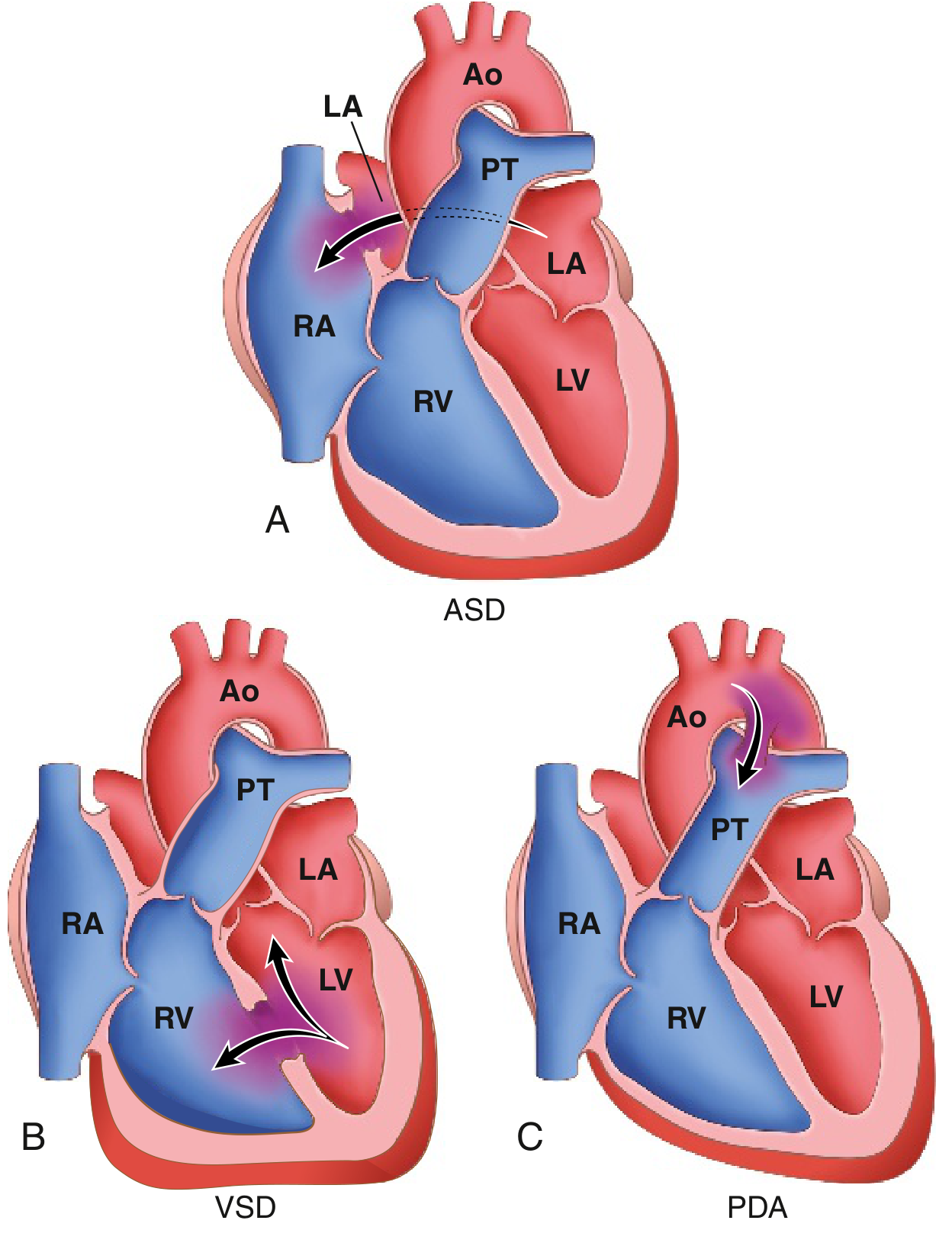
1. Atrial Septal Defect (ASD)
- Abnormal fixed opening in the atrial septum allowing unrestricted blood flow between atria
- Ostium secundum type accounts for ~90% of ASDs - smooth-walled defect near the foramen ovale
- ASDs increase right ventricular and pulmonary outflow volumes
- Contrast with Patent Foramen Ovale (PFO): in 20% of normal individuals the fetal foramen ovale fails to fuse, allowing transient right-to-left flow during straining - can cause paradoxical embolism
- Robbins & Kumar Basic Pathology, p. 348-349
2. Ventricular Septal Defect (VSD)
- Most common congenital heart defect overall
- Causes both increased pulmonary blood flow AND pressure (higher-pressure left-to-right shunt)
- Small VSDs may close spontaneously; large VSDs require surgical or catheter-based closure
- Carries risk of Eisenmenger syndrome if uncorrected
3. Patent Ductus Arteriosus (PDA)
- Failure of the ductus arteriosus (connecting aorta to pulmonary artery in fetal circulation) to close after birth
- Causes aorta-to-pulmonary artery shunting
- Recognized by a classic continuous "machinery" murmur
- Managed with indomethacin (NSAIDs promote closure) or surgical ligation
Acyanotic Obstructive Lesions (no shunt)
- Pulmonary stenosis, aortic stenosis/atresia, and coarctation of the aorta - obstruct blood flow without initial shunting
- Coarctation of aorta: narrowing of the aorta, males affected 2x more often (though females with Turner syndrome frequently have it). Two forms:
- Preductal ("infantile") - hypoplasia of aortic arch proximal to a PDA
- Postductal ("adult") - discrete ridge-like infolding adjacent to the ligamentum arteriosum
- Robbins & Kumar Basic Pathology, p. 350
CYANOTIC (Right-to-Left Shunts) - "The Five Ts"
Cyanotic CHDs cause mixing of deoxygenated and oxygenated blood or direct right-to-left shunting. Clinically apparent cyanosis requires 3-5 mg/dL of deoxygenated hemoglobin (O2 sat ~70-80%). - Tintinalli's Emergency Medicine, p. 862
The five Ts are:
| Disease | Key Feature |
|---|---|
| Tetralogy of Fallot (ToF) | Most common cyanotic CHD after infancy; ~10% of all CHD |
| Transposition of Great Arteries (TGA) | 5-8% of CHD; most common presenting in the newborn period |
| Truncus Arteriosus | Single arterial trunk from both ventricles |
| Tricuspid anomalies | Tricuspid atresia, Ebstein's anomaly |
| Total Anomalous Pulmonary Venous Return (TAPVR) | All pulmonary veins drain into right heart |
Tetralogy of Fallot (ToF)
The four components:
- Large VSD
- Right ventricular outflow obstruction (pulmonic stenosis, valvular or supravalvular)
- Overriding aorta (aorta sits over the VSD)
- Right ventricular hypertrophy
The degree of cyanosis depends on severity of right ventricular outflow obstruction. Severe pulmonic stenosis creates a right-to-left shunt with decreased pulmonary blood flow. The feared complication is "tet spells" - hypercyanotic episodes triggered by crying, feeding, or defecation. Auscultation: loud systolic ejection murmur at lower-left sternal border, loud single S2, aortic ejection click. - Tintinalli's Emergency Medicine, p. 863
Transposition of Great Arteries (TGA)
- Aorta arises from the right ventricle; pulmonary artery from the left ventricle (discordant connection)
- Creates two separate, parallel circulations - incompatible with postnatal life unless a shunt (VSD, PFO, or PDA) allows oxygenated blood to reach the aorta
- VSD present in 1/3 of cases
- Dominant manifestation is cyanosis from birth
- Requires emergent surgical repair within the first few days of life (arterial switch operation)
- Robbins & Kumar Basic Pathology, p. 349
Key Comparison: Acyanotic vs. Cyanotic
| Feature | Acyanotic (L→R shunt) | Cyanotic (R→L shunt) |
|---|---|---|
| Initial cyanosis | Absent | Present from birth |
| Pulmonary blood flow | Increased | Decreased (usually) |
| Risk | Eisenmenger syndrome later | Hypoxia, polycythemia |
| Examples | ASD, VSD, PDA, coarctation | ToF, TGA, truncus arteriosus |
Genetic Associations Worth Noting
- Trisomy 21 (Down syndrome) - ASD, VSD, atrioventricular canal defect
- Turner syndrome (45, X) - Coarctation of aorta, bicuspid aortic valve
- Noonan syndrome - Pulmonary stenosis, hypertrophic cardiomyopathy
- DiGeorge syndrome (22q11 deletion) - Truncus arteriosus, interrupted aortic arch, ToF
- Maternal rubella - PDA, pulmonary artery stenosis, ASD
Sources: Robbins & Kumar Basic Pathology; Tintinalli's Emergency Medicine; Braunwald's Heart Disease
This is a shared conversation. Sign in to Orris to start your own chat.