Infective Endocarditis

Reading File
Finding Sources
Searching Images

infective endocarditis vegetation heart valve
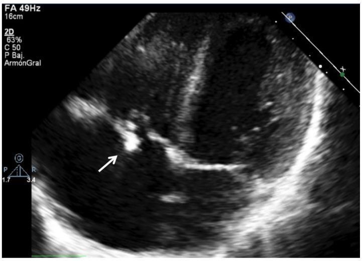
This diagnostic image is a 2D transthoracic echocardiogram (TTE) in a pediatric patient, focusing on the right heart chambers. The image demonstrates a prominent, hyperechoic (bright) irregular mass attached to the tricuspid valve leaflet, as indicated by the white arrow. This echogenic structure is characteristic of a vegetation, a hallmark of infective endocarditis. The valve leaflets themselves appear thickened and irregular, which disrupts normal coaptation. The clinical context involves a 16-year-old male with a history of congenital heart disease (restrictive perimembranous VSD) and tricuspid regurgitation, now presenting with Viridans Streptococcus endocarditis. The image illustrates the typical ultrasonographic presentation of right-sided infective endocarditis, showing how the vegetation appears as a distinct, reflective cluster against the darker, less echogenic blood-filled chambers and myocardium. This finding is a major Duke criterion for the diagnosis of infective endocarditis.
This diagnostic image is a 2D transthoracic echocardiogram (TTE) in a pediatric patient, focusing on the right heart chambers. The image demonstrates a prominent, hyperechoic (bright) irregular mass attached to the tricuspid valve leaflet, as indicated by the white arrow. This echogenic structure is characteristic of a vegetation, a hallmark of infective endocarditis. The valve leaflets themselves appear thickened and irregular, which disrupts normal coaptation. The clinical context involves a 16-year-old male with a history of congenital heart disease (restrictive perimembranous VSD) and tricuspid regurgitation, now presenting with Viridans Streptococcus endocarditis. The image illustrates the typical ultrasonographic presentation of right-sided infective endocarditis, showing how the vegetation appears as a distinct, reflective cluster against the darker, less echogenic blood-filled chambers and myocardium. This finding is a major Duke criterion for the diagnosis of infective endocarditis.

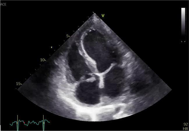
Transthoracic echocardiogram (TTE) in an apical four-chamber view demonstrating infective endocarditis of the tricuspid valve. The diagnostic image shows a prominent, irregularly shaped, and highly echogenic mass (vegetation) attached to the tricuspid valve leaflets. The vegetation exhibits a heterogeneous texture and appears to be broad-based with a possible stalk-like extension protruding into the right atrium. Notable features include thickening of the valve leaflets and evidence of valvular distortion or prolapse, characteristic of active endocarditis. The surrounding right heart chambers are visible, providing anatomical context for the right-sided valvular pathology. This image serves as a clinical example of the ultrasonic appearance of vegetations, crucial for the diagnosis of right-sided infective endocarditis, often associated with intravenous drug use or indwelling cardiac devices.
Transthoracic echocardiogram (TTE) in an apical four-chamber view demonstrating infective endocarditis of the tricuspid valve. The diagnostic image shows a prominent, irregularly shaped, and highly echogenic mass (vegetation) attached to the tricuspid valve leaflets. The vegetation exhibits a heterogeneous texture and appears to be broad-based with a possible stalk-like extension protruding into the right atrium. Notable features include thickening of the valve leaflets and evidence of valvular distortion or prolapse, characteristic of active endocarditis. The surrounding right heart chambers are visible, providing anatomical context for the right-sided valvular pathology. This image serves as a clinical example of the ultrasonic appearance of vegetations, crucial for the diagnosis of right-sided infective endocarditis, often associated with intravenous drug use or indwelling cardiac devices.

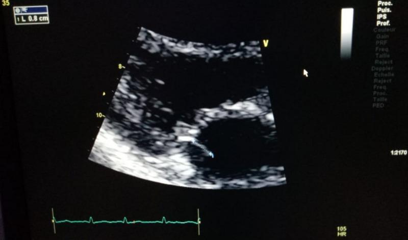
This diagnostic image is a transthoracic echocardiogram (TTE) in a parasternal long-axis view, demonstrating a case of native mitral valve infective endocarditis. The image shows the heart's internal structures with varying echogenicity: the myocardium appears relatively hypoechoic (darker gray), while the mitral valve leaflets are more echogenic (brighter). A distinct, highly echogenic, linear structure representing a vegetation is visible attached to the atrial surface of a mitral valve leaflet. On-screen calipers measure this vegetation at approximately 0.8 cm in length. The vegetation exhibits an irregular morphology and is located centrally in the valvular apparatus between the left atrium and left ventricle. An electrocardiogram (ECG) tracing is visible at the bottom of the frame for temporal orientation during the cardiac cycle. This visual evidence serves as a primary diagnostic criterion for infective endocarditis, illustrating the characteristic appearance of mobile vegetations on cardiac valves via ultrasound imaging.
This diagnostic image is a transthoracic echocardiogram (TTE) in a parasternal long-axis view, demonstrating a case of native mitral valve infective endocarditis. The image shows the heart's internal structures with varying echogenicity: the myocardium appears relatively hypoechoic (darker gray), while the mitral valve leaflets are more echogenic (brighter). A distinct, highly echogenic, linear structure representing a vegetation is visible attached to the atrial surface of a mitral valve leaflet. On-screen calipers measure this vegetation at approximately 0.8 cm in length. The vegetation exhibits an irregular morphology and is located centrally in the valvular apparatus between the left atrium and left ventricle. An electrocardiogram (ECG) tracing is visible at the bottom of the frame for temporal orientation during the cardiac cycle. This visual evidence serves as a primary diagnostic criterion for infective endocarditis, illustrating the characteristic appearance of mobile vegetations on cardiac valves via ultrasound imaging.

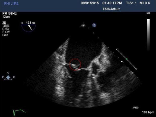
This diagnostic image is a 2D Transesophageal Echocardiogram (TEE) in an adult patient, focused on the mitral valve apparatus. The image shows a cross-sectional view of the heart chambers where the mitral valve leaflets are visible. Circled in red is a distinct, hyperechoic, and irregular mobile mass attached to the atrial side of a mitral valve leaflet, highly characteristic of infective endocarditis vegetation. The surrounding cardiac structures show typical ultrasound echogenicity, though there is some posterior acoustic shadowing that limits visualization of the distal myocardial segments. The primary clinical significance is the demonstration of valvular vegetation in the setting of suspected systemic infection, providing a key diagnostic finding for infective endocarditis. The image is optimized for cardiology and infectious disease education, illustrating the morphological appearance of pathological valvular attachments.

This diagnostic image is a 2D Transesophageal Echocardiogram (TEE) in an adult patient, focused on the mitral valve apparatus. The image shows a cross-sectional view of the heart chambers where the mitral valve leaflets are visible. Circled in red is a distinct, hyperechoic, and irregular mobile mass attached to the atrial side of a mitral valve leaflet, highly characteristic of infective endocarditis vegetation. The surrounding cardiac structures show typical ultrasound echogenicity, though there is some posterior acoustic shadowing that limits visualization of the distal myocardial segments. The primary clinical significance is the demonstration of valvular vegetation in the setting of suspected systemic infection, providing a key diagnostic finding for infective endocarditis. The image is optimized for cardiology and infectious disease education, illustrating the morphological appearance of pathological valvular attachments.

Searching PubMed

"infective endocarditis"[MeSH Terms] AND "management"

Reading File
Reading File
Reading File
Reading File
Reading File
Reading File
Reading File
Searching Images

Janeway lesions Osler nodes Roth spots infective endocarditis peripheral stigmata

Finding Sources
Finding Sources
Reading File
Reading File
Reading File
Finding Sources
Your credits are exhausted. You can purchase a top-up or subscribe to a plan to continue using Orris. If you're already subscribed, your daily credits will reset tomorrow.
This is a shared conversation. Sign in to Orris to start your own chat.