
Acute pte evaluation
Reading File
Acute pulmonary thromboembolism (PTE) evaluation involves risk stratification, clinical scoring, imaging, and lab workup. Let me pull from the textbook library and current literature simultaneously.
Searching sources...
Finding Sources
Searching PubMed
"pulmonary embolism"[MeSH Terms] AND "diagnosis"[MeSH Terms]
Good sources identified. Now reading the most relevant blocks in parallel - Rosen's Emergency Medicine (ideal for acute evaluation), Murray & Nadel's (comprehensive respiratory), and a paragraph search for Wells/PESI scoring.
Reading File
Finding Sources
Finding Sources
Good content. Now reading Rosen's full PE section and also getting the Tintinalli diagnostic algorithm with the Wells score flowchart image.
Reading File
Reading File
Excellent - very detailed content from Rosen's and Tintinalli's. Let me now get the Wells score criteria table, PERC rule, and PESI/risk stratification data, plus the diagnostic flowchart image.
Reading File
Reading File
Loading Image
Excellent - I now have the diagnostic algorithm flowchart, Wells score table, PERC details, and comprehensive clinical content. Let me get the PERC table, risk stratification/PESI, and a couple more key sections.
Reading File
Finding Sources
Reading File
Reading File
I now have comprehensive, authoritative content for a thorough response. Let me also get the ECG image of acute cor pulmonale and the CTPA image.
Loading Image
Loading Image
I now have everything needed for a comprehensive, well-illustrated answer. Here is the full response:
Acute Pulmonary Thromboembolism (PTE) - Evaluation
Overview
PE can present anywhere on a spectrum from asymptomatic to sudden cardiovascular collapse. Evaluation follows a structured pathway: recognize suspicion → estimate pretest probability (PTP) → selective testing → imaging when indicated → risk stratify the confirmed diagnosis.
1. Clinical Presentation
The clinical presentation is highly variable. Any ED visit involving dyspnea, chest pain, palpitations, syncope, or unexplained malaise could represent PE - but this does not mean every such patient requires full workup.
| Symptom | Frequency in PE |
|---|---|
| Dyspnea | 75-80% (most common) |
| Chest pain (pleuritic or vague) | ~67% |
| Cough / hemoptysis | Less common; suggests pulmonary infarction |
| Unilateral leg swelling | <30%, but relatively specific when present |
| Syncope | <5% of syncope patients have PE, but it can be the sole presentation |
| Fever | Usually low-grade; >38.6°C suggests infection instead |
Key point: the most common symptom (dyspnea) is absent in 20-25% of patients with PE. Do not rule out PE simply because a classic symptom is missing. - Rosen's Emergency Medicine, 9e
2. Step 1 - Determine Pretest Probability (PTP)
This is the single most important first step. It determines whether and what testing is needed.
Wells Score for PE
| Clinical Feature | Points |
|---|---|
| Previous PE or DVT | 1.5 |
| Heart rate >100 bpm | 1.5 |
| Recent surgery or immobilization (within 4 weeks) | 1.5 |
| Clinical signs of DVT (calf swelling/tenderness) | 3 |
| Hemoptysis | 1 |
| Active cancer (treated within 6 months or palliative) | 1 |
| Alternative diagnosis less likely than PE | 3 |
Interpretation:
- Score 0-1: Low probability (~3-5%)
- Score 2-6: Moderate probability (~20-30%)
- Score >6: High probability (~60-80%)
- Binary: ≤4 = PE unlikely; >4 = PE likely
Note: The "alternative diagnosis" criterion gives this tool a subjective element that empowers clinical judgment - this is a feature, not a bug. - Rosen's Emergency Medicine, 9e
Revised Geneva Score (fully objective - no subjective items)
Includes: age >65, prior DVT/PE, surgery/fracture <1 month, active cancer, unilateral lower limb pain, hemoptysis, HR 75-94 or ≥95, pain on deep palpation of lower limb + unilateral edema.
Gestalt
Unstructured clinical gestalt (low <15%, moderate 15-40%, high >40%) is also validated and may actually be the most accurate method among experienced clinicians.
3. Step 2 - Diagnostic Algorithm
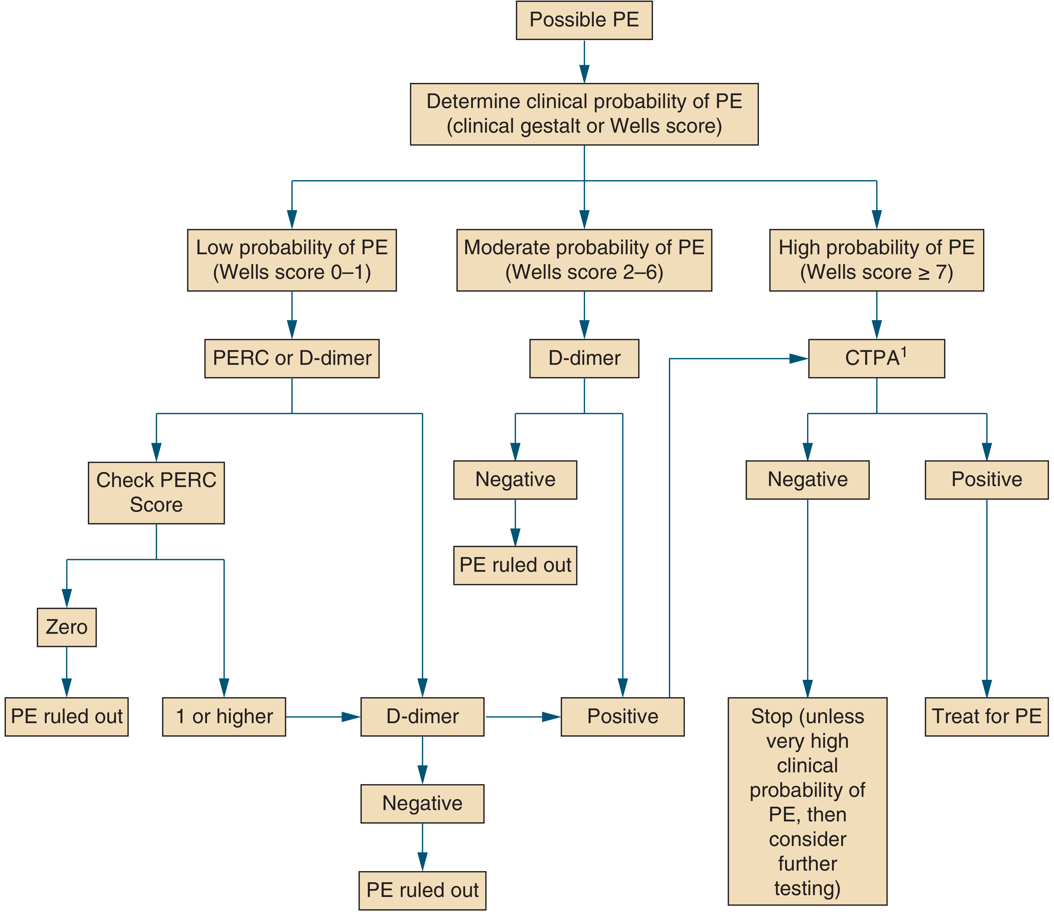
Fig. Proposed algorithm for evaluation of suspected PE - Symptom to Diagnosis, 4e
PERC Rule (PE Rule-Out Criteria)
For patients with low gestalt PTP, apply PERC. If all 8 criteria are met (score = 0), PE can be excluded without further testing:
- Age < 50
- Pulse < 100
- SpO2 > 94%
- No unilateral leg swelling
- No hemoptysis
- No recent trauma or surgery
- No prior PE/DVT
- No hormone use (OCP/HRT)
ACEP, ACP, and ASH guidelines all endorse the PERC rule. - Tintinalli's Emergency Medicine, 9e
4. Step 3 - D-Dimer Testing
D-dimer is a sensitive but non-specific screening test. It works by detecting fibrin degradation products from clot lysis.
- Sensitivity: 95-98% for PE
- Specificity: 40-55%
- NPV: 99-100% when PTP is low-to-moderate
Use D-dimer when PTP is non-high (Wells ≤6 or gestalt <40%). A negative result excludes PE without imaging.
Age-Adjusted D-Dimer Threshold
Standard cutoff: 500 ng/mL. In patients ≥50 years, use:
Age × 10 ng/mL as the threshold for positivity
This increases the proportion of patients who avoid CTPA by 10-20% in those >70 years, while maintaining >95% sensitivity. - Rosen's Emergency Medicine, 9e
YEARS Algorithm
Uses 3 questions (DVT signs, hemoptysis, PE most likely diagnosis):
- 0 YEARS criteria: D-dimer threshold raised to 1000 ng/mL
- ≥1 YEARS criteria: use standard 500 ng/mL threshold
- Validated in multicenter study; reduced CTPA by 14%. Also adapted for pregnancy.
Causes of False-Positive D-Dimer
Age >70, pregnancy, active malignancy, surgery within prior week, liver disease, rheumatoid arthritis, infections, trauma.
Causes of False-Negative D-Dimer
Symptoms >5 days, small clots, isolated calf thrombosis, lipemia, subacute/chronic PE.
5. Step 4 - Pulmonary Vascular Imaging
Order imaging when PTP is high or D-dimer is positive.
CT Pulmonary Angiography (CTPA) - First-Line
- Sensitivity/Specificity: 90-95% on multidetector CT
- PE appears as hypodense filling defects in contrast-opacified pulmonary arteries
- "Saddle PE" = clot straddling the bifurcation of main pulmonary arteries
- PE described by most proximal location (main, lobar, segmental, subsegmental)
- "Massive PE" is a physiologic/hemodynamic diagnosis, not a CT-size diagnosis; preferred term is "high-risk PE"
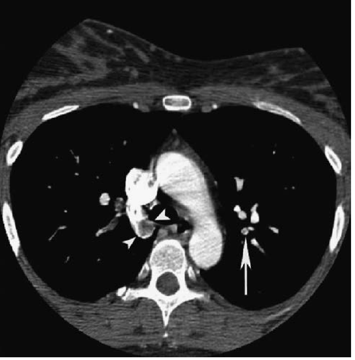
Axial CTPA showing circular filling defect in right middle lobar pulmonary artery (arrowheads) and segmental artery filling defect (arrow) - Tintinalli's Emergency Medicine, 9e
V/Q Scan
- Alternative to CTPA (preferred in: pregnancy, contrast allergy, renal failure)
- Normal perfusion scan: ~100% sensitive for excluding PE
- High-probability result (≥2 wedge-shaped defects with normal ventilation): >80% probability of PE
- All other results are non-diagnostic
- V/Q SPECT has pooled sensitivity 96%, specificity 97% - superior to planar V/Q
Bedside Ultrasound
- DVT found on lower limb US = equivalent to a PE diagnosis → initiate treatment
- Emergency physician-performed: 86-96% sensitive, 93-97% specific vs. radiology US
- Negative US does NOT exclude PE (fewer than 50% of PE patients have detectable DVT)
- Point-of-care echo: RV:LV ratio >1:1 in diastole raises suspicion for PE (and worsens prognosis)
MRI
- Zero radiation - considered for pregnant patients
- Pooled sensitivity ~75%, specificity ~80% → not first-line
6. ECG Findings in Acute PE
ECG is non-specific but can provide supportive evidence, especially for high-risk PE:
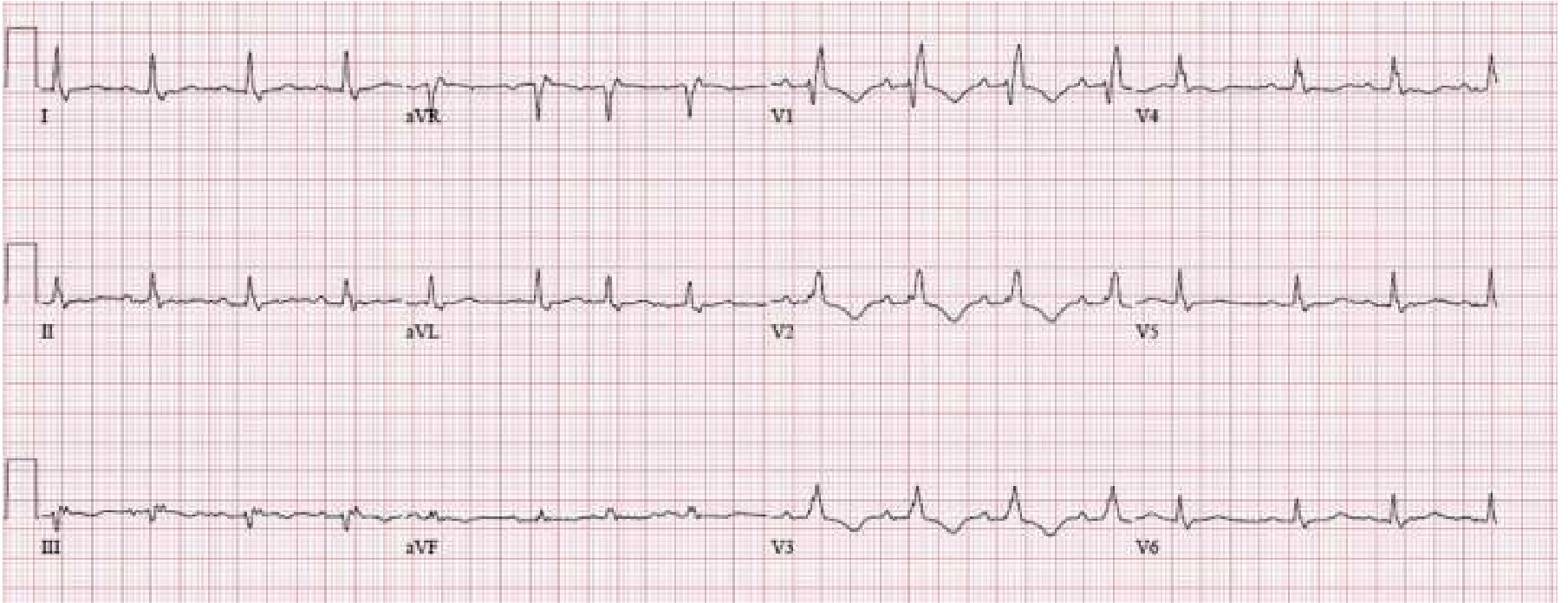
ECG showing S1Q3T3 pattern, new complete RBBB, and deep T-wave inversions V1-V3 - signs of acute RV strain/cor pulmonale. These findings indicate right ventricular dysfunction and carry poor prognosis. - Rosen's Emergency Medicine, 9e
Common ECG findings in PE (none pathognomonic):
- Sinus tachycardia (most common)
- S1Q3T3: S wave in lead I, Q wave and T-wave inversion in lead III
- New RBBB
- T-wave inversions V1-V4
- Right axis deviation
7. Risk Stratification of Confirmed PE
Once PE is confirmed, risk stratify to guide management:
AHA/ESC Classification
| Category | Definition | 30-day Mortality |
|---|---|---|
| High-Risk (Massive) | Hemodynamically unstable (SBP <90, or drop ≥40 mmHg for >15 min, or need for vasopressors) | 15-50% |
| Intermediate-Risk (Submassive) | Hemodynamically stable + RV dysfunction (echo/CTPA) and/or elevated troponin/BNP | 3-15% |
| Low-Risk | Hemodynamically stable, no RV dysfunction, normal biomarkers | <1-3% |
PESI and Simplified PESI (sPESI)
The PESI is the most extensively validated tool for predicting 30-day mortality:
| Predictor | Original PESI | sPESI |
|---|---|---|
| Age | Age in years | +1 if >80 |
| Male sex | +10 | - |
| Cancer | +30 | +1 |
| Heart failure | +10 | +1 (combined) |
| Chronic lung disease | +10 | (combined) |
| HR ≥110/min | +20 | +1 |
| SBP <100 mmHg | +30 | +1 |
| RR ≥30/min | +20 | - |
| Temp <36°C | +20 | - |
| Altered mental status | +60 | - |
| SpO2 <90% | +20 | +1 |
PESI Classes:
- Class I (≤65 pts): 0-1.6% mortality (very low)
- Class II (66-85): 1.7-3.5% (low)
- Class III (86-105): 3.2-7.1% (moderate)
- Class IV (106-125): 4-11.4% (high)
- Class V (>125): 10-24.5% (very high)
sPESI = 0: Low risk. sPESI ≥1: High risk. - Sabiston Textbook of Surgery, 7e
Biomarkers for Risk Stratification
- Troponin I/T: RV myocardial injury marker; elevated = worse prognosis even in hemodynamically stable patients
- BNP/NT-proBNP: RV pressure overload marker; elevated = unfavorable prognosis
- Echo findings: RV:LV ratio >1.0, TAPSE <15 mm = poor prognosis
8. Risk Stratification → Treatment Matrix
| Risk | Criteria | Action |
|---|---|---|
| Low | Hemodynamically stable, no RV dysfunction | DOAC (preferred), consider discharge (Hestia criteria) |
| Intermediate-low | Stable + PESI high + troponin negative | Anticoagulate, admit, monitor |
| Intermediate-high | Stable + RV dysfunction + troponin positive | Anticoagulate (heparin), activate PERT, monitored bed/ICU, consider thrombolysis |
| High | Hemodynamically unstable | UFH, PERT activation, systemic thrombolysis or thromboembolectomy, ICU |
PERT (Pulmonary Embolism Response Team): Multidisciplinary team activated for intermediate-high and high-risk PE; includes emergency medicine, pulmonology, cardiology, and cardiac surgery. - Rosen's Emergency Medicine, 9e
Quick Reference Summary
Possible PE?
    ↓
Assess PTP (Wells / Gestalt)
    ↓
Low PTP → PERC: all 0? → Stop (no PE)
               ≥1? → D-dimer
    ↓
Moderate PTP → D-dimer
               Negative → PE excluded
               Positive → CTPA
    ↓
High PTP → CTPA directly
    ↓
CTPA positive → Risk stratify (hemodynamics + RV + troponin + PESI)
    → Treat accordingly
Sources: Rosen's Emergency Medicine 9e | Tintinalli's Emergency Medicine 9e | Sabiston Textbook of Surgery 7e | Symptom to Diagnosis 4e | Murray & Nadel's Respiratory Medicine
This is a shared conversation. Sign in to Orris to start your own chat.