
Does post spinal headache occur day 3 post operatively
post dural puncture headache onset timing day 3 post spinal anesthesia
Post-Dural Puncture Headache (PDPH) on Day 3 Post-Operatively
Onset Timing - What the Evidence Says
| Timeframe | % of PDPH Cases |
|---|---|
| Within 48 hours | ~66% |
| Within 3 days | >90% |
| Up to 5-7 days | Remaining cases |
| Beyond 7 days | Very rare (atypical, warrants investigation) |
Classic Features to Confirm the Diagnosis
- Postural (orthostatic) headache - worsens when sitting/standing, relieved by lying flat
- Frontal or occipital location
- Associated symptoms: nausea, vomiting, neck stiffness, tinnitus, photophobia, diplopia
Key Clinical Points
- Day 3 presentation is typical and expected - do not dismiss it
- If headache onset is >5 days after the procedure, consider alternative diagnoses (cerebral venous thrombosis, meningitis, subdural haematoma) and consider neuroimaging per the 2023 international consensus guidelines on PDPH
- PDPH carries a significantly elevated risk of cerebral venous thrombosis and subdural haematoma (adjusted OR 19.0) and bacterial meningitis (adjusted OR 39.7) - so proper recognition and management is important
- Spontaneous resolution occurs in ~72% by 7 days and 87% by 6 months
- Epidural blood patch remains the definitive treatment if conservative measures fail
Summary
If patient has history of migraine what medicine can beer given she has headache and is npo
PDPH treatment NPO patient IV medications migraine history post spinal headache management
Medications for PDPH in an NPO Patient with Migraine History
First-Line: IV Analgesics & Anti-emetics
| Drug | Route | Dose | Notes |
|---|---|---|---|
| Paracetamol (Acetaminophen) | IV | 1g IV q6h | Safe, first-line analgesic |
| Ketorolac | IV/IM | 15-30mg IV/IM q6h | NSAID; avoid if bleeding risk post-op |
| Metoclopramide | IV | 10mg IV q8h | Anti-emetic + mild analgesic for migraine; often used as adjunct in migraine protocols |
| Ondansetron | IV | 4-8mg IV q8h | For nausea/vomiting associated with PDPH or migraine |
Migraine-Specific IV Option
| Drug | Route | Dose | Notes |
|---|---|---|---|
| Metoclopramide | IV | 10-20mg IV | Dopamine antagonist - actually one of the most effective IV treatments for acute migraine. Can help PDPH-related and migraine-type headache simultaneously |
| Prochlorperazine | IV/IM | 12.5mg IM or 10mg IV | Phenothiazine anti-emetic; excellent for acute migraine, off-label for PDPH |
| Sumatriptan | SC | 6mg SC | Only if the headache is purely migrainous. Use with caution in PDPH - triptans work by vasoconstriction but evidence for PDPH is poor/insufficient (Cochrane review). Suitable if you believe this is migraine predominating |
| Valproate sodium | IV | 300-400mg IV over 30 min | Used for refractory acute migraine; note: avoid in pregnancy/women of childbearing age |
PDPH-Specific: IV Caffeine
| Drug | Route | Dose | Notes |
|---|---|---|---|
| Caffeine sodium benzoate | IV | 500mg IV over 1 hour (can repeat once) | The most studied pharmacological treatment specifically for PDPH. Cerebral vasoconstriction reduces headache. 58% of North American anaesthesiologists use it. Can be given IV when NPO |
IV Corticosteroids (Adjunct)
| Drug | Route | Dose | Notes |
|---|---|---|---|
| Hydrocortisone | IV | 200mg IV loading, then 100mg q8h | Evidence from one RCT showing reduced PDPH severity. Avoid if infection/LP was done for infectious reasons |
| Dexamethasone | IV | 8mg IV | Sometimes used as adjunct; insufficient evidence for PDPH specifically, but useful for migraine |
Opioids (Reserve / PRN)
| Drug | Route | Notes |
|---|---|---|
| Morphine / Fentanyl | IV | For breakthrough severe pain; avoid as first-line due to nausea/sedation |
What to AVOID
- Sumatriptan oral / Ergotamine - not possible (NPO, and ergotamine avoided post-op)
- Triptans as primary PDPH treatment - insufficient evidence; do not replace EBP with them
- Aspirin/oral NSAIDs - NPO restriction
Definitive Treatment: Epidural Blood Patch (EBP)
Practical Suggested Order (NPO + Migraine + PDPH)
- IV Paracetamol 1g - immediate, safe
- IV Metoclopramide 10mg - works for both PDPH-related and migraine-type headache, also controls nausea
- IV Caffeine sodium benzoate 500mg over 1 hour - PDPH-specific
- IV Ketorolac 15-30mg - if no bleeding contraindication
- If migraine component prominent: SC Sumatriptan 6mg or IV Prochlorperazine
- If severe/not responding: Epidural blood patch
Eczema treatment
atopic dermatitis eczema treatment guidelines 2024 2025
atopic dermatitis eczema treatment stepwise approach diagram
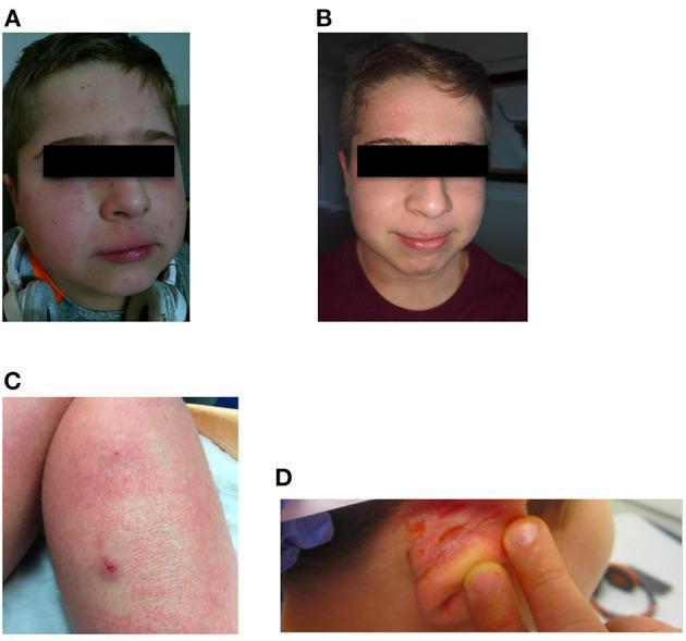
A multi-panel clinical photograph collection documenting atopic dermatitis (eczema) and treatment response. Panels A and B present a 'before and after' comparison of a pediatric patient's face. Panel A shows significant midfacial erythema, rough skin texture, and inflammatory papules on the cheeks and perioral region. Panel B demonstrates a marked reduction in inflammation and erythema with smoother skin texture following treatment with dupilumab. Panel C displays the right leg with diffuse, reticular erythema, xerosis, and a discrete inflammatory papule. Panel D shows the right ear with intense erythema, thickened skin, and visible fissuring or linear erosions. The images illustrate the clinical manifestations of severe eczema and its dermatological improvement under monoclonal antibody therapy (dupilumab), highlighting changes in skin morphology, color, and integrity across multiple anatomical regions.
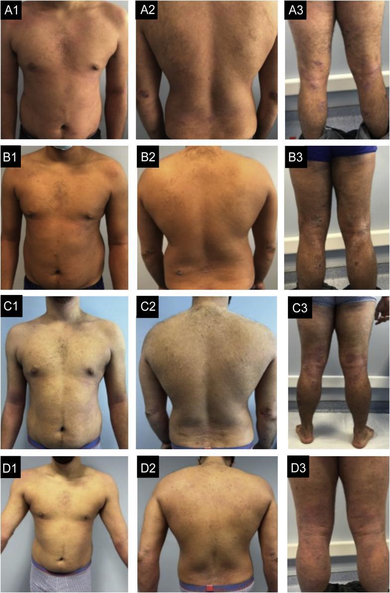
Clinical photograph series documenting the longitudinal progression of severe atopic dermatitis in a male patient across four stages of treatment (A1-D3). The images are organized in a grid showing the anterior trunk, posterior trunk, and posterior lower limbs. (Row A) Basal state: Exhibits widespread severe eczema characterized by extensive moderate erythema and edema on the trunk, with nummular-pattern lesions on the limbs showing intense oozing, crusting, and linear excoriations. (Row B) Post-dupilumab (6 months): Shows minimal clinical improvement with persistent erythema and scaling. (Row C) Post-baricitinib (6 months): Demonstrates lack of response with new inflammatory lesions appearing on the feet. (Row D) Post-tralokinumab (4 months): Displays significant global improvement; the trunk is nearly clear, while the limbs show resolution of excoriations and marked reduction in edema and lichenification. This comparative series illustrates treatment resistance and subsequent successful therapeutic intervention in refractory atopic dermatitis.
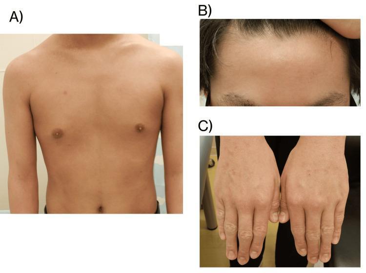
This composite clinical photograph displays the dermatologic status of a 14-year-old male patient following four weeks of upadacitinib treatment for atopic dermatitis. Image A provides an anterior view of the torso (chest and abdomen), showing significant resolution of previously active eczema, with even skin tone and absence of acute inflammatory lesions, lichenification, or excoriations. Image B is a close-up of the forehead and hairline, demonstrating clear skin without erythema or scaling. Image C shows the dorsal aspect of both hands; while the skin appears largely improved, there is evidence of mild residual xerosis (dryness), accentuated skin markings, and faint post-inflammatory pigmentary changes over the metacarpophalangeal joints. These images illustrate a positive clinical response to JAK inhibitor therapy, correlated with a low Eczema Area and Severity Index (EASI) score of 0.6, indicating near-complete skin clearance. The visual evidence serves as an educational example of therapeutic progression and disease management in pediatric atopic dermatitis.
Eczema (Atopic Dermatitis) - Treatment
Core Principles
Step 1 - All Patients: Skin Care & Emollients (Foundation)
- Emollients/moisturisers are the cornerstone of therapy - applied several times daily (oils, ointments, creams, lotions)
- In mild cases, emollients alone can be as effective as topical steroids
- Avoid known triggers: soaps, detergents, wool, heat, sweat, stress, certain foods
- Luke-warm baths/showers; pat dry gently; apply emollient immediately after bathing
Step 2 - Mild to Moderate: Topical Therapies
Topical Corticosteroids (TCS) - First-Line for Flares
| Potency | Examples | Use |
|---|---|---|
| Mild | Hydrocortisone 1% | Face, eyelids, skin folds, infants |
| Moderate | Clobetasone butyrate 0.05%, Betamethasone valerate 0.025% | Body in children |
| Potent | Betamethasone valerate 0.1%, Mometasone furoate | Trunk/limbs in adults |
| Very potent | Clobetasol propionate 0.05% | Thick lichenified plaques, short courses only |
- Fluorinated corticosteroids must NOT be used on the face (risk of cutaneous atrophy)
- Use milder preparations (triamcinolone 0.025%) for face and flexures
- Apply once or twice daily during flares; taper to lowest effective potency
Topical Calcineurin Inhibitors (TCIs) - Steroid-Sparing
- Tacrolimus ointment (0.03% children, 0.1% adults) and Pimecrolimus cream 1%
- Preferred for face, eyelids, skin folds where steroids cause atrophy
- Can be used as proactive/maintenance therapy 2-3x per week to prevent flares
Topical PDE-4 Inhibitors
- Crisaborole 2% ointment - for mild-moderate AD in patients ≥3 months
- Non-steroidal; useful when TCS or TCIs are not tolerated
Newest Topical (2025 Approved)
- Tapinarof cream (Vtama) - approved Dec 2025 for adults and children ≥2 years - a novel aryl hydrocarbon receptor (AhR) agonist
Step 3 - Moderate to Severe: Systemic Therapies
Biologics (Preferred Systemic Agents - AAD Strongly Recommended)
| Drug | Class | Dose/Route | Notes |
|---|---|---|---|
| Dupilumab (Dupixent) | Anti-IL-4Rα | SC every 2 weeks | First approved biologic; gold standard for moderate-severe AD; approved from 6 months of age |
| Tralokinumab (Adbry) | Anti-IL-13 | SC every 2 weeks | Adults; good for dupilumab failures |
| Lebrikizumab (Ebglyss) | Anti-IL-13 | SC every 2 weeks | Adults + children ≥12 yrs; approved Sept 2024 |
| Nemolizumab (Nemluvio) | Anti-IL-31Rα | SC every 4 weeks | Approved late 2024; particularly targets itch |
JAK Inhibitors (Oral - AAD Strongly Recommended)
| Drug | Target | Age |
|---|---|---|
| Upadacitinib (Rinvoq) | JAK1 | ≥12 years |
| Abrocitinib (Cibinqo) | JAK1 | ≥12 years |
| Baricitinib (Olumiant) | JAK1/2 | Adults |
- JAK inhibitors are fast-acting (improvement within days)
- Screen for TB, hepatitis B, VZV vaccination before starting
- Monitor for thrombosis, serious infections
Conventional Immunosuppressants (Conditionally Recommended - AAD)
- Cyclosporine - fastest acting; use for short-term control (≤2 years); monitor BP/renal function
- Methotrexate - weekly dosing; cheap; folate supplementation required
- Azathioprine - check TPMT activity before starting
- Mycophenolate mofetil - slower onset; good tolerability
The AAD 2025 guidelines strongly recommend against long-term systemic corticosteroids for AD due to rebound, side effects, and lack of disease modification.
Step 4 - Adjunctive Therapies
Phototherapy (Conditionally Recommended)
- Narrowband UVB (NB-UVB) - most commonly used; 3x/week
- UVA1 - for acute flares
- Suitable when topical therapy fails and biologics unavailable
Antihistamines
- Sedating antihistamines (e.g. hydroxyzine, chlorphenamine) at night for itch-related sleep disturbance
- Non-sedating antihistamines have limited benefit for AD itch specifically
Managing Secondary Infection
- Atopic dermatitis patients are highly susceptible to Staphylococcus aureus colonisation and infection
- Infected eczema: topical or oral antibiotics (flucloxacillin/cefalexin for staph); short course
- Eczema herpeticum (HSV): systemic aciclovir - a dermatological emergency
Clinical Images
Quick Summary by Severity
| Severity | Treatment |
|---|---|
| Mild | Emollients + mild TCS PRN |
| Moderate | Emollients + moderate/potent TCS + TCI (face) ± crisaborole |
| Severe | Add dupilumab / JAK inhibitor; or cyclosporine/MTX if unavailable |
| Refractory severe | Switch biologic; tralokinumab, lebrikizumab, nemolizumab |
| Infected | Antibiotics; eczema herpeticum → aciclovir |