Here are your complete MBBS 1st Year Biochemistry notes covering your entire syllabus:
MBBS 1st Year Biochemistry - Complete Notes
SECTION 1: CALCIUM METABOLISM
Calcium - Normal Values & Distribution
- Total serum calcium: 9-11 mg/dL (2.25-2.75 mmol/L)
- 50% ionized (active), 40% protein-bound (mainly albumin), 10% complexed
- Corrected Ca = Measured Ca + 0.8 × (4 - serum albumin)
Regulation of Calcium
Three hormones regulate calcium:
| Hormone | Source | Effect on Ca |
|---|
| PTH | Chief cells of parathyroid | ↑ Ca (↑ bone resorption, ↑ renal reabsorption, ↑ 1,25-VitD) |
| Vitamin D (1,25-dihydroxycholecalciferol) | Kidney (1-hydroxylation) | ↑ Ca (↑ gut absorption, ↑ bone resorption) |
| Calcitonin | Parafollicular C cells of thyroid | ↓ Ca (↓ bone resorption) |
Hypercalcemia
Causes (CHIMPANZEES mnemonic):
- C - Cancer (most common cause in hospitalized patients - PTHrP)
- H - Hyperparathyroidism (most common outpatient cause - primary HPT)
- I - Immobility
- M - Milk-alkali syndrome
- P - Paget's disease
- A - Addison's disease
- N - Neoplasms
- Z - Zollinger-Ellison
- E - Excess Vitamin D
- E - Excess Vitamin A
- S - Sarcoidosis/Thiazides
Clinical features: "Bones, Stones, Groans, Psychic Moans"
- Bones: bone pain, pathological fractures
- Stones: renal calculi (calcium oxalate/phosphate)
- Groans: nausea, vomiting, constipation, anorexia
- Psychic Moans: confusion, psychosis, lethargy, coma
ECG: Shortened QT interval
Treatment: IV saline (first line) → bisphosphonates → calcitonin → dialysis
Hypocalcemia
Causes:
- Hypoparathyroidism (post-thyroidectomy most common)
- Vitamin D deficiency (rickets in children, osteomalacia in adults)
- Pseudohypoparathyroidism (resistance to PTH - Albright hereditary osteodystrophy)
- Hypomagnesemia (impairs PTH secretion)
- Chronic renal failure (↓ 1-hydroxylation)
Clinical features:
- Neuromuscular excitability - tetany, paresthesias, cramps
- Chvostek's sign - facial twitch on tapping facial nerve
- Trousseau's sign - carpal spasm with BP cuff inflated (more specific)
- ECG: Prolonged QT interval
- Cataracts, seizures, laryngospasm, bronchospasm
Tetany
Tetany = spontaneous repetitive firing of motor nerves due to hypocalcemia (or hypomagnesemia, alkalosis).
Mechanism: Low ionized Ca²⁺ → reduces threshold for nerve excitation → repetitive depolarization
Types:
- Hypocalcemic tetany - low Ca²⁺
- Alkalotic tetany - alkalosis reduces ionized Ca (more Ca binds albumin)
- Hypomagnesemic tetany - Mg²⁺ required for PTH secretion
Signs: Chvostek, Trousseau, carpopedal spasm, laryngospasm, Erb's sign (+ve at 6mA)
SECTION 2: IRON METABOLISM
Iron in the Body
- Total body iron: ~4g in males, ~2.5g in females
- Distribution: Hemoglobin (65%), Ferritin/Hemosiderin (30%), Myoglobin (4%), Enzymes (1%)
- Daily requirement: Men 1mg/day absorbed; Women 1.5mg/day; Pregnancy 3mg/day
Iron Absorption
- Site: Duodenum and upper jejunum
- Form absorbed: Fe²⁺ (ferrous) - Vitamin C helps reduce Fe³⁺ to Fe²⁺
- Facilitated by: Vitamin C, gastric acid, meat
- Inhibited by: Phytates, oxalates, tannins (tea), antacids, high pH
Steps:
- Luminal Fe³⁺ reduced to Fe²⁺ by Duodenal Cytochrome b (Dcytb)
- Fe²⁺ enters enterocyte via DMT-1 (divalent metal transporter-1)
- Inside cell: stored as ferritin OR transferred to blood via Ferroportin
- Fe²⁺ oxidized to Fe³⁺ by Hephaestin (ferroxidase)
- Fe³⁺ binds Transferrin in blood (2 iron atoms per transferrin)
Mucosal Block Theory
- After a large iron load, enterocytes become "saturated" with iron stored as ferritin
- These iron-laden enterocytes block further iron absorption for 2-3 days (lifespan of enterocyte)
- When enterocyte is shed, iron is lost in feces
- Mechanism: Hepcidin (liver-produced, APR protein) downregulates ferroportin → reduces iron export from enterocytes → "mucosal block"
- Hepcidin is the MASTER regulator of iron homeostasis
Hepcidin
- Produced by liver in response to: iron overload, inflammation, infection
- Hepcidin → binds ferroportin → internalization/degradation of ferroportin → iron trapped in enterocytes and macrophages
- In iron deficiency: hepcidin falls → more ferroportin → more iron absorption
Transport and Storage
- Transferrin: plasma transport protein; TIBC = total iron binding capacity
- Ferritin: storage form (liver, spleen, bone marrow); reflects iron stores
- Hemosiderin: insoluble, degraded ferritin - seen in iron overload
- Normal values: Serum iron 60-150 μg/dL; Ferritin 12-300 ng/mL; TIBC 250-370 μg/dL
- Transferrin saturation = (Serum Fe / TIBC) × 100 = normally 30%
Iron Deficiency Anemia (IDA)
Stages:
- Iron depletion - Ferritin ↓, serum Fe normal
- Iron-deficient erythropoiesis - Ferritin ↓↓, TIBC ↑, transferrin saturation ↓
- Iron deficiency anemia - Hb ↓, microcytic hypochromic RBCs
Lab: ↓ Hb, ↓ MCV, ↓ MCH, ↓ serum iron, ↑ TIBC, ↓ ferritin, ↑ RDW
Clinical: Koilonychia (spoon nails), angular cheilitis, glossitis, Plummer-Vinson syndrome (web in esophagus)
Wilson Disease (Copper - for comparison)
- AR disorder - ATP7B gene mutation (chromosome 13)
- Impaired copper excretion into bile → accumulation in liver, brain, cornea, kidney
- Kayser-Fleischer rings in cornea (gold-brown, pathognomonic)
- Ceruloplasmin is low (but this is a plasma copper carrier, not iron!)
- Hepatic: cirrhosis, fulminant hepatic failure
- Neuropsychiatric: tremor, dysarthria, personality change
- Treatment: D-penicillamine, zinc acetate, trientine
Hemosiderosis
- Definition: Excess iron deposition (as hemosiderin) WITHOUT tissue damage
- Hemochromatosis = iron overload WITH organ damage
Primary (Hereditary) Hemochromatosis:
- AR, HFE gene mutation (C282Y most common)
- Excessive iron absorption despite adequate stores
- Deposits in: liver (cirrhosis), pancreas (bronze diabetes), heart (cardiomyopathy), joints, skin (bronze pigmentation), gonads
- Classic triad: Cirrhosis + diabetes + bronze skin = "Bronze diabetes"
- Diagnosis: Transferrin saturation >45%, ferritin markedly elevated, liver biopsy, HFE gene testing
- Treatment: Phlebotomy (preferred), deferasirox/deferoxamine
SECTION 3: Na⁺ AND K⁺ REGULATION IN KIDNEY
Sodium Handling
- Freely filtered at glomerulus; ~99% reabsorbed
- Proximal tubule (PT): 67% reabsorbed (Na-glucose, Na-amino acid cotransporters; Na/H exchanger)
- Loop of Henle (thick ascending limb): 25% - NKCC2 cotransporter (target of loop diuretics)
- Distal convoluted tubule (DCT): 5% - NCC (thiazide target); regulated by aldosterone
- Collecting duct: 3% - ENaC (aldosterone regulated); aquaporin-2 (ADH regulated)
Hormonal regulation:
| Hormone | Source | Effect on Na | Effect on K |
|---|
| Aldosterone | Adrenal cortex (zona glomerulosa) | ↑ reabsorption in CD | ↑ excretion |
| ANP/BNP | Heart atria/ventricles | ↓ reabsorption | - |
| ADH (vasopressin) | Posterior pituitary | ↑ water reabsorption (not Na) | - |
Potassium Handling
- 90% reabsorbed in PT and loop of Henle (passive, with Na)
- Fine-tuned in collecting duct by aldosterone
- Aldosterone → ↑ principal cell Na reabsorption → more negative lumen → K⁺ secreted
- K⁺ is excreted by principal cells via ROMK channels
- K⁺ is reabsorbed by intercalated cells via H/K-ATPase
Hyponatremia (Na⁺ < 135 mEq/L)
Classification by osmolality:
-
Hypotonic hyponatremia (most common type):
- Hypovolemic: Diarrhea, vomiting, diuretics, Addison's → ↑ ADH (appropriate)
- Euvolemic: SIADH, hypothyroidism, psychogenic polydipsia → ↑ ADH (inappropriate)
- Hypervolemic: CHF, cirrhosis, nephrotic syndrome → ↑ ADH (appropriate)
-
Isotonic hyponatremia (Pseudohyponatremia): Hyperlipidemia, hyperproteinemia (no real change in plasma Na)
-
Hypertonic hyponatremia: Hyperglycemia (glucose draws water → dilutes Na); mannitol
Symptoms: Headache, nausea, confusion, seizures, coma (severe)
Treatment:
- Chronic/asymptomatic: fluid restrict (SIADH), treat underlying cause
- Severe/symptomatic: Hypertonic saline (3%)
- Danger: Overcorrect too fast → Central Pontine Myelinolysis (CPM/ODS)
- Safe correction rate: ≤8-10 mEq/L per 24 hours
Pseudohypernatremia
- Na appears falsely elevated in lab due to decreased water fraction in plasma
- Actually rare - more common issue is pseudohyponatremia
- Can occur with very high lipids/proteins if certain methods used
- True hypernatremia = Na⁺ > 145 mEq/L
Hypernatremia (Na⁺ > 145 mEq/L)
Causes:
- Water loss (insensible, diarrhea, DI - central/nephrogenic)
- Sodium gain (hypertonic saline, hyperaldosteronism)
- Inadequate intake
Clinical: Thirst, lethargy, seizures, brain hemorrhage (cerebral dehydration)
Treatment: Free water (oral or D5W IV); correct slowly (max 10-12 mEq/L per day)
Hypokalemia (K⁺ < 3.5 mEq/L)
Causes:
- GI loss: vomiting, diarrhea
- Renal loss: diuretics (loop, thiazide), hyperaldosteronism, RTA, Bartter/Gitelman syndrome
- Transcellular shift: insulin, β2-agonists, alkalosis
- Decreased intake
Clinical:
- Muscle weakness, cramps, paralysis (flaccid)
- Cardiac: U waves on ECG, flat T waves, arrhythmias
- Hypokalemic nephropathy (polyuria)
- Metabolic alkalosis
Treatment: KCl replacement; IV for severe (<2.5 or symptomatic)
Hyperkalemia (K⁺ > 5.5 mEq/L)
Causes:
- Renal failure (most common)
- ACE inhibitors, K-sparing diuretics (spironolactone)
- Addison's disease (↓ aldosterone)
- Cell lysis: rhabdomyolysis, hemolysis, tumor lysis
- Acidosis (H⁺ shifts into cells, K⁺ shifts out)
- Pseudohyperkalemia: prolonged tourniquet, hemolysis of sample
Clinical:
- Muscle weakness
- ECG changes (in order): Peaked T waves → wide QRS → sine wave → VF/asystole
- Paralysis
Treatment (CBDIGK):
- Calcium gluconate - membrane stabilization (immediate, 5 min)
- Bicarbonate - shifts K into cells
- Dextrose + Insulin - shifts K into cells (onset 20-30 min)
- Inhaled β2-agonist (albuterol)
- Glucose + Insulin (same as D)
- K elimination: Kayexalate (sodium polystyrene) or Patiromer; Dialysis (definitive)
SECTION 4: pH REGULATION
Normal pH: 7.35-7.45
Respiratory Mechanism
- CO₂ + H₂O ⇌ H₂CO₃ ⇌ H⁺ + HCO₃⁻ (carbonic anhydrase)
- Lungs regulate CO₂
- Acidosis: Hyperventilate → ↓ pCO₂ → ↑ pH (compensation for metabolic acidosis)
- Alkalosis: Hypoventilate → ↑ pCO₂ → ↓ pH (compensation for metabolic alkalosis)
- Speed: Very fast (minutes)
- Henderson-Hasselbalch: pH = 6.1 + log([HCO₃⁻] / 0.03 × pCO₂)
Renal Mechanism
Three mechanisms by kidney:
-
Bicarbonate reabsorption (Proximal Tubule)
- H⁺ secreted into lumen via Na/H exchanger
- H⁺ + HCO₃⁻ → H₂CO₃ → CO₂ + H₂O (luminal carbonic anhydrase)
- CO₂ enters cell → HCO₃⁻ reformed → absorbed into blood
- Net effect: HCO₃⁻ is "reclaimed"
-
Titratable acid excretion (Distal Tubule)
- H⁺ secreted into lumen
- Combines with HPO₄²⁻ → H₂PO₄⁻ (titratable acid)
- Excreted in urine (can excrete ~30 mEq/day this way)
-
Ammonium excretion (Distal Tubule/Collecting Duct)
- Glutamine → NH₃ + HCO₃⁻ (regenerated bicarbonate into blood!)
- NH₃ + H⁺ → NH₄⁺ (trapped in tubular lumen → excreted)
- Most important in chronic acidosis (can increase 10-fold)
- Speed: Slow (days) but powerful
Acid-Base Disorders
| Disorder | pH | Primary Change | Compensation |
|---|
| Metabolic Acidosis | ↓ | ↓ HCO₃⁻ | ↓ pCO₂ (hyperventilate) |
| Metabolic Alkalosis | ↑ | ↑ HCO₃⁻ | ↑ pCO₂ (hypoventilate) |
| Respiratory Acidosis | ↓ | ↑ pCO₂ | ↑ HCO₃⁻ (kidney) |
| Respiratory Alkalosis | ↑ | ↓ pCO₂ | ↓ HCO₃⁻ (kidney) |
Compensatory Mechanisms (Winter's Formula and Rules)
- Metabolic acidosis → Respiratory compensation: Expected pCO₂ = 1.5 × HCO₃⁻ + 8 (±2) (Winter's formula)
- Metabolic alkalosis: Expected pCO₂ = 0.7 × HCO₃⁻ + 21 (±2)
- Acute Resp. Acidosis: HCO₃⁻ ↑ by 1 for every 10 ↑ pCO₂
- Chronic Resp. Acidosis: HCO₃⁻ ↑ by 3.5 for every 10 ↑ pCO₂
- Acute Resp. Alkalosis: HCO₃⁻ ↓ by 2 for every 10 ↓ pCO₂
- Chronic Resp. Alkalosis: HCO₃⁻ ↓ by 5 for every 10 ↓ pCO₂
Metabolic Acidosis
Causes using Anion Gap:
Anion Gap (AG)
- AG = Na⁺ - (Cl⁻ + HCO₃⁻) = normally 8-12 mEq/L
- Represents unmeasured anions (proteins, phosphate, sulfate)
High AG Metabolic Acidosis (MUDPILES mnemonic):
| Letter | Cause |
|---|
| M | Methanol |
| U | Uremia |
| D | DKA |
| P | Propylene glycol/Paraldehyde |
| I | Isoniazid/Iron |
| L | Lactic acidosis |
| E | Ethylene glycol |
| S | Salicylates |
Normal AG (Hyperchloremic) Metabolic Acidosis (DURHAM):
- D - Diarrhea (loss of HCO₃⁻)
- U - Ureteral diversion
- R - RTA (renal tubular acidosis)
- H - Hyperalimentation
- A - Addison's disease
- M - medications (carbonic anhydrase inhibitors)
Delta-Delta Ratio (for mixed disorders): ΔAG / ΔHCO₃⁻
- <0.4: Pure non-gap metabolic acidosis
- 0.4-0.8: Mixed high-gap + non-gap acidosis
- 1-2: Pure high-gap acidosis
-
2: Mixed high-gap acidosis + metabolic alkalosis
Metabolic Alkalosis
Causes:
- Chloride-responsive (urine Cl <20): vomiting, NG suction, loop/thiazide diuretics → Treat with NaCl
- Chloride-resistant (urine Cl >20): Hyperaldosteronism (Conn's), Cushing's, Bartter, Gitelman → Treat underlying cause
Respiratory Acidosis
- Causes: Hypoventilation - COPD, obesity hypoventilation, neuromuscular disease, sedation, obstructive sleep apnea
- Compensation: Kidney retains HCO₃⁻
Respiratory Alkalosis
- Causes: Hyperventilation - anxiety, pain, PE, early salicylate toxicity, liver failure, high altitude, pregnancy
- Compensation: Kidney excretes HCO₃⁻
Mixed Acid-Base Disorders
Suspect when compensation doesn't match expected:
- Examples:
- DKA + vomiting: High-AG met. acidosis + met. alkalosis
- COPD + diuretics: Resp. acidosis + met. alkalosis
- Sepsis + renal failure: Lactic acidosis + uremic acidosis
- Triple disorder: Met. acidosis + met. alkalosis + resp. disorder
SECTION 5: ENZYMES
Factors Affecting Enzyme Activity
1. Substrate Concentration - Michaelis-Menten Kinetics
- At low [S]: rate increases linearly (first order)
- At high [S]: rate plateaus at Vmax (zero order)
- Km = [S] when velocity = Vmax/2
- Low Km = high affinity for substrate
- Lineweaver-Burk plot (double reciprocal): x-intercept = -1/Km, y-intercept = 1/Vmax
2. Temperature
- Rate doubles for every 10°C rise (Q10 = 2)
- Optimal ~37°C for human enzymes
- Above optimal → protein denaturation → activity ↓
- Below optimal → reduced kinetic energy → activity ↓
3. pH
- Each enzyme has an optimal pH
- Pepsin: pH 1-2; Salivary amylase: pH 6.8; Trypsin/most enzymes: pH 7-8; Alkaline phosphatase: pH 9
- Extreme pH changes denature enzyme by altering ionization of active site residues
Enzyme Regulation
Short-Term Regulation
-
Allosteric regulation (most important - 5 marks)
- Effectors bind to allosteric site (not active site)
- Conformational change → ↑ or ↓ activity
- Positive allosteric effectors: Activate enzyme (e.g., AMP activates PFK-1)
- Negative allosteric effectors: Inhibit enzyme (e.g., ATP, citrate inhibit PFK-1)
- Shows sigmoidal kinetics (cooperativity)
- Example: Hemoglobin (O₂ binding cooperativity), Phosphofructokinase-1
- ATCase (aspartate transcarbamoylase) - classic allosteric enzyme
- Allosteric enzymes: R state (relaxed, active) and T state (tense, inactive)
-
Covalent modification (3 marks)
- Phosphorylation/dephosphorylation (most common)
- Glycogen phosphorylase: phosphorylated = ACTIVE; glycogen synthase: phosphorylated = INACTIVE
- Kinases add phosphate; Phosphatases remove phosphate
- Protein kinase A (PKA) activated by cAMP
- Other covalent modifications: adenylation, methylation, acetylation
-
Feedback inhibition (ultra-short control)
- End product inhibits first committed enzyme of pathway
- Example: CTP inhibits ATCase (pyrimidine synthesis)
Long-Term Regulation
-
Enzyme Induction (Transcriptional)
- Substrate or hormone induces gene expression → more enzyme protein
- Time scale: hours to days
- Example: Cytochrome P450 induced by barbiturates, rifampicin, phenytoin, ethanol
- Example: Glucokinase induced by insulin
-
Enzyme Repression
- End product represses transcription of enzyme genes
- Example: Heme inhibits ALA synthase (first enzyme of heme synthesis)
- When heme accumulates → ALA synthase gene repressed
Enzyme Classification (6 major classes - IUB system)
| Class | Reaction Catalyzed | Key Example |
|---|
| 1. Oxidoreductases | Oxidation-reduction (electron transfer) | LDH, succinate dehydrogenase, catalase |
| 2. Transferases | Transfer of functional groups | Aminotransferases (ALT, AST), kinases |
| 3. Hydrolases | Hydrolysis | Lipase, amylase, pepsin, trypsin |
| 4. Lyases | Addition/removal to double bonds (non-hydrolytic) | Aldolase, decarboxylases, citrate synthase |
| 5. Isomerases | Intramolecular rearrangements | Phosphoglucose isomerase, mutases |
| 6. Ligases (Synthetases) | Bond formation using ATP | Acetyl-CoA carboxylase, aminoacyl-tRNA synthetase |
Enzyme Inhibition
Reversible Inhibition
1. Competitive Inhibition
- Inhibitor structurally similar to substrate → competes for active site
- Inhibitor binds active site reversibly
- Vmax unchanged; Km increases
- Can be overcome by ↑ substrate concentration
- Lineweaver-Burk: Lines intersect on y-axis (same Vmax)
- Examples: Methotrexate (DHFR), Statins (HMG-CoA reductase), Sulfonamides (PABA/DHPS)
2. Uncompetitive Inhibition
- Inhibitor binds ONLY enzyme-substrate complex (ES complex)
- Both Vmax and Km decrease (proportionally)
- Lines on Lineweaver-Burk are parallel
3. Mixed (Non-competitive) Inhibition
- Inhibitor can bind either E or ES complex (at allosteric site)
- Vmax decreases; Km unchanged or changes
- Pure non-competitive: Vmax ↓, Km unchanged
- Example: Heavy metals (Pb, Hg) inhibiting enzymes
Irreversible Inhibition
- Covalent bond formation with enzyme → permanent inactivation
- Cannot be reversed by ↑ substrate
- Suicide inhibitors (also called: mechanism-based inhibitors or suicide substrates or Kcat inhibitors or enzyme-activated irreversible inhibitors)
- Active substrate analogs activated by the enzyme's own active site mechanism to form covalent bond
- Examples: Aspirin (irreversibly acetylates COX), Allopurinol → oxypurinol (inhibits xanthine oxidase), Fluorouracil (inhibits thymidylate synthase), Penicillin (inhibits transpeptidase), Organophosphates (inhibit AChE)
- Other names for suicide inhibition: Mechanism-based inhibition, Kcat inhibition, suicide inactivation
Difference: Competitive vs. Non-competitive
| Feature | Competitive | Non-competitive |
|---|
| Inhibitor binding site | Active site | Allosteric site |
| Effect on Vmax | No change | Decreased |
| Effect on Km | Increased | Unchanged (pure) |
| Reversible by [S]? | Yes | No |
| Lineweaver-Burk | Intersect on y-axis | Intersect on x-axis |
Isoenzymes (Isozymes)
- Multiple forms of same enzyme, same reaction, different structure (different subunits)
- Encoded by different genes or different post-translational modifications
Applied aspects:
LDH (Lactate Dehydrogenase)
- 5 isoforms (LDH-1 to LDH-5) - made of H and M subunits (tetramers)
- LDH-1 (H₄): Heart and RBCs - elevated in MI and hemolysis
- LDH-5 (M₄): Liver and skeletal muscle - elevated in hepatitis
- LDH-1 > LDH-2 (flip) = myocardial infarction
CK (Creatine Kinase)
- CK-MM: Skeletal muscle
- CK-MB: Myocardium - marker of MI (rises in 4-8h, peaks 12-24h, normalizes 48-72h)
- CK-BB: Brain
Alkaline Phosphatase
- Liver isoform, bone isoform, placenta isoform (Regan's enzyme)
- Regan's enzyme = placental ALP isoenzyme found in some cancers (lung, ovary) - tumor marker
- MCQ tip: Regan's enzyme is heat-stable (bone ALP is heat-labile; liver ALP intermediate)
Amylase
- Salivary amylase (AMY1), Pancreatic amylase (AMY2)
- Acute Pancreatitis: Serum amylase rises within 2-12h, peaks 12-72h, returns to normal in 3-5 days; Lipase more specific and stays elevated longer
Cardiac Markers Summary
| Marker | Rises | Peaks | Normalizes | Notes |
|---|
| Troponin I/T | 3-6h | 12-24h | 7-10 days | Most sensitive/specific for MI |
| CK-MB | 4-8h | 12-24h | 48-72h | Used for reinfarction |
| Myoglobin | 1-3h | 6-9h | 24h | First to rise, not specific |
| LDH-1 | 12-24h | 48-72h | 8-14 days | Historical |
Cellular Markers
| Enzyme/Marker | Organ | Clinical Use |
|---|
| ALT (GPT) | Liver (specific) | Hepatitis, liver damage |
| AST (GOT) | Liver, heart, muscle | Less specific |
| ALP | Liver, bone | Cholestasis, Paget's |
| GGT | Liver | Alcohol use, drug-induced |
| Amylase/Lipase | Pancreas | Pancreatitis |
| CPK-MB | Heart | MI |
| Troponin I/T | Heart | MI |
| PSA | Prostate | Prostate cancer |
| AFP | Liver | HCC |
| Acid phosphatase | Prostate | Prostate cancer (historical) |
| 5'-Nucleotidase | Liver | Liver disease |
Coenzymes and Cofactors
- Cofactor: Non-protein component required for enzyme activity
- Inorganic: metal ions (Zn²⁺, Fe²⁺, Mg²⁺, Cu²⁺)
- Organic: coenzymes
- Coenzyme is a type of cofactor (organic cofactor)
- Coenzyme: Organic, small molecules (often vitamin-derived); loosely bound = cosubstrate; tightly bound = prosthetic group
Key Coenzymes (MCQ targets):
| Coenzyme | Vitamin precursor | Function | Enzyme example |
|---|
| TPP (Thiamine Pyrophosphate) | Vitamin B1 (Thiamine) | Oxidative decarboxylation of α-keto acids; transketolase | Pyruvate DH, α-KG DH, BCKD, Transketolase (HMP) |
| FAD/FMN | Vitamin B2 (Riboflavin) | Electron/H carrier (oxidoreductase) | Succinate DH, Acyl-CoA DH |
| NAD⁺/NADP⁺ | Vitamin B3 (Niacin) | Electron/H carrier | Most dehydrogenases; NADP in HMP, fatty acid synthesis |
| CoA (Coenzyme A) | Vitamin B5 (Pantothenic acid) | Acyl group transfer | Acetyl-CoA, Succinyl-CoA |
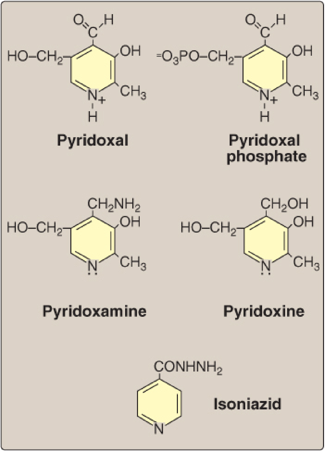
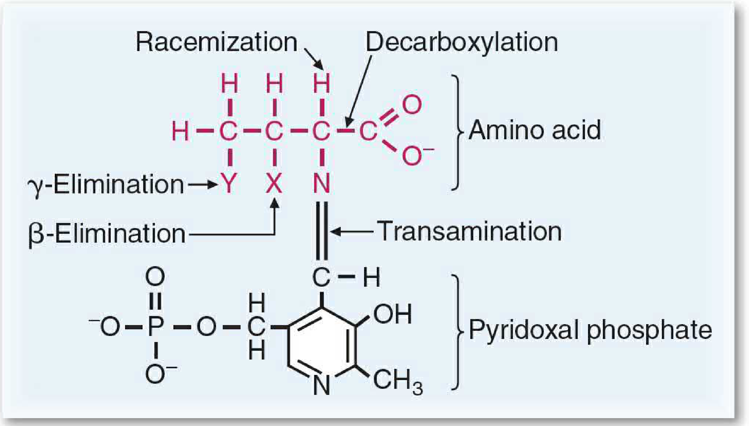
| PLP (Pyridoxal Phosphate) | Vitamin B6 (Pyridoxine) | Amino acid metabolism - transamination, decarboxylation, racemization, deamination | ALT, AST, amino acid decarboxylases |
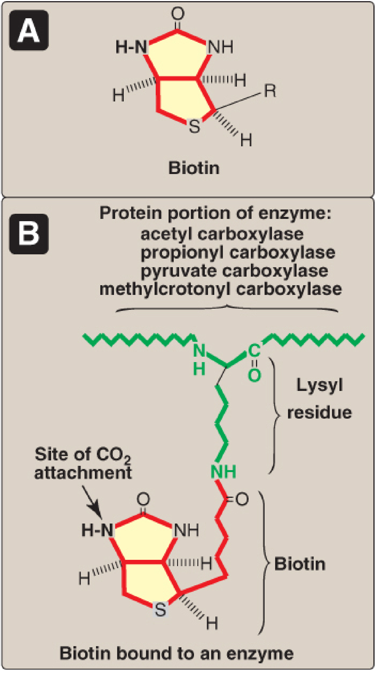
| Biotin | Vitamin B7 (Biotin) | CO₂ transfer (carboxylation) | Pyruvate carboxylase, ACC, PCCase, MCC |
| THF (Tetrahydrofolate) | Vitamin B9 (Folate) | 1-carbon transfer (methyl, methylene, formyl groups) | Thymidylate synthase, purine synthesis, homocysteine methylation |
| Cobalamin (B12) | Vitamin B12 | Methyl transfer; isomerization of methylmalonyl-CoA | Methionine synthase, Methylmalonyl-CoA mutase |
| Lipoic acid | - | Acyl transfer (oxidative decarboxylation complex) | Pyruvate DH complex |
| CoQ (Ubiquinone) | - | Electron carrier in ETC | Complex I → CoQ → Complex III |
Acyl-transferring coenzymes: CoA (acetyl/acyl transfer), Lipoic acid (in PDH complex)
SECTION 6: CARBOHYDRATE METABOLISM
Glucose Transporters
GLUT Transporters (Facilitated diffusion, Na-independent)
| Transporter | Location | Key Features |
|---|
| GLUT-1 | RBCs, brain, placenta | Basal glucose uptake; always expressed |
| GLUT-2 | Liver, pancreatic β-cells, kidney, intestine | High Km (low affinity) - glucose sensor in β-cells |
| GLUT-3 | Neurons | High affinity (low Km) |
| GLUT-4 | Skeletal muscle, adipose tissue, heart | Insulin-stimulated (translocation from intracellular vesicles to membrane) |
| GLUT-5 | Small intestine, testes | Fructose transporter |
| GLUT-12 | Heart, prostate, small intestine | Insulin-regulated (like GLUT-4) |
SGLT (Sodium-Glucose Cotransporters - Active transport, Na-dependent)
| Transporter | Location | Function |
|---|
| SGLT-1 | Small intestine (mainly), kidney (S3) | Absorbs glucose + galactose from gut (high affinity, low capacity); 2 Na⁺ per glucose |
| SGLT-2 | Kidney proximal tubule (S1, S2) | Reabsorbs ~90% filtered glucose (low affinity, high capacity); 1 Na⁺ per glucose |
SGLT-2 inhibitors (gliflozins - dapagliflozin, empagliflozin): Used in T2DM; block renal glucose reabsorption → glycosuria
Glycolysis
Site: Cytoplasm; occurs in all cells
Key steps:
- Glucose → Glucose-6-phosphate (Hexokinase/Glucokinase - ATP used; irreversible)
- G-6-P → F-6-P (Phosphoglucose isomerase)
- F-6-P → F-1,6-bisphosphate (Phosphofructokinase-1/PFK-1 - ATP used; RATE-LIMITING STEP)
- F-1,6-bisP → DHAP + Glyceraldehyde-3-P (Aldolase)
- G-3-P → 1,3-bisphosphoglycerate (G-3-P dehydrogenase - NAD⁺ → NADH)
- 1,3-BPG → 3-PG (Phosphoglycerate kinase - ATP generated, substrate-level)
- 3-PG → 2-PG (Mutase)
- 2-PG → Phosphoenolpyruvate (Enolase)
- PEP → Pyruvate (Pyruvate kinase - ATP generated; irreversible)
Net yield: 2 ATP, 2 NADH, 2 pyruvate per glucose
Irreversible enzymes (regulatory): Hexokinase, PFK-1, Pyruvate kinase
Allosteric regulation of PFK-1:
- Activated by: AMP, ADP, F-2,6-bisphosphate (most potent activator)
- Inhibited by: ATP, citrate, H⁺
Gluconeogenesis
Site: Liver (mainly), kidney cortex
4 Non-carbohydrate precursors:
- Amino acids (glucogenic) - e.g., alanine (Cori/glucose-alanine cycle), glutamine
- Lactate (Cori cycle: muscle → liver → glucose → muscle)
- Glycerol (from triglyceride hydrolysis)
- Propionate (from odd-chain fatty acid oxidation; as propionyl-CoA → succinyl-CoA → enters TCA)
Note: Even-chain fatty acids CANNOT contribute to gluconeogenesis (acetyl-CoA cannot be converted to OAA directly in mammals)
4 Key bypass enzymes (overcome irreversible glycolytic steps):
| Glycolytic Enzyme (irreversible) | Gluconeogenic Bypass Enzyme |
|---|
| Pyruvate kinase | Pyruvate carboxylase + PEPCK |
| PFK-1 | Fructose-1,6-bisphosphatase (FBPase-1) |
| Hexokinase/Glucokinase | Glucose-6-phosphatase |
- Pyruvate carboxylase (mitochondria): Pyruvate + CO₂ + ATP → OAA (biotin cofactor)
- PEPCK (mitochondria/cytoplasm): OAA + GTP → PEP + CO₂
- Fructose-1,6-bisphosphatase: F-1,6-BP → F-6-P
- Glucose-6-phosphatase (ER, liver/kidney only): G-6-P → Glucose (allows glucose export)
Regulation:
- Glucagon + Glucocorticoids stimulate gluconeogenesis
- Insulin inhibits gluconeogenesis
- PEPCK is induced by glucagon (via cAMP)
Glycogen Metabolism
Structure: α1-4 linkages in main chain; α1-6 linkages at branch points; Glycogenin is primer protein
Glycogen Synthesis:
- Glucose → G-6-P (Hexokinase)
- G-6-P → G-1-P (Phosphoglucomutase)
- G-1-P + UTP → UDP-Glucose (UDP-glucose pyrophosphorylase - driven by PPi hydrolysis)
- UDP-Glucose → Glycogen (Glycogen synthase; adds to existing chain)
- Branching enzyme (α1-4 → α1-6 transglycosylase): adds branch points
Glycogenolysis:
- Glycogen → G-1-P (Glycogen phosphorylase + Pi; cleaves α1-4 bonds)
- Debranching enzyme: transfers 3 of 4 residues from branch to main chain; then α1-6 glucosidase releases free glucose
- G-1-P → G-6-P (Phosphoglucomutase)
- G-6-P → Glucose (Glucose-6-phosphatase; only liver and kidney can do this)
Regulation:
| State | Glycogen Synthase | Glycogen Phosphorylase |
|---|
| Fed (Insulin) | Active (dephosphorylated) | Inactive |
| Fasted (Glucagon/Epi) | Inactive (phosphorylated) | Active (phosphorylated) |
Glycogen Storage Diseases (GSDs):
| Disease | Enzyme defect | Glycogen accumulated | Features |
|---|
| Von Gierke (Type I) | Glucose-6-phosphatase | Normal structure, liver | Hepatomegaly, hypoglycemia, hyperlipidemia, lactic acidosis |
| Pompe (Type II) | Lysosomal α-1,4-glucosidase (acid maltase) | All organs | Cardiomegaly, muscle weakness; only GSD with lysosomal involvement |
| Cori (Type III) | Debranching enzyme | Short branches, liver/muscle | Hepatomegaly, fasting hypoglycemia, myopathy |
| Anderson (Type IV) | Branching enzyme | Long unbranched chains | Cirrhosis |
| McArdle (Type V) | Muscle phosphorylase | Muscle | Muscle cramps with exercise, no rise in lactate |
| Hers (Type VI) | Liver phosphorylase | Liver | Mild hepatomegaly |
Fructose Metabolism
Two pathways:
-
In muscle/adipose: Fructose + ATP → Fructose-6-phosphate (Hexokinase, low affinity)
-
In liver (main pathway):
- Fructose + ATP → Fructose-1-phosphate (Fructokinase, very active - bypasses PFK-1 regulation!)
- F-1-P → DHAP + Glyceraldehyde (Aldolase B)
- Glyceraldehyde → G-3-P (Triokinase)
- Then enters glycolysis/lipogenesis
Clinical:
- Essential fructosuria: Fructokinase deficiency; benign; fructose in urine
- Hereditary Fructose Intolerance (HFI): Aldolase B deficiency; F-1-P accumulates → inhibits glycogenolysis and gluconeogenesis → severe hypoglycemia; liver failure; AR
- Dietary treatment: eliminate fructose, sucrose, sorbitol
HMP Pathway (Hexose Monophosphate / Pentose Phosphate Pathway)
Site: Cytoplasm; mainly liver, RBCs, adipose, adrenal cortex, mammary gland
Two phases:
Oxidative phase (irreversible):
- G-6-P → 6-Phosphogluconolactone → 6-PG → Ribulose-5-P
- Enzyme: G6PD (glucose-6-phosphate dehydrogenase) - rate-limiting
- Product: 2 NADPH (reductive power) per glucose
Non-oxidative phase (reversible):
- Interconversion of sugars using Transketolase (TPP cofactor) and Transaldolase
- Products: Ribose-5-phosphate (for nucleotide synthesis), glycolytic intermediates
Significance of HMP pathway:
- NADPH production: For fatty acid synthesis, steroid synthesis, glutathione reduction, antioxidant defense, cytochrome P450, NADPH oxidase (phagocytes)
- Ribose-5-phosphate: For nucleotide/nucleic acid synthesis
- Active in: RBCs (NADPH for GSH → protect against oxidative damage), liver, adrenal (steroid synthesis)
- G6PD deficiency: NADPH ↓ → GSH ↓ → RBC hemolysis upon oxidative stress (primaquine, dapsone, fava beans) → hemolytic anemia; X-linked recessive; most common RBC enzyme deficiency
Polyol Pathway (Sorbitol Pathway)
- Occurs in tissues with insulin-independent glucose uptake (lens, retina, peripheral nerves, kidney glomerulus, Schwann cells)
- Glucose → Sorbitol (Aldose reductase; NADPH) → Fructose (Sorbitol dehydrogenase; NAD⁺)
- In hyperglycemia: Excess sorbitol accumulates (can't escape cell easily)
- Consequences: Osmotic damage → cataracts, peripheral neuropathy, retinopathy, nephropathy (diabetic complications)
- Aldose reductase inhibitors (epalrestat): Under investigation to prevent diabetic complications
Regulation of Blood Glucose
Normal fasting: 70-100 mg/dL; 2h postprandial <140 mg/dL
Hormones:
| Hormone | Effect | Mechanism |
|---|
| Insulin | ↓ glucose (anabolic) | ↑ GLUT-4 translocation; ↑ glycolysis; ↑ glycogen synthesis; ↑ lipogenesis; ↓ gluconeogenesis |
| Glucagon | ↑ glucose (catabolic) | ↑ glycogenolysis (liver); ↑ gluconeogenesis; ↓ glycolysis |
| Epinephrine | ↑ glucose (fight/flight) | ↑ glycogenolysis (liver + muscle); ↑ lipolysis |
| Cortisol | ↑ glucose (stress) | ↑ gluconeogenesis (induces PEPCK); anti-insulin |
| Growth hormone | ↑ glucose | Anti-insulin effects |
Glucose-lowering drugs: Insulin, Metformin (↑ AMPK → ↓ gluconeogenesis), Sulfonylureas (↑ insulin secretion), SGLT-2 inhibitors, GLP-1 agonists
Alcohol Metabolism
Pathway:
- Ethanol → Acetaldehyde (Alcohol dehydrogenase/ADH; NAD⁺ → NADH) - mainly in cytosol
- Acetaldehyde → Acetate (Aldehyde dehydrogenase/ALDH; NAD⁺ → NADH) - mitochondria
- Acetate → Acetyl-CoA (in peripheral tissues)
MEOS (Microsomal Ethanol Oxidizing System): CYP2E1; induced by chronic alcohol; uses NADPH; produces ROS
Effects of High NADH/NAD⁺ ratio (acute toxicity):
- ↓ Gluconeogenesis → Hypoglycemia (OAA → malate; pyruvate → lactate)
- ↑ Lactic acid → Lactic acidosis (high AG)
- ↑ Lipogenesis → Fatty liver (↑ NADH → ↑ glycerol-3-P + ↑ acetyl-CoA → ↑ TAG synthesis)
- ↑ Ketogenesis → Alcoholic ketoacidosis
- Inhibition of TCA cycle
- ↑ Urate production → Gout
Chronic toxicity:
- Fatty liver → Alcoholic hepatitis → Cirrhosis
- Wernicke-Korsakoff (thiamine deficiency)
- Pancreatitis, cardiomyopathy, peripheral neuropathy
Fatty Liver (Hepatic Steatosis):
- Most common alcohol-related liver disease
- Mechanism: ↑ NADH → ↑ TAG synthesis; ↑ acetyl-CoA → ↑ fatty acid synthesis; ↓ fatty acid oxidation; ↓ VLDL export
- Reversible with abstinence
SECTION 7: MUCOPOLYSACCHARIDES AND LYSOSOMAL STORAGE DISEASES
Mucopolysaccharides (Glycosaminoglycans - GAGs)
- Definition: Unbranched polysaccharides of repeating disaccharide units (amino sugar + uronic acid)
- Always negatively charged → bind cations and water → form gels
- Found in ECM, mostly as proteoglycans (GAG chains covalently linked to core protein)
Types:
| GAG | Sulfation | Location |
|---|
| Hyaluronic acid | No | Synovial fluid, vitreous, ECM |
| Chondroitin sulfate | Yes | Cartilage, bone, skin |
| Dermatan sulfate | Yes | Skin, tendons |
| Heparan sulfate | Yes | Basement membrane, cell surface |
| Keratan sulfate | Yes | Cornea, cartilage |
| Heparin | Yes | Mast cells (anti-coagulant) |
Mucopolysaccharidoses (MPS)
| Disease | Enzyme Defect | GAG accumulated | Features |
|---|
| Hurler syndrome (MPS I-H) | α-L-Iduronidase | Dermatan + Heparan sulfate | AR; Mental retardation, coarse facies, corneal clouding, hepatosplenomegaly, gargoylism; Autosomal RECESSIVE; NO corneal clouding in Hunter |
| Hunter syndrome (MPS II) | Iduronate-2-sulfatase | Dermatan + Heparan sulfate | X-linked recessive (only X-linked MPS); NO corneal clouding; mild form possible |
| Sanfilippo (MPS III) | Various (4 subtypes) | Heparan sulfate | Severe mental retardation, mild somatic features |
| Morquio (MPS IV) | Galactosamine-6-sulfatase or β-Galactosidase | Keratan sulfate | Skeletal dysplasia, normal intelligence, odontoid hypoplasia |
| Maroteaux-Lamy (MPS VI) | N-Acetylgalactosamine-4-sulfatase | Dermatan sulfate | Normal intelligence |
| Sly (MPS VII) | β-Glucuronidase | Dermatan + Heparan + Chondroitin | Variable |
Key MCQ: Hurler vs Hunter: Both accumulate dermatan + heparan; Hurler is AR with corneal clouding; Hunter is X-linked without corneal clouding.
Galactosemia
- AR disorder; deficiency of one of 3 enzymes:
- Classic: Galactose-1-phosphate uridyl transferase (GALT) - most severe
- Galactokinase deficiency (mild, cataracts only)
- UDP-galactose-4-epimerase deficiency
Classic Galactosemia:
- Galactose-1-phosphate accumulates → toxic to liver, brain, kidney
- Presents in newborns after breast feeding or lactose-containing formula
- Features: Jaundice, cataracts, hepatomegaly, E. coli sepsis, intellectual disability, renal tubular acidosis
- Reducing substance in urine (positive Benedict's, Clinitest; negative glucose oxidase strip)
- Treatment: Eliminate lactose and galactose from diet immediately
SECTION 8: LIPID METABOLISM
Fatty Acid Oxidation
Alpha (α) Oxidation
- Site: Peroxisomes
- Oxidizes fatty acids at the α-carbon (C2 position)
- Used for phytanic acid (branched-chain fatty acid from diet; C3 has methyl group blocking β-oxidation)
- Refsum disease: Deficiency of phytanoyl-CoA hydroxylase → phytanic acid accumulates → retinitis pigmentosa, peripheral neuropathy, cerebellar ataxia
Beta (β) Oxidation
Site: Mitochondrial matrix
Activation: Fatty acid + CoA + ATP → Acyl-CoA (Acyl-CoA synthetase; in outer mitochondrial membrane)
Entry into mitochondria:
- Long-chain FA: Requires Carnitine shuttle (Carnitine acyl transferase I - rate-limiting, inhibited by malonyl-CoA)
- Medium/Short chain: Directly enter
Steps (repeated for each cycle):
- Acyl-CoA → Trans-Δ²-Enoyl-CoA (Acyl-CoA dehydrogenase; FAD → FADH₂)
- Enoyl-CoA → L-3-Hydroxyacyl-CoA (Enoyl-CoA hydratase)
- 3-OH-Acyl-CoA → 3-Ketoacyl-CoA (3-Hydroxyacyl-CoA dehydrogenase; NAD⁺ → NADH)
- 3-Ketoacyl-CoA → Acetyl-CoA + (n-2)Acyl-CoA (Thiolase/β-ketothiolase)
ATP yield from palmitate (C16:0):
- 7 cycles of β-oxidation → 7 FADH₂ + 7 NADH + 8 Acetyl-CoA
- 8 Acetyl-CoA × 10 ATP = 80 ATP
- 7 FADH₂ × 1.5 ATP = 10.5 ATP
- 7 NADH × 2.5 ATP = 17.5 ATP
- Total: 108 - 2 (activation) = 106 net ATP
Odd-chain fatty acids: Yield Propionyl-CoA in final cycle
- Propionyl-CoA → Methylmalonyl-CoA (Propionyl-CoA carboxylase; biotin) → Succinyl-CoA (Methylmalonyl-CoA mutase; B12 cofactor) → TCA cycle
- B12 deficiency → Methylmalonic aciduria
Unsaturated fatty acids: Need extra enzymes (isomerase, reductase); yield slightly less ATP
De Novo Synthesis of Fatty Acids
Site: Cytoplasm (liver, adipose, mammary gland)
Key enzyme: Acetyl-CoA Carboxylase (ACC) - rate-limiting enzyme
- Acetyl-CoA + CO₂ + ATP → Malonyl-CoA (biotin cofactor)
- Activated by: insulin, citrate
- Inhibited by: glucagon, palmitoyl-CoA, AMPK
Fatty Acid Synthase (FAS) complex - multienzyme
- ACP (Acyl Carrier Protein) holds intermediates
- Acetyl-ACP + Malonyl-ACP → elongation by 2 carbons
- Each cycle: 1 NADPH (ketoreduction) + 1 NADPH (enoyl reduction) = 2 NADPH used
- Product: Palmitate (C16:0) after 7 cycles
Citrate shuttle: Acetyl-CoA from mitochondria → Citrate (crosses membrane) → Cleaved by ATP-citrate lyase in cytoplasm → Acetyl-CoA + OAA
NADPH for synthesis from: HMP pathway and Malic enzyme (malate → pyruvate + NADPH)
Triglyceride Synthesis (Triacylglycerol/TAG)
- Glycerophosphate pathway (main):
- Glycerol-3-phosphate + 2 Fatty acyl-CoA → Phosphatidic acid (PA)
- PA + dephosphorylation → Diacylglycerol (DAG)
- DAG + Fatty acyl-CoA → TAG (DGAT enzyme)
- Occurs in: liver, adipose, intestine
- Glycerol-3-phosphate source: glycolysis (DHAP) in most tissues; glycerol in liver (glycerol kinase)
Ketone Body Metabolism (Ketogenesis)
Site of synthesis: Liver mitochondria (liver CANNOT use ketone bodies!)
Steps:
- 2 Acetyl-CoA → Acetoacetyl-CoA (Thiolase)
- Acetoacetyl-CoA + Acetyl-CoA → HMG-CoA (HMG-CoA synthase - rate-limiting)
- HMG-CoA → Acetoacetate + Acetyl-CoA (HMG-CoA lyase)
- Acetoacetate → β-hydroxybutyrate (β-OH-butyrate dehydrogenase; NADH)
- Acetoacetate → Acetone (spontaneous decarboxylation; smell of ketones)
Ketone bodies: Acetoacetate, β-hydroxybutyrate, Acetone
Conditions favoring ketogenesis: Starvation, DKA, low-carb diet, alcoholism, prolonged exercise
Stimulated by: ↑ Acetyl-CoA, ↑ fat delivery to liver, ↓ malonyl-CoA (malonyl-CoA inhibits carnitine transferase → less FA entry → ketogenesis regulated!)
Ketolysis (Ketone Utilization)
Site: Extrahepatic tissues - brain, heart, skeletal muscle, kidney
Steps:
- β-Hydroxybutyrate → Acetoacetate (β-OH-butyrate dehydrogenase; NAD⁺)
- Acetoacetate + Succinyl-CoA → Acetoacetyl-CoA + Succinate (Succinyl-CoA:3-oxoacid CoA transferase / SCOT - also called thiophorase; ABSENT in liver → liver cannot use ketones)
- Acetoacetyl-CoA → 2 Acetyl-CoA (Thiolase) → TCA cycle
Ketosis vs. Ketoacidosis:
- Ketosis: physiological, mild ketone elevation (starvation, low-carb)
- Ketoacidosis: pathological, severe (DKA, alcoholic KA); pH <7.3, high AG
Ketosis Summary (causes):
- ↑ FA mobilization (starvation, insulin deficiency)
- ↑ FA delivery to liver
- ↑ β-oxidation → ↑ Acetyl-CoA
- OAA consumed (gluconeogenesis) → less Acetyl-CoA enters TCA
- Net: ↑ Acetyl-CoA → ↑ HMG-CoA → ↑ ketone bodies
Cholesterol Metabolism
Synthesis:
- Site: Liver (mainly), intestine, adrenal, gonads
- Rate-limiting enzyme: HMG-CoA reductase (in ER)
- HMG-CoA → Mevalonate (HMG-CoA reductase; 2 NADPH)
- Mevalonate → Squalene → Lanosterol → Cholesterol
- Inhibited by: statins (competitive inhibitors of HMG-CoA reductase), cholesterol itself
Regulation of HMG-CoA reductase:
- Insulin: activates (dephosphorylation)
- Glucagon/AMPK: inhibits (phosphorylation)
- Oxysterols: decrease SREBP-2 transcription → ↓ HMG-CoA reductase
Causes of Hypercholesterolemia:
- Familial hypercholesterolemia (FH): LDL receptor mutation (AR or AD) → ↑↑ LDL
- Diet high in saturated fat/cholesterol
- Hypothyroidism (↓ LDL receptor)
- Nephrotic syndrome (↑ VLDL synthesis)
- Diabetes mellitus
- Drugs (steroids, thiazides)
- Cholestasis
- Obesity
Steroids from cholesterol:
- Cholesterol → Pregnenolone (CYP11A1; side-chain cleavage) - first step
- Pathways diverge to:
- Glucocorticoids (cortisol) - zona fasciculata
- Mineralocorticoids (aldosterone) - zona glomerulosa
- Sex hormones (estrogen, testosterone, progesterone) - gonads/zona reticularis
- Bile acids (primary: cholic acid, chenodeoxycholic acid) - liver
- Vitamin D (7-dehydrocholesterol → cholecalciferol in skin)
Lipoproteins (HDL, LDL, Chylomicrons)
Structure: Core (TAG, cholesterol esters) + Shell (phospholipids, free cholesterol, apoproteins)
Classification:
| Lipoprotein | Source | Main Lipid | Key Apolipoproteins | Function |
|---|
| Chylomicrons | Small intestine | TAG (diet) | Apo B-48, Apo C-II, Apo E | Transport dietary fat from gut to tissues |
| VLDL | Liver | TAG (endogenous) | Apo B-100, Apo C-II, Apo E | Transport endogenous fat to tissues |
| IDL | From VLDL | Cholesterol+TAG | Apo B-100, Apo E | Intermediate; taken up by liver or → LDL |
| LDL | From IDL | Cholesterol esters | Apo B-100 | Deliver cholesterol to tissues ("bad") |
| HDL | Liver + intestine | Protein, phospholipid | Apo A-I, Apo A-II | Reverse cholesterol transport ("good") |
Chylomicron Metabolism:
- Dietary fat packaged with Apo B-48 in enterocytes
- Released into lymph → thoracic duct → blood
- Apo C-II activates Lipoprotein Lipase (LPL) on capillary endothelium → hydrolyzes TAG → FFA released to tissues
- Chylomicron remnant (depleted of TAG, retains Apo E) → taken up by liver (Apo E receptor)
LDL Metabolism:
- VLDL → IDL (LPL removes TAG; Apo C-II transferred to HDL)
- IDL → LDL (Hepatic lipase removes more TAG; Apo E removed; only Apo B-100 remains)
- LDL → Cells via LDL receptor (Apo B-100 binds LDL receptor; receptor-mediated endocytosis)
- Cholesterol ester hydrolyzed → free cholesterol → inhibits HMG-CoA reductase; activates ACAT; downregulates LDL receptor
HDL (Reverse Cholesterol Transport):
- Nascent HDL (disc-shaped, Apo A-I) secreted from liver and intestine
- Picks up cholesterol from peripheral tissues via ABCA1 transporter
- LCAT (lecithin-cholesterol acyl transferase; activated by Apo A-I) esterifies cholesterol → core → HDL becomes spherical
- Cholesterol esters transferred to VLDL/LDL via CETP (cholesteryl ester transfer protein)
- HDL → liver via SR-B1 receptor (selective lipid uptake)
- High HDL = protective against atherosclerosis
Eicosanoids
- Derived from: 20-carbon polyunsaturated fatty acids (mainly arachidonic acid - C20:4, ω-6)
- Arachidonic acid released from membrane phospholipids by Phospholipase A₂ (activated by hormones, trauma; inhibited by glucocorticoids via lipocortin)
Types:
| Type | Enzyme | Key Products | Effects |
|---|
| Prostaglandins (PG) | COX-1, COX-2 | PGE₂, PGI₂ (prostacyclin), PGF₂α | Pain, fever, inflammation; PGI₂ = vasodilator/anti-platelet |
| Thromboxanes (TX) | COX pathway | TXA₂ | Platelet aggregation, vasoconstriction |
| Leukotrienes (LT) | 5-Lipoxygenase | LTB₄, LTC₄, LTD₄, LTE₄ (SRS-A) | Inflammation; LTB₄ = neutrophil chemotaxis; LTC4/D4/E4 = bronchoconstriction |
| Lipoxins | Lipoxygenase | LXA₄ | Anti-inflammatory |
Aspirin:
- Irreversibly acetylates COX-1 and COX-2 (serine residue in active site)
- = Suicide inhibitor of COX
- In platelets (no nucleus → can't make new COX) → permanent inhibition → ↓ TXA₂ → ↓ platelet aggregation
- Anti-platelet effect lasts platelet lifetime (7-10 days)
- Also ↓ PGI₂ but endothelial cells can regenerate COX
Phospholipids Classification
Definition: Glycerophospholipids or sphingomyelin; contain phosphate ester
Glycerophospholipids:
- Backbone: Glycerol-3-phosphate
- Position 1: Saturated FA; Position 2: Unsaturated FA
- Position 3: Phosphate + head group
| Phospholipid | Head group | Function |
|---|
| Phosphatidylcholine (Lecithin) | Choline | Most abundant; lung surfactant; VLDL component |
| Phosphatidylethanolamine (Cephalin) | Ethanolamine | Brain, clotting |
| Phosphatidylserine | Serine | Apoptosis signal (flips to outer leaflet) |
| Phosphatidylinositol | Inositol | PIP₂ → IP₃ + DAG (signaling); anchor for GPI proteins |
| Phosphatidylglycerol | Glycerol | Mitochondria, cardiolipin |
| Cardiolipin | 2 phosphatidylglycerol | Inner mitochondrial membrane; antigen in syphilis (VDRL) |
| Plasmalogen | Vinyl ether linkage | Heart, muscle, brain |
| PAF (Platelet Activating Factor) | Choline, ether linkage | Platelet activation, inflammation |
Sphingomyelin:
- Backbone: Sphingosine (not glycerol)
- Myelin sheath; stored in lysosomes
- Niemann-Pick disease: Sphingomyelinase deficiency → sphingomyelin accumulation
PUFA (Polyunsaturated Fatty Acids) Significance
- Essential fatty acids (EFA): Linoleic acid (C18:2, ω-6) and α-Linolenic acid (C18:3, ω-3) - cannot be synthesized in humans
- ω-6 series: Linoleic → Arachidonic acid (precursor of pro-inflammatory eicosanoids)
- ω-3 series: α-Linolenic → EPA (C20:5) → DHA (C22:6)
- Anti-inflammatory (EPA → anti-inflammatory eicosanoids)
- DHA: brain development, retina function
- ↓ Triglycerides, ↓ cardiovascular risk
EFA deficiency: Dermatitis (scaly skin), poor wound healing, poor growth, infertility, susceptibility to infection
SECTION 9: ELECTRON TRANSPORT CHAIN (ETC)
Site: Inner mitochondrial membrane (IMM)
Complexes:
| Complex | Name | Coenzymes | Inhibitors |
|---|
| I | NADH: CoQ oxidoreductase | FMN, Fe-S | Rotenone, Amytal |
| II | Succinate: CoQ oxidoreductase | FAD, Fe-S | Carboxin, malonate (succinate dehydrogenase) |
| III | CoQ: Cytochrome c oxidoreductase (Cytochrome bc1) | Cyt b, Cyt c₁, Fe-S | Antimycin A |
| IV | Cytochrome c oxidase (Cytochrome aa₃) | Cyt a, Cyt a₃, Cu | Cyanide, CO, azide, H₂S |
| V | ATP synthase (F₀F₁) | - | Oligomycin (F₀), Venturicidin |
Electron flow: NADH → Complex I → CoQ → Complex III → Cyt c → Complex IV → O₂
Proton pumping (Mitchell's Chemiosmotic Theory):
- Complexes I, III, IV pump H⁺ from matrix to intermembrane space
- Proton gradient (electrochemical gradient) drives ATP synthase (Complex V)
- F₀ subunit: proton channel; F₁ subunit: ATP synthesis
- P/O ratio: NADH = 2.5 ATP; FADH₂ = 1.5 ATP
Uncouplers:
- Dissipate proton gradient → heat generated instead of ATP
- Examples: 2,4-Dinitrophenol (DNP), Thermogenin (UCP-1) in brown adipose tissue (BAT - for thermogenesis in neonates), Carbonyl cyanide m-chlorophenylhydrazone (CCCP)
- Aspirin in high doses, thyroid hormones
SECTION 10: COLLAGEN AND ECM
Collagen Structure
- Most abundant protein in human body (~30% of total protein)
- Triple helix of 3 α-chains; Gly-X-Y repeat sequence (Gly every 3rd position - essential!)
- X and Y are often Proline and Hydroxyproline
- Hydroxyproline and hydroxylysine stabilize the triple helix via H-bonds
Collagen Types (Collagen Classification)
| Type | Location | Features |
|---|
| I | Bone, skin, tendons, dentin, cornea, sclera | Most abundant; thick fibers |
| II | Cartilage, vitreous humor | Thin fibers |
| III | Skin, blood vessels, uterus | Reticular fibers; found with type I |
| IV | Basement membrane | Meshwork, no fibers |
| V | Fetal tissue, placenta | - |
Collagen Synthesis
Intracellular:
- Preprocollagen mRNA → Preprocollagen (ribosome)
- Signal peptide cleaved → Procollagen
- Hydroxylation of Pro → Hydroxyproline (Prolyl hydroxylase; requires Vitamin C and Fe²⁺/O₂)
- Hydroxylation of Lys → Hydroxylysine (Lysyl hydroxylase; requires Vit C)
- Glycosylation of hydroxylysine (Gal, Glu added)
- Triple helix formed (Procollagen) - C-propeptide initiates
Extracellular:
7. Procollagen secreted → N and C propeptides cleaved by Procollagen peptidase → Tropocollagen
8. Tropocollagen self-assembles into fibrils
9. Cross-linking: Lysyl oxidase (Cu²⁺-dependent) oxidizes lysine/hydroxylysine → Aldehyde → forms covalent cross-links (allysine) → mature collagen fiber
Vitamin C deficiency → Scurvy: Impaired hydroxylation → unstable collagen → bleeding gums, poor wound healing, perifollicular hemorrhage, corkscrew hairs, hemarthrosis
Copper deficiency: ↓ Lysyl oxidase → weak collagen (similar to Menkes disease - X-linked Cu deficiency)
Proteoglycans and Glycosaminoglycans
ECM Definition: The extracellular matrix (ECM) is a highly organized network of macromolecules secreted by cells into the surrounding space; provides structural support, regulates cell behavior, cell-cell communication, and tissue homeostasis.
ECM components:
- Fibrous proteins: Collagen (tensile strength), Elastin (elasticity), Fibronectin (cell adhesion - RGD motif binds integrins), Laminin (basement membrane)
- Proteoglycans: Core protein + GAG chains; fill space, resist compression, bind growth factors, filter molecules
- Glycoproteins: Fibronectin, Laminin, Nidogen
Proteoglycans:
- Core protein with covalently attached GAG chains
- Examples: Aggrecan (cartilage), Decorin (skin/tendons), Perlecan (basement membrane), Syndecan (cell surface), Versican
- Functions of proteoglycans/GAGs:
- Water retention (highly hydrophilic → resist compression)
- Structural support of ECM
- Bind growth factors (bFGF, VEGF) and protect them from degradation
- Cell adhesion and migration
- Filter size and charge-based (glomerular basement membrane)
- Anticoagulant (heparan sulfate/heparin activates AT-III)
QUICK MCQ SUMMARY TABLES
Key "First" / "Only" / "Most" Facts
| Fact | Answer |
|---|
| Rate-limiting enzyme of glycolysis | PFK-1 |
| Rate-limiting enzyme of gluconeogenesis | PEPCK (or Fructose-1,6-bisphosphatase) |
| Rate-limiting enzyme of fatty acid synthesis | Acetyl-CoA Carboxylase (ACC) |
| Rate-limiting enzyme of cholesterol synthesis | HMG-CoA Reductase |
| Rate-limiting enzyme of HMP pathway | G6PD |
| Rate-limiting enzyme of β-oxidation | Carnitine acyltransferase-I (CPTI) |
| Rate-limiting enzyme of TCA cycle | Isocitrate Dehydrogenase |
| Rate-limiting enzyme of urea cycle | Carbamoyl phosphate synthetase I |
| Enzyme deficient in Hurler syndrome | α-L-Iduronidase |
| Enzyme deficient in Hunter syndrome | Iduronate sulfatase |
| X-linked MPS | Hunter syndrome |
| MPS with corneal clouding | Hurler (NOT Hunter) |
| GSD with lysosomal involvement | Pompe disease |
| GSD with no rise in lactate with exercise | McArdle (Type V) |
| Coenzyme for oxidative decarboxylation | TPP (Thiamine pyrophosphate) |
| Coenzyme for carboxylation reactions | Biotin |
| Coenzyme for transamination | PLP (B6) |
| Coenzyme for 1-C transfer | THF (Folate) |
| Suicide inhibitor for platelets | Aspirin |
| Other name for suicide inhibition | Mechanism-based / Kcat inhibition |
| LDL receptor defect disease | Familial Hypercholesterolemia |
| Regan's enzyme | Placental isoform of ALP (found in lung/ovary cancer) |
| Tissue that cannot use ketone bodies | Liver (lacks SCOT) |
| GLUT-4 is stimulated by | Insulin |
| SGLT-2 inhibitors target | Renal proximal tubule glucose reabsorption |
| Color of hemosiderin on Perls stain | Blue (Prussian blue) |
| Mucosal block mediator | Hepcidin |
| Key feature: Hurler vs Hunter | Hurler = corneal clouding; Hunter = no clouding, X-linked |
This covers your entire syllabus. These notes are organized for MBBS 1st year biochemistry exams with high-yield MCQ points, key enzymes, clinical correlations, and comparison tables. Study tip: focus especially on the allosteric regulation section (5 marks), enzyme inhibition comparisons (diagrams 3.27/3.29), and metabolic pathway integration.All notes are complete above. Here's a quick navigation guide to what's covered:
What's Covered - Index
| Section | Topics |
|---|
| 1. Calcium | Regulation (PTH/VitD/Calcitonin), Hypercalcemia (causes + CHIMPANZEES), Hypocalcemia, Tetany + signs |
| 2. Iron | Absorption + pathway (DMT1/ferroportin), Mucosal block theory + Hepcidin, Iron deficiency anemia, Wilson disease, Hemosiderosis/Hemochromatosis |
| 3. Na⁺/K⁺ Kidney | Tubular handling, Aldosterone/ADH, Hypo/hypernatremia + Pseudohypernatremia, Hypo/hyperkalemia with ECG changes + treatment |
| 4. pH Regulation | Respiratory + Renal mechanisms, All 4 acid-base disorders, Winter's formula, Compensation rules, Anion Gap (MUDPILES), Mixed disorders, Delta-delta ratio |
| 5. Enzymes | Michaelis-Menten, Temp/pH effects, Allosteric regulation (5m), Covalent modification, Induction (CYP450) + Repression (heme synthesis), Enzyme classification (6 classes), Competitive vs Non-competitive (comparison table), Suicide inhibition (other names), Isoenzymes - LDH/CK/ALP/Amylase, Regan's enzyme, Coenzymes table (TPP/FAD/NAD/CoA/PLP/Biotin/Folate) |
| 6. Carbohydrates | GLUT 1-5/12, SGLT 1/2, Glycolysis (key enzymes), Gluconeogenesis (4 precursors + 4 bypass enzymes), Glycogen synthesis/lysis, Glycogen storage diseases (Type I-VI), Fructose metabolism + HFI, HMP pathway + significance + G6PD deficiency, Polyol pathway + diabetic complications, Blood glucose regulation, Alcohol metabolism + fatty liver |
| 7. Mucopolysaccharides | GAGs classification, MPS (Hurler vs Hunter - key comparison), Galactosemia |
| 8. Lipids | Alpha oxidation (Refsum), Beta oxidation (steps + ATP yield), Propionyl-CoA metabolism (B12), De novo synthesis (ACC/FAS), TAG synthesis, Ketogenesis (HMG-CoA), Ketolysis (SCOT), Ketosis summary, Cholesterol synthesis + steroids, Causes of hypercholesterolemia, Lipoprotein metabolism (chylomicrons/LDL/HDL), Eicosanoids + Aspirin, Phospholipid classification, PUFA significance |
| 9. ETC | All 5 complexes + inhibitors, Chemiosmotic theory, Uncouplers |
| 10. Collagen/ECM | Collagen types, Synthesis steps (Vit C role), Lysyl oxidase (Cu), ECM definition, Proteoglycan functions |
| Quick MCQ Table | All high-yield one-liners |