
Whipples procedure steps
Whipple procedure pancreaticoduodenectomy surgical steps
https://www.sciencedirect.com/science/article/pii/S258984502…
https://www.ncbi.nlm.nih.gov/books/NBK560747
Whipple Procedure (Pancreaticoduodenectomy) - Operative Steps
Overview
PHASE 1 - Preoperative & Entry
- In patients with elevated CA 19-9 or questionable lesions on imaging, diagnostic laparoscopy is performed first to exclude occult peritoneal or liver metastases before committing to laparotomy.
- Upper midline incision (standard). A bilateral subcostal (chevron) incision is used selectively in obese patients or those with prior lower abdominal surgery.
- The umbilical ligament is divided and dissected back to the umbilical fissure of the liver. This pedicled flap is preserved for later coverage of the gastroduodenal artery (GDA) stump.
- Full abdominal exploration to confirm no metastatic disease (liver, peritoneum, lymph nodes).
- A self-retaining retractor (e.g., Thompson Surgical System) is placed.
PHASE 2 - Resection (6 Steps per Evans et al.)
Step 1 - Entry into the lesser sac and SMV identification
- The lesser sac is entered through the gastrocolic ligament.
- The hepatic flexure of the colon is taken down.
- The gastrocolic trunk is divided.
- The inferior border of the pancreatic body is identified and the visceral peritoneum incised from left to right toward the SMV.
- The infrapancreatic superior mesenteric vein (SMV) is carefully exposed at the inferior border of the pancreatic neck, near the uncinate process.
- If venous resection is anticipated or the patient is morbidly obese, a Cattell-Braasch maneuver (mobilization of the entire right colon and small bowel mesentery root) may be performed.
Step 2 - Kocher maneuver
- The peritoneum lateral to the duodenum is incised.
- The duodenum and pancreatic head are mobilized off the retroperitoneum (Kocher maneuver), exposing the inferior vena cava (IVC) and left renal vein.
- This allows palpation for vascular involvement and access to the SMA origin.
- Note: Resectability is now primarily determined by preoperative imaging, not intraoperative palpation.
Step 3 - Cholecystectomy and bile duct division
- The gallbladder is mobilized from the liver, and the cystic duct and artery are ligated and divided.
- The common hepatic duct is circumferentially dissected and divided just above the cystic duct entry point.
- The bile duct is then dissected inferiorly toward the duodenum; inferior traction on the distal bile duct exposes the anterior portal vein.
- The GDA is ligated and divided at its origin from the hepatic artery after confirming hepatic artery patency (a test clamp is applied with confirmation of a hepatic artery pulse).
Step 4 - Gastric or duodenal division
- Classic Whipple: The antrum is divided and the distal stomach resected.
- Pylorus-preserving (PPPD): The duodenum is divided 2 cm distal to the pylorus with a GI stapler.
Step 5 - Jejunal division
- The jejunum is divided approximately 10 cm distal to the ligament of Treitz using a GI stapler.
- The jejunal mesentery is divided at the serosa/mesentery junction using an energy device - great care is taken here to avoid inadvertent SMA ligation as the bowel passes posterior to the SMA.
- The duodenum and proximal jejunum are then delivered under the mesenteric vessels from left to right.
Step 6 - Pancreatic neck transection and uncinate process dissection
- A retropancreatic tunnel is created anterior to the portal vein behind the pancreatic neck using a blunt-tipped clamp.
- Hemostatic sutures are placed on both the superior and inferior sides of the planned transection line.
- The pancreatic neck is divided with electrocautery anterior to the portal vein (cautery is avoided near the pancreatic duct).
- The SMV is mobilized; small venous branches from the uncinate process draining into the portal vein/SMV are ligated individually.
- The SMV is retracted medially, exposing the SMA beneath it.
- A periadventitial dissection plane is developed along the SMA from caudal to cranial - all fibrofatty, lymphatic, and perineural tissue to the right and anterior to the SMA is taken with the specimen (critical for oncological clearance).
- Multiple inferior pancreaticoduodenal arteries (IPDA) from the SMA are individually suture-ligated.
- The uncinate process is freed from behind the SMV and SMA, completing the resection. The specimen is removed.
PHASE 3 - Reconstruction
1. Pancreaticojejunostomy (PJ) - most proximal
- Duct-to-mucosa (end-to-side): The pancreatic duct is sutured directly to the jejunal mucosa with an inner layer of interrupted absorbable sutures. An outer layer approximates the pancreatic capsule to the seromuscular layer of the jejunum. This is the preferred technique for soft pancreas or small duct.
- Dunking (invagination) anastomosis: The pancreatic stump is intussuscepted (dunked) into the end of the jejunum - can be terminoterminal or terminolateral.
- A pancreaticogastrostomy (PG) is an alternative to PJ, anastomosing the pancreatic remnant to the posterior stomach wall.
2. Hepaticojejunostomy (HJ) - 10-15 cm distal to PJ
- The common hepatic duct stump is anastomosed end-to-side to the jejunum with a single layer of interrupted absorbable sutures.
3. Gastrojejunostomy or Duodenojejunostomy - most distal
- Classic Whipple: A gastrojejunostomy is created (side-to-side or end-to-side) approximately 40-60 cm distal to the HJ - usually antecolic to avoid compression.
- PPPD: A duodenojejunostomy is created, restoring pyloric continuity.
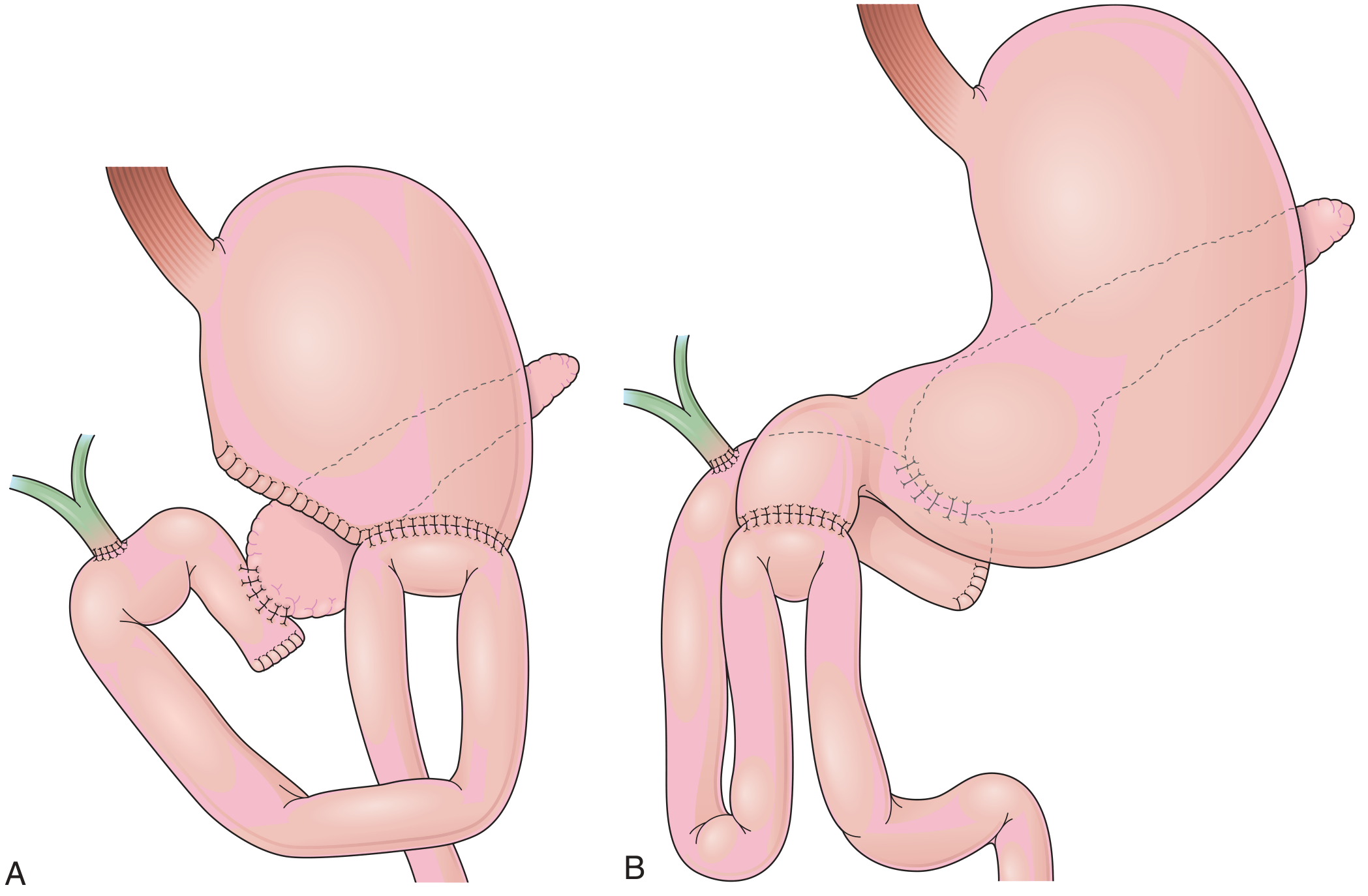
PHASE 4 - Closure
- Abdominal drains are placed near the pancreatic and biliary anastomoses (selective vs. routine placement remains debated; a randomized multicenter trial supported drain placement for patients who develop postoperative leaks).
- The umbilical ligament pedicle flap may be sutured over the GDA stump to protect against postoperative hemorrhage from stump blowout.
- Fascial and skin closure.
Classic vs. Pylorus-Preserving: Key Differences
| Feature | Classic Whipple | PPPD |
|---|---|---|
| Gastric division | Antrum resected | Duodenum divided 2 cm distal to pylorus |
| Gastric anastomosis | Gastrojejunostomy | Duodenojejunostomy |
| Oncologic margin | Slightly wider | Equivalent for most tumors |
| Delayed gastric emptying | Less common | More common historically |
Key Complications to Know
- Postoperative pancreatic fistula (POPF) - most common major complication; results from pancreaticojejunostomy leak. Graded A/B/C by ISGPF criteria.
- Delayed gastric emptying (DGE) - especially with PPPD.
- Post-pancreatectomy hemorrhage - may be from GDA stump or pseudoaneurysm.
- Bile leak from hepaticojejunostomy.
- Wound infection / intra-abdominal abscess.
When to do pppd and classical
pylorus preserving pancreaticoduodenectomy versus classic Whipple
PMID: 39757070
PPPD vs. Classic Whipple - When to Choose Each
PPPD (Pylorus-Preserving Pancreaticoduodenectomy)
- Tumor is in the head of the pancreas, uncinate process, or ampulla, and is not encroaching on the pylorus or first part of the duodenum
- The proximal duodenum and pylorus are free of tumor - clear resection margins can be achieved while preserving the pylorus
- Periampullary tumors (ampullary carcinoma, distal cholangiocarcinoma, duodenal adenocarcinoma of D2/D3) where D1 is not involved
- Benign or premalignant lesions (chronic pancreatitis, large symptomatic cysts, IPMN of the head) - preservation of gastric function is especially valued
- The goal is to avoid postgastrectomy symptoms (dumping syndrome, alkaline reflux gastritis) by preserving antral and pyloric function
- Avoids postgastrectomy symptoms (dumping, alkaline reflux)
- Better preservation of GI function and nutritional status long term
- No difference in overall survival or recurrence vs. classic Whipple (well established across multiple RCTs and meta-analyses)
- No difference in pancreatic fistula rates
Classic Whipple (Standard Pancreaticoduodenectomy)
| Indication | Rationale |
|---|---|
| Tumor encroaches on D1 (first part of duodenum) or the distal stomach | A clear resection margin cannot be achieved with PPPD; antral resection is required for R0 |
| FAP (Familial Adenomatous Polyposis) with diffuse duodenal polyposis | Entire duodenum including D1 must be removed |
| Tumor invades the pylorus or gastric antrum directly | Pylorus preservation is oncologically unsafe |
| Gastric outlet obstruction from tumor | Antral resection required |
| Lymph node involvement along the lesser curvature or in the perigastric region | En bloc antral resection improves nodal clearance |
| Surgeon preference / specific anatomical factors | Historically some surgeons prefer classic; equivalent oncologic outcomes |
- Ensures wider proximal margin in tumors close to pylorus
- May reduce risk of delayed gastric emptying (DGE) in some series - though this is debated
What the Evidence Shows
Summary Decision Table
| Scenario | Choice |
|---|---|
| Pancreatic head cancer, pylorus uninvolved | PPPD (standard) |
| Ampullary or periampullary cancer, D1 free | PPPD (standard) |
| Chronic pancreatitis / benign lesion | PPPD preferred |
| Tumor at/near pylorus or antrum | Classic Whipple |
| FAP with duodenal polyposis | Classic Whipple |
| D1 involved or margin threatened | Classic Whipple |
| Gastric outlet obstruction from tumor | Classic Whipple |
Bailey and Love's 28e summarizes it clearly: "PPPD is the standard resection for tumours of the pancreatic head or ampulla. The Whipple procedure is now reserved for situations in which the entire duodenum has to be removed (e.g. FAP) or in which the tumour encroaches on the first part of the duodenum or distal stomach and PPPD would not achieve a clear resection margin."
Diagnotic molecular pathology
What aspect of diagnostic molecular pathology do you need? · What level of detail do you need?
Everything to write as short note
Diagnostic Molecular Pathology - Short Note
Definition
- Diagnosis and tumour classification
- Prognostic prediction
- Identifying hereditary cancer risk
- Determining treatment (theranostics)
- Identifying residual disease after treatment
Basic Methods
1. In Situ Hybridisation (ISH)
- Uses a labelled oligonucleotide probe targeting a specific RNA or DNA sequence
- Allows visualisation of the presence, absence and location of a nucleic acid sequence in tissue sections
- Visualisation by: autoradiography, fluorescence (FISH), or bright-field (CISH) microscopy
- FISH (Fluorescence ISH): detects gene amplifications, translocations, and deletions
- CISH (Chromogenic ISH): combines ISH + IHC; common alternative to FISH for HER2 amplification detection
- Key applications: HER2 amplification (breast, gastric, CRC), EBV, CMV, high-risk HPV detection
2. Polymerase Chain Reaction (PCR)
- Amplifies DNA, yielding millions of copies from a single target sequence
- RT-PCR (Reverse Transcriptase PCR): amplifies RNA by first converting to cDNA
- Real-time PCR (qPCR): quantifies DNA/RNA in real time
- Can be performed on fresh or formalin-fixed paraffin-embedded (FFPE) tissue
- Applications:
- Mutational analysis (e.g. KRAS, BRAF mutations)
- Clonality testing (lymphoid tumours)
- Microsatellite instability (MSI) testing
- Fusion gene detection (e.g. BCR-ABL)
- Infectious agent detection
3. Next-Generation Sequencing (NGS)
- Performs massively parallel sequencing - simultaneously examines millions of DNA/RNA fragments
- Works on FFPE tissue
- Detects: point mutations, small insertions/deletions (indels), copy number variants (CNVs), gene fusions
- Evaluates 20-500 genes in a single assay
- Greater analytical sensitivity than Sanger sequencing - can detect low-frequency alleles
- Targeted NGS panels identify multiple known mutations in one test
- Also measures Tumour Mutation Burden (TMB)
4. Sanger Sequencing / Pyrosequencing
- Traditional sequencing method; useful for targeted single-gene analysis
- Less sensitive than NGS but widely available and reliable for known hotspot mutations
Types of Genetic Abnormalities Detected
| Type | Description | Examples |
|---|---|---|
| Point mutations | Single nucleotide change in DNA; germline or somatic | TP53, KRAS mutations |
| Indels | Insertions/deletions causing frameshift | KIT mutations |
| Fusion genes | From translocations or deletions; chimeric proteins | BCR-ABL t(9;22) in CML; BCL2 t(14;18) in follicular lymphoma; TMPRSS2-ERG in prostate cancer |
| Gene amplification | Increased copy number → overexpression | HER2 in breast/gastric cancer |
| Microsatellite instability (MSI) | Instability of short tandem repeats due to MMR deficiency | Lynch syndrome, sporadic CRC |
| Epigenetic changes | DNA methylation, histone modification silencing genes | MLH1 hypermethylation in sporadic CRC |
Mismatch Repair (MMR) and MSI
- MMR genes (MLH1, MSH2, MSH6, PMS2) correct replication errors
- Loss of MMR function → MSI-H (microsatellite instability-high) / dMMR (deficient MMR)
- Causes:
- Germline mutation = Lynch syndrome (autosomal dominant; risk of CRC, endometrial, other cancers at early age)
- Sporadic somatic methylation of MLH1 (more common, ~85%)
- Prevalence: ~15% of CRCs, ~30% of endometrial carcinomas
- IHC (preferred initial test in most centres) - loss of staining = gene abnormality
- PCR-based microsatellite testing
- NGS
- Phenotypic classification (e.g. medullary CRC is typically MSI-H; right-sided, mucinous)
- Better prognosis overall
- Predicts response to immune checkpoint inhibitors (ICIs)
- No response to 5-fluorouracil
- Screens for Lynch syndrome
Detection Methods - Summary Table
| Abnormality | Preferred Method |
|---|---|
| Point mutations / small indels | NGS, PCR |
| Gene fusions | FISH, NGS, PCR |
| Gene amplifications | FISH, NGS |
| Tumour mutation burden (TMB) | NGS |
| MSI/dMMR | IHC (initial), PCR, NGS |
| Viral genomes (EBV, HPV, CMV) | ISH |
Rule: IHC is often a useful and sufficient initial test for many of these abnormalities before molecular testing.
Theranostics - Molecular Changes and Drug Therapy
| Molecular Finding | Target Drug |
|---|---|
| HER2 amplification (breast, gastric, CRC) | Trastuzumab, pertuzumab |
| KRAS/BRAF wild-type (CRC) | Anti-EGFR antibodies (cetuximab, panitumumab) |
| BRAF V600E | BRAF inhibitors (vemurafenib, dabrafenib) |
| ALK, RET, NTRK, ROS1, FGFR2 fusions | Tyrosine kinase inhibitors |
| MSI-H / high TMB | Immune checkpoint inhibitors (pembrolizumab, nivolumab) |
| BCR-ABL t(9;22) | Imatinib and other TKIs |
| PD-L1 expression | ICIs |
Indications for Molecular Analysis of Tumour Tissue
- Diagnosis and classification
- Selection of therapy
- Prognosis
- Staging
- Monitoring disease burden
- Screening for germline mutations
- Confirmation of neoplasia (e.g. clonality testing in lymphoma)
Key Pathways Disrupted in Tumorigenesis
- Signal transduction / proliferation: KRAS, EGFR, BRAF, growth factor receptors
- Cell cycle control: p16 (CDKN2A), RB
- DNA repair: BRCA1/2, MMR genes
- Apoptosis: BCL2, TP53
Practical Notes
- Pathologist assesses tumour cell content (%), cellularity, and necrosis before molecular analysis
- Microdissection (conventional or laser-assisted) improves tumour DNA yield from FFPE sections
- Germline vs. somatic testing: germline mutations are present in every cell (e.g. BRCA1/2, Lynch syndrome); somatic mutations are confined to tumour cells
Convert to pdf
ls /tmp/workspace/ 2>/dev/null || mkdir -p /tmp/workspace && echo "ready"
~/diagnostic_molecular_pathology.py
from reportlab.lib.pagesizes import A4
from reportlab.lib.styles import getSampleStyleSheet, ParagraphStyle
from reportlab.lib.units import cm
from reportlab.lib import colors
from reportlab.platypus import (
SimpleDocTemplate, Paragraph, Spacer, Table, TableStyle,
HRFlowable, KeepTogether
)
from reportlab.lib.enums import TA_LEFT, TA_CENTER, TA_JUSTIFY
OUTPUT = "/tmp/workspace/Diagnostic_Molecular_Pathology.pdf"
doc = SimpleDocTemplate(
OUTPUT,
pagesize=A4,
leftMargin=2*cm, rightMargin=2*cm,
topMargin=2.2*cm, bottomMargin=2*cm,
title="Diagnostic Molecular Pathology",
author="Orris Medical"
)
styles = getSampleStyleSheet()
# Custom styles
HEADING1 = ParagraphStyle("H1", parent=styles["Normal"],
fontSize=16, fontName="Helvetica-Bold", spaceAfter=6,
spaceBefore=14, textColor=colors.HexColor("#1a3a5c"))
HEADING2 = ParagraphStyle("H2", parent=styles["Normal"],
fontSize=12, fontName="Helvetica-Bold", spaceAfter=4,
spaceBefore=10, textColor=colors.HexColor("#2e6da4"))
HEADING3 = ParagraphStyle("H3", parent=styles["Normal"],
fontSize=11, fontName="Helvetica-Bold", spaceAfter=3,
spaceBefore=8, textColor=colors.HexColor("#1a5276"))
BODY = ParagraphStyle("Body", parent=styles["Normal"],
fontSize=9.5, fontName="Helvetica", spaceAfter=4,
spaceBefore=2, leading=14, alignment=TA_JUSTIFY)
BULLET = ParagraphStyle("Bullet", parent=styles["Normal"],
fontSize=9.5, fontName="Helvetica", spaceAfter=2,
spaceBefore=1, leading=13, leftIndent=14, bulletIndent=4)
SMALL = ParagraphStyle("Small", parent=styles["Normal"],
fontSize=8, fontName="Helvetica-Oblique", spaceAfter=3,
textColor=colors.HexColor("#555555"))
TABLE_HEADER = ParagraphStyle("TH", parent=styles["Normal"],
fontSize=9, fontName="Helvetica-Bold", textColor=colors.white, leading=12)
TABLE_CELL = ParagraphStyle("TC", parent=styles["Normal"],
fontSize=9, fontName="Helvetica", leading=12)
def h1(text): return Paragraph(text, HEADING1)
def h2(text): return Paragraph(text, HEADING2)
def h3(text): return Paragraph(text, HEADING3)
def body(text): return Paragraph(text, BODY)
def bullet(text): return Paragraph(f"• {text}", BULLET)
def sp(h=4): return Spacer(1, h)
def hr(): return HRFlowable(width="100%", thickness=0.8, color=colors.HexColor("#2e6da4"), spaceAfter=4, spaceBefore=4)
def thin_hr(): return HRFlowable(width="100%", thickness=0.4, color=colors.HexColor("#aaaaaa"), spaceAfter=3, spaceBefore=3)
def make_table(headers, rows, col_widths=None):
data = [[Paragraph(h, TABLE_HEADER) for h in headers]]
for row in rows:
data.append([Paragraph(str(c), TABLE_CELL) for c in row])
t = Table(data, colWidths=col_widths, repeatRows=1)
t.setStyle(TableStyle([
("BACKGROUND", (0,0), (-1,0), colors.HexColor("#2e6da4")),
("ROWBACKGROUNDS", (0,1), (-1,-1), [colors.HexColor("#f0f5fa"), colors.white]),
("GRID", (0,0), (-1,-1), 0.4, colors.HexColor("#b0c4de")),
("VALIGN", (0,0), (-1,-1), "MIDDLE"),
("LEFTPADDING", (0,0), (-1,-1), 5),
("RIGHTPADDING", (0,0), (-1,-1), 5),
("TOPPADDING", (0,0), (-1,-1), 4),
("BOTTOMPADDING", (0,0), (-1,-1), 4),
]))
return t
story = []
# ── TITLE ──────────────────────────────────────────────
story.append(sp(6))
story.append(Paragraph("DIAGNOSTIC MOLECULAR PATHOLOGY", ParagraphStyle(
"Title", parent=styles["Normal"], fontSize=20, fontName="Helvetica-Bold",
alignment=TA_CENTER, textColor=colors.HexColor("#1a3a5c"), spaceAfter=4)))
story.append(Paragraph("Short Note", ParagraphStyle(
"Sub", parent=styles["Normal"], fontSize=12, fontName="Helvetica-Oblique",
alignment=TA_CENTER, textColor=colors.HexColor("#2e6da4"), spaceAfter=2)))
story.append(Paragraph("Source: Bailey & Love's 28e | Henry's Clinical Diagnosis 24e", ParagraphStyle(
"Src", parent=styles["Normal"], fontSize=8, fontName="Helvetica-Oblique",
alignment=TA_CENTER, textColor=colors.HexColor("#777777"), spaceAfter=8)))
story.append(hr())
story.append(sp(6))
# ── 1. DEFINITION ──────────────────────────────────────
story.append(h1("1. Definition"))
story.append(body(
"Diagnostic molecular pathology refers to multiple tests that assess <b>molecules "
"(DNA, RNA, and proteins) in tissue</b> to provide information useful for:"
))
for item in [
"Diagnosis and tumour classification",
"Prognostic prediction",
"Identifying hereditary cancer risk",
"Determining treatment (theranostics)",
"Identifying residual disease after treatment",
]:
story.append(bullet(item))
story.append(body("<i>Note: Immunohistochemistry (IHC) is conventionally considered a separate category.</i>"))
story.append(sp(4))
# ── 2. BASIC METHODS ───────────────────────────────────
story.append(h1("2. Basic Methods"))
# ISH
story.append(h2("2.1 In Situ Hybridisation (ISH)"))
story.append(body(
"Uses a <b>labelled oligonucleotide probe</b> targeting a specific RNA or DNA sequence, allowing "
"visualisation of presence, absence, and location of nucleic acid sequences in tissue sections."
))
story.append(body("<b>Variants:</b>"))
for item in [
"<b>FISH</b> (Fluorescence ISH) – fluorescence microscopy; detects gene amplifications, translocations, deletions",
"<b>CISH</b> (Chromogenic ISH) – bright-field; combines ISH + IHC; common alternative to FISH for HER2 detection",
"<b>Radioactive ISH</b> – autoradiography (less common now)",
]:
story.append(bullet(item))
story.append(body("<b>Key applications:</b> HER2 amplification (breast, gastric, CRC), EBV, CMV, high-risk HPV detection"))
story.append(sp(4))
# PCR
story.append(h2("2.2 Polymerase Chain Reaction (PCR)"))
story.append(body(
"Amplifies DNA, yielding millions of copies from a single target sequence. "
"Can be performed on <b>fresh or formalin-fixed paraffin-embedded (FFPE)</b> tissue."
))
story.append(body("<b>Variants:</b>"))
for item in [
"<b>RT-PCR</b> (Reverse Transcriptase PCR) – amplifies RNA by converting to cDNA first",
"<b>Real-time PCR (qPCR)</b> – quantifies DNA/RNA; distinct from RT-PCR despite similar abbreviation",
]:
story.append(bullet(item))
story.append(body("<b>Applications:</b> Mutational analysis (KRAS, BRAF), clonality testing in lymphoma, MSI testing, "
"fusion gene detection (BCR-ABL), infectious agent detection"))
story.append(sp(4))
# NGS
story.append(h2("2.3 Next-Generation Sequencing (NGS)"))
story.append(body(
"Performs <b>massively parallel sequencing</b> — simultaneously examines millions of DNA/RNA fragments. "
"Works on FFPE tissue. Evaluates <b>20–500 genes in a single assay</b>."
))
story.append(body("<b>Detects:</b> point mutations, small indels, copy number variants (CNVs), gene fusions, tumour mutation burden (TMB)"))
story.append(body("<b>Advantages over Sanger:</b> greater sensitivity (detects low-frequency alleles), higher throughput, "
"simultaneous multi-gene analysis"))
story.append(sp(4))
# Sanger
story.append(h2("2.4 Sanger Sequencing / Pyrosequencing"))
story.append(body(
"Traditional targeted single-gene sequencing. Less sensitive than NGS but reliable for known hotspot mutations. "
"Still widely used for specific variant confirmation."
))
story.append(sp(6))
# ── 3. GENETIC ABNORMALITIES ───────────────────────────
story.append(h1("3. Types of Genetic Abnormalities Detected"))
story.append(make_table(
["Type", "Description", "Examples"],
[
["Point mutations", "Single nucleotide change; germline or somatic", "TP53, KRAS mutations"],
["Indels", "Insertions/deletions → frameshift", "KIT mutations"],
["Fusion genes", "From translocations/deletions; chimeric proteins", "BCR-ABL t(9;22) in CML; BCL2 t(14;18) in FL; TMPRSS2-ERG in prostate cancer"],
["Gene amplification", "Increased copy number → overexpression", "HER2 in breast/gastric/CRC"],
["MSI", "MMR deficiency → short tandem repeat instability", "Lynch syndrome, sporadic CRC (MLH1 methylation)"],
["Epigenetic changes", "DNA methylation/histone modification silencing genes", "MLH1 hypermethylation in sporadic CRC"],
],
col_widths=[3.2*cm, 7.5*cm, 6.3*cm]
))
story.append(sp(8))
# ── 4. MMR / MSI ───────────────────────────────────────
story.append(h1("4. Mismatch Repair (MMR) and Microsatellite Instability (MSI)"))
story.append(body(
"MMR genes (<b>MLH1, MSH2, MSH6, PMS2</b>) correct DNA replication errors. "
"Loss of MMR function → <b>MSI-H</b> (microsatellite instability-high) / <b>dMMR</b> (deficient MMR)."
))
story.append(h3("Causes:"))
for item in [
"<b>Germline mutation</b> = <b>Lynch syndrome</b> (autosomal dominant; risk of CRC, endometrial + other cancers at early age)",
"<b>Sporadic somatic methylation of MLH1</b> (more common, ~85% of MSI-H cases)",
]:
story.append(bullet(item))
story.append(h3("Prevalence:"))
for item in ["~15% of colorectal carcinomas (CRC)", "~30% of endometrial carcinomas"]:
story.append(bullet(item))
story.append(h3("Testing for MSI/MMR:"))
for item in [
"<b>IHC</b> – preferred initial test; loss of staining = gene abnormality",
"<b>PCR-based microsatellite testing</b>",
"<b>NGS</b>",
]:
story.append(bullet(item))
story.append(h3("Clinical Value of MSI-H:"))
story.append(make_table(
["Clinical Use", "Detail"],
[
["Phenotypic classification", "Medullary CRC typically MSI-H; right-sided, mucinous histology, BRAF V600E mutation"],
["Prognosis", "MSI-H = better overall prognosis"],
["Therapy prediction", "Responds to immune checkpoint inhibitors (ICIs); no response to 5-fluorouracil"],
["Germline screening", "Screens for Lynch syndrome"],
],
col_widths=[5*cm, 12*cm]
))
story.append(sp(8))
# ── 5. DETECTION METHOD SUMMARY ───────────────────────
story.append(h1("5. Detection Methods — Summary"))
story.append(make_table(
["Abnormality", "Preferred Method(s)"],
[
["Point mutations / small indels", "NGS, PCR"],
["Gene fusions", "FISH, NGS, PCR"],
["Gene amplifications", "FISH, NGS"],
["Tumour mutation burden (TMB)", "NGS"],
["MSI / dMMR", "IHC (initial), PCR, NGS"],
["Viral genomes (EBV, HPV, CMV)", "ISH"],
],
col_widths=[9*cm, 8*cm]
))
story.append(body("<i>Rule: IHC is often a useful and sufficient initial test for many abnormalities before molecular testing.</i>"))
story.append(sp(8))
# ── 6. THERANOSTICS ───────────────────────────────────
story.append(h1("6. Theranostics — Molecular Changes and Drug Therapy"))
story.append(body(
"An increasingly common reason for molecular testing is predicting drug response "
"('theranostics') — particularly for targeted therapies and immune checkpoint inhibitors (ICIs)."
))
story.append(make_table(
["Molecular Finding", "Tumour Type", "Target Drug"],
[
["HER2 amplification", "Breast, gastric, CRC", "Trastuzumab, pertuzumab"],
["KRAS/BRAF wild-type", "CRC", "Anti-EGFR (cetuximab, panitumumab)"],
["BRAF V600E", "Melanoma, CRC, others", "BRAF inhibitors (vemurafenib, dabrafenib)"],
["ALK, RET, NTRK, ROS1, FGFR2 fusions", "NSCLC, others", "Tyrosine kinase inhibitors (TKIs)"],
["MSI-H / high TMB", "Multiple", "ICIs (pembrolizumab, nivolumab)"],
["BCR-ABL t(9;22)", "CML", "Imatinib and other TKIs"],
["PD-L1 expression", "Multiple", "ICIs"],
],
col_widths=[5.5*cm, 4.5*cm, 7*cm]
))
story.append(h3("Tumours Responding to ICIs:"))
for t in ["Malignant melanoma", "Non-small cell lung cancer (NSCLC)", "Small cell lung cancer (SCLC)",
"Breast carcinoma", "Urothelial carcinoma", "Hepatocellular carcinoma"]:
story.append(bullet(t))
story.append(sp(8))
# ── 7. INDICATIONS ────────────────────────────────────
story.append(h1("7. Indications for Molecular Analysis of Tumour Tissue"))
for i, item in enumerate([
"Diagnosis and classification",
"Selection of therapy",
"Prognosis",
"Staging",
"Monitoring disease burden",
"Screening for germline mutations",
"Confirmation of neoplasia (e.g. clonality testing in lymphoma)",
], 1):
story.append(Paragraph(f"{i}. {item}", BULLET))
story.append(sp(8))
# ── 8. PATHWAYS ───────────────────────────────────────
story.append(h1("8. Key Pathways Disrupted in Tumorigenesis"))
story.append(make_table(
["Pathway", "Key Genes"],
[
["Signal transduction / proliferation", "KRAS, EGFR, BRAF, growth factor receptors"],
["Cell cycle control", "p16 (CDKN2A), RB"],
["DNA repair", "BRCA1/2, MMR genes (MLH1, MSH2, MSH6, PMS2)"],
["Apoptosis", "BCL2, TP53"],
],
col_widths=[7*cm, 10*cm]
))
story.append(sp(8))
# ── 9. PRACTICAL NOTES ────────────────────────────────
story.append(h1("9. Practical Notes"))
for item in [
"The pathologist assesses <b>tumour cell content (%)</b>, cellularity, and necrosis before molecular analysis",
"<b>Microdissection</b> (conventional or laser-assisted) improves tumour DNA yield from FFPE sections",
"<b>Germline mutations</b> are present in every cell of the body (e.g. BRCA1/2, Lynch syndrome MMR mutations)",
"<b>Somatic mutations</b> are confined to tumour cells only (e.g. acquired KRAS, BRAF mutations)",
"Adequate tumour DNA quality and quantity are essential — samples with high necrosis or low cellularity may fail",
]:
story.append(bullet(item))
story.append(sp(10))
story.append(thin_hr())
story.append(Paragraph(
"Sources: Bailey & Love's Short Practice of Surgery 28e, Ch. 11 | "
"Henry's Clinical Diagnosis and Management by Laboratory Methods 24e",
SMALL))
doc.build(story)
print(f"PDF created: {OUTPUT}")
cd /tmp/workspace && python diagnostic_molecular_pathology.py
Diagnostic Molecular Pathology
PDF Document · PDF
- Definition
- Basic Methods (ISH, PCR, NGS, Sanger)
- Types of Genetic Abnormalities
- MMR / MSI (with clinical value table)
- Detection Methods summary table
- Theranostics and drug targets
- Indications for molecular analysis
- Key pathways disrupted in tumorigenesis
- Practical notes
Tep and taap short note
TEP and TAPP - Short Note
(Laparoscopic Inguinal Hernia Repair)
Background
- TEP - Totally Extraperitoneal Repair
- TAPP - Transabdominal Preperitoneal Repair
Common Features of Both Procedures
- Patient in Trendelenburg position; surgeon stands contralateral to the hernia
- General anaesthesia required (patients cannot tolerate abdominal insufflation while awake)
- Mesh size typically 10 × 15 cm, placed to cover the entire myopectineal orifice
- Mesh is usually not fixed (or selectively fixed) to reduce chronic pain
- Can repair both unilateral and bilateral hernias in the same sitting
- IEHS guidelines: Grade A recommendation that TEP and TAPP are preferred over Lichtenstein repair for recurrent hernias after prior open anterior repair
TEP - Totally Extraperitoneal Repair
Definition
Steps
- Infraumbilical curvilinear incision (contralateral to hernia side)
- Dissect subcutaneous tissue down to anterior rectus sheath → incise fascia transversely
- Rectus muscle fibres retracted laterally to expose posterior rectus sheath
- Dissecting balloon (or blunt laparoscope) advanced toward pubic symphysis and inflated slowly under direct vision to develop the preperitoneal space
- Three trocars placed in midline:
- 10/12 mm Hassan trocar at umbilicus
- 5 mm trocar at 1/3 distance from umbilicus to pubic symphysis
- 5 mm trocar at 2/3 distance from umbilicus to pubic symphysis
- Pneumopreperitoneum (CO₂) to 15 mmHg (not pneumoperitoneum)
- Preperitoneal fat dissected bluntly; inferior epigastric vessels identified and preserved
- Hernia sac reduced; cord structures skeletonised
- Mesh placed over myopectineal orifice; surgeon holds inferior mesh edge during slow desufflation to prevent peritoneal flap herniation below mesh
- Anterior rectus sheath closed with interrupted suture; skin closed
Key Technical Points
- If peritoneal rent occurs → close immediately with absorbable endoloop or intracorporeal suture; otherwise field of view is compromised and small bowel obstruction risk rises
- If significant pneumoperitoneum develops → Veress needle at Palmer's point to desufflate peritoneal gas
- No need for peritoneal closure (peritoneum never opened)
Advantages
- No intraperitoneal entry → no risk of visceral injury from ports
- No port-site hernia through an iatrogenic peritoneal defect
- No risk of intraperitoneal adhesions
- Useful when prior surgery makes TAPP difficult (adhesions within peritoneum avoided)
Disadvantages / Contraindications
- Smaller working space - steeper learning curve
- Prior pelvic surgery, pelvic radiation, or lower midline incisions → preperitoneal space may be scarred, making safe access difficult
- Peritoneal tear complicates the procedure significantly
TAPP - Transabdominal Preperitoneal Repair
Definition
Steps
- Pneumoperitoneum to 15 mmHg via Veress needle or Hasson (open) technique
- Three trocars placed:
- 10 mm camera port: midline supra- or infraumbilical
- Two 5 mm working trocars: lateral, slightly inferior to umbilical port (avoiding inferior epigastric vessels)
- Patient placed in Trendelenburg → pelvis inspected
- Key landmarks visualised: bladder, median and medial umbilical ligaments, external iliac vessels, inferior epigastric vessels
- Peritoneal incision made at medial umbilical ligament, 3-4 cm superior to hernia defect, carried laterally to ASIS
- For bilateral repair: bilateral incisions with a midline bridge preserved (to protect patent urachus)
- Inferior peritoneal edge retracted; preperitoneal space dissected to expose spermatic cord
- Direct hernia sac: inverted and fixed to Cooper's ligament (prevents haematoma/seroma)
- Indirect hernia sac: grasped, elevated superiorly, space below developed bluntly; sac dissected from cord, cord skeletonised
- Mesh placed (10 × 15 cm) to cover myopectineal orifice
- Peritoneum closed over the mesh (staples, tacks, or suture) - critical to prevent mesh contact with bowel
- Desufflation, trocar removal, skin closure
Robotic TAPP
- All robotic inguinofemoral hernia repairs are performed as TAPP
- Camera port placed at least 2 cm supraumbilical (longer instruments need more space)
- Two 8 mm robotic trocars in right and left midclavicular lines, 8-10 cm apart
Advantages
- Wide operative field - no technical space constraints
- Contralateral hernia can be identified without additional dissection of preperitoneal space
- Better for bilateral hernias, large defects, and when scarring makes anterior approach challenging
- Intraperitoneal perspective easier for surgeons learning the technique
- Preferred approach for robotic surgery
Disadvantages / Contraindications
- Peritoneal cavity entered → risk of visceral/vascular injury from trocar/instruments
- Risk of port-site hernia through peritoneal defect
- Extensive adhesiolysis may be required in patients with multiple prior abdominal surgeries
- Peritoneum must be closed over mesh - technically important step
Critical Anatomy in the Preperitoneal Space
| Zone | Location | Content |
|---|---|---|
| Zone 1 | Lateral to internal inguinal ring / spermatic vessels | Psoas muscle posteriorly; ASIS laterally |
| Zone 2 | Medial to inferior epigastric vessels | Site of direct hernias |
| Zone 3 | Between zones 1 and 2 | Inferior epigastric vessels, internal inguinal ring, spermatic cord, external iliac vessels |
Two Danger Triangles - MUST AVOID
- Boundaries: vas deferens (medial), testicular vessels (lateral), peritoneal fold (inferior)
- Contains: external iliac artery and vein - injury is catastrophic
- Boundaries: iliopubic tract (superior), testicular vessels (medial), peritoneal fold (inferior)
- Contains: lateral femoral cutaneous nerve, genitofemoral nerve, femoral nerve - injury causes chronic groin/thigh pain
Rule: No tacks, staples, or sutures lateral to the testicular vessels or below the iliopubic tract
TEP vs. TAPP - Comparison Table
| Feature | TEP | TAPP |
|---|---|---|
| Peritoneal entry | No | Yes |
| Working space | Smaller (preperitoneal only) | Larger (intraperitoneal view) |
| Learning curve | Steeper | Less steep |
| Visceral injury risk | Lower | Higher (trocars in abdomen) |
| Port-site hernia | No | Possible |
| Adhesion risk | None | Possible |
| Bilateral repair | Yes (no extra steps) | Yes (bilateral incisions) |
| Peritoneal closure needed | No | Yes (over mesh) |
| Prior pelvic surgery | Difficult (scarred preperitoneal space) | Better tolerated |
| Robotic platform | Not standard | Preferred |
| Recurrent hernia (after open) | Both equally recommended (IEHS Grade A) | Both equally recommended |
| Anaesthesia | GA required | GA required |
Indications for Laparoscopic Repair (TEP/TAPP)
- Primary inguinal hernia (unilateral or bilateral)
- Bilateral inguinal hernias - laparoscopic superior to open (Grade A evidence)
- Recurrent hernia after prior open anterior repair - TEP/TAPP preferred over open
- Hernia repair concurrent with another laparoscopic procedure (e.g. prostatectomy) without contamination
- Femoral hernias (both techniques access the femoral canal well)
Contraindications
- Inability to tolerate general anaesthesia
- For TEP: Prior pelvic surgery/radiation or lower midline incisions (scarred preperitoneal space)
- For TAPP: Multiple prior abdominal surgeries with extensive adhesions
- Unstable cardiovascular disease
- Uncontrolled coagulopathy
Complications Specific to Laparoscopic Repair
| Complication | Notes |
|---|---|
| Injury to triangle of doom | External iliac vessel injury - rare but catastrophic |
| Injury to triangle of pain | Chronic groin/thigh neuralgia |
| Seroma | Most common; usually self-limiting |
| Peritoneal rent (TEP) | Must be repaired to maintain working space |
| Port-site hernia | More common with TAPP |
| Mesh migration/folding | During desufflation in TEP if not held |
| Recurrence | ~1-2%; similar to open Lichtenstein |
| Conversion to open | Required in ~1-3% |
Generate a PDF of this TEP/TAPP short note
ls /tmp/workspace/
~/tep_tapp.py
from reportlab.lib.pagesizes import A4
from reportlab.lib.styles import getSampleStyleSheet, ParagraphStyle
from reportlab.lib.units import cm
from reportlab.lib import colors
from reportlab.platypus import (
SimpleDocTemplate, Paragraph, Spacer, Table, TableStyle,
HRFlowable, KeepTogether
)
from reportlab.lib.enums import TA_LEFT, TA_CENTER, TA_JUSTIFY
OUTPUT = "/tmp/workspace/TEP_TAPP_Short_Note.pdf"
doc = SimpleDocTemplate(
OUTPUT, pagesize=A4,
leftMargin=2*cm, rightMargin=2*cm,
topMargin=2.2*cm, bottomMargin=2*cm,
title="TEP and TAPP - Laparoscopic Inguinal Hernia Repair",
author="Orris Medical"
)
# ── Styles ────────────────────────────────────────────
TITLE_S = ParagraphStyle("TitleS", fontSize=18, fontName="Helvetica-Bold",
alignment=TA_CENTER, textColor=colors.HexColor("#1a3a5c"), spaceAfter=4)
SUB_S = ParagraphStyle("SubS", fontSize=11, fontName="Helvetica-Oblique",
alignment=TA_CENTER, textColor=colors.HexColor("#2e6da4"), spaceAfter=2)
SRC_S = ParagraphStyle("SrcS", fontSize=8, fontName="Helvetica-Oblique",
alignment=TA_CENTER, textColor=colors.HexColor("#777777"), spaceAfter=8)
H1_S = ParagraphStyle("H1S", fontSize=13, fontName="Helvetica-Bold",
textColor=colors.HexColor("#1a3a5c"), spaceBefore=12, spaceAfter=5)
H2_S = ParagraphStyle("H2S", fontSize=11, fontName="Helvetica-Bold",
textColor=colors.HexColor("#2e6da4"), spaceBefore=9, spaceAfter=4)
H3_S = ParagraphStyle("H3S", fontSize=10, fontName="Helvetica-Bold",
textColor=colors.HexColor("#1a5276"), spaceBefore=7, spaceAfter=3)
BODY_S = ParagraphStyle("BodyS", fontSize=9.5, fontName="Helvetica",
leading=14, spaceAfter=4, spaceBefore=2, alignment=TA_JUSTIFY)
BULL_S = ParagraphStyle("BullS", fontSize=9.5, fontName="Helvetica",
leading=13, spaceAfter=2, spaceBefore=1, leftIndent=14, bulletIndent=4)
NOTE_S = ParagraphStyle("NoteS", fontSize=8.5, fontName="Helvetica-Oblique",
textColor=colors.HexColor("#444444"), spaceAfter=4, spaceBefore=2,
leftIndent=10, leading=12)
SMALL_S = ParagraphStyle("SmallS", fontSize=8, fontName="Helvetica-Oblique",
textColor=colors.HexColor("#555555"), spaceAfter=3)
TH_S = ParagraphStyle("THS", fontSize=9, fontName="Helvetica-Bold",
textColor=colors.white, leading=12)
TC_S = ParagraphStyle("TCS", fontSize=9, fontName="Helvetica", leading=12)
TC_B_S = ParagraphStyle("TCBS", fontSize=9, fontName="Helvetica-Bold", leading=12)
def h1(t): return Paragraph(t, H1_S)
def h2(t): return Paragraph(t, H2_S)
def h3(t): return Paragraph(t, H3_S)
def body(t):return Paragraph(t, BODY_S)
def bull(t):return Paragraph(f"• {t}", BULL_S)
def note(t):return Paragraph(f"<i>{t}</i>", NOTE_S)
def sp(h=4):return Spacer(1, h)
def hr(): return HRFlowable(width="100%", thickness=0.8,
color=colors.HexColor("#2e6da4"), spaceAfter=4, spaceBefore=4)
def thr(): return HRFlowable(width="100%", thickness=0.4,
color=colors.HexColor("#aaaaaa"), spaceAfter=3, spaceBefore=3)
def table(headers, rows, widths=None):
data = [[Paragraph(h, TH_S) for h in headers]]
for row in rows:
cells = []
for c in row:
if isinstance(c, str) and c.startswith("<b>"):
cells.append(Paragraph(c, TC_B_S))
else:
cells.append(Paragraph(str(c), TC_S))
data.append(cells)
t = Table(data, colWidths=widths, repeatRows=1)
t.setStyle(TableStyle([
("BACKGROUND", (0,0), (-1,0), colors.HexColor("#2e6da4")),
("ROWBACKGROUNDS", (0,1), (-1,-1),
[colors.HexColor("#eef4fb"), colors.white]),
("GRID", (0,0), (-1,-1), 0.4, colors.HexColor("#b0c4de")),
("VALIGN", (0,0), (-1,-1), "MIDDLE"),
("LEFTPADDING", (0,0), (-1,-1), 5),
("RIGHTPADDING",(0,0), (-1,-1), 5),
("TOPPADDING", (0,0), (-1,-1), 4),
("BOTTOMPADDING",(0,0),(-1,-1), 4),
]))
return t
W = 17*cm # usable width
story = []
# ══ TITLE BLOCK ══════════════════════════════════════════
story += [sp(6),
Paragraph("TEP & TAPP", TITLE_S),
Paragraph("Laparoscopic Inguinal Hernia Repair — Short Note", SUB_S),
Paragraph("Sources: Schwartz's Principles of Surgery 11e | Fischer's Mastery of Surgery 8e | Mulholland & Greenfield's Surgery 7e", SRC_S),
hr(), sp(4)]
# ══ 1. BACKGROUND ════════════════════════════════════════
story += [h1("1. Background")]
story.append(body(
"Laparoscopic inguinal hernia repairs have become widely popular due to noninferiority "
"to open repair, improved cosmesis, and faster recovery. The two principal laparoscopic "
"techniques are <b>TEP (Totally Extraperitoneal Repair)</b> and "
"<b>TAPP (Transabdominal Preperitoneal Repair)</b>. "
"Both involve dissection of the preperitoneal space and placement of a prosthetic mesh "
"over the myopectineal orifice, but differ in how that space is accessed."
))
for b in [
"Over <b>20 million</b> inguinal hernias repaired worldwide annually",
"Lifetime risk: <b>27% in men</b>, 3% in women",
"<b>General anaesthesia</b> required for both (patients cannot tolerate abdominal insufflation awake)",
"IEHS guidelines: <b>Grade A recommendation</b> — TEP and TAPP preferred over Lichtenstein for recurrent hernias after prior open anterior repair",
]:
story.append(bull(b))
story.append(sp(4))
# ══ 2. TEP ═══════════════════════════════════════════════
story += [h1("2. TEP — Totally Extraperitoneal Repair"), h2("Definition")]
story.append(body(
"Access to the preperitoneal space <b>without entering the peritoneal cavity</b>. "
"Dissection occurs entirely between the peritoneum and the anterior abdominal wall."
))
story.append(h2("Operative Steps"))
steps_tep = [
("1", "Infraumbilical curvilinear incision (contralateral to hernia side)"),
("2", "Dissect to anterior rectus sheath → incise fascia transversely; retract rectus muscle laterally"),
("3", "Advance dissecting balloon (or blunt laparoscope) toward pubic symphysis; inflate slowly under direct vision to develop preperitoneal space"),
("4", "Three midline trocars: 10/12 mm Hassan at umbilicus; 5 mm at 1/3 and 5 mm at 2/3 distance from umbilicus to pubic symphysis"),
("5", "Pneumopreperitoneum (CO₂) to 15 mmHg (NOT pneumoperitoneum)"),
("6", "Blunt dissection of preperitoneal fat; identify and preserve inferior epigastric vessels"),
("7", "Reduce hernia sac; skeletonise cord structures"),
("8", "Place 10×15 cm mesh over myopectineal orifice"),
("9", "Slow desufflation under direct vision — hold inferior mesh edge to prevent peritoneal flap herniation below mesh"),
("10","Close anterior rectus sheath with interrupted suture; skin closure"),
]
tep_table_data = [["Step", "Action"]]
for s, a in steps_tep:
tep_table_data.append([Paragraph(s, TC_B_S), Paragraph(a, TC_S)])
t = Table(tep_table_data, colWidths=[1.2*cm, W-1.2*cm], repeatRows=1)
t.setStyle(TableStyle([
("BACKGROUND", (0,0), (-1,0), colors.HexColor("#2e6da4")),
("ROWBACKGROUNDS", (0,1), (-1,-1), [colors.HexColor("#eef4fb"), colors.white]),
("GRID", (0,0), (-1,-1), 0.4, colors.HexColor("#b0c4de")),
("VALIGN", (0,0), (-1,-1), "TOP"),
("LEFTPADDING", (0,0), (-1,-1), 5),
("RIGHTPADDING", (0,0), (-1,-1), 5),
("TOPPADDING", (0,0), (-1,-1), 4),
("BOTTOMPADDING", (0,0), (-1,-1), 4),
("FONTNAME", (0,0), (-1,0), "Helvetica-Bold"),
("TEXTCOLOR", (0,0), (-1,0), colors.white),
]))
story.append(t)
story.append(sp(4))
story.append(h2("Key Technical Points"))
for b in [
"<b>Peritoneal rent</b>: close immediately with absorbable endoloop or intracorporeal suture; unrepaired rent compromises field and risks small bowel obstruction",
"<b>Significant pneumoperitoneum</b>: insert Veress needle at Palmer's point to desufflate",
"No peritoneal closure required (peritoneum never formally opened)",
]:
story.append(bull(b))
story.append(h2("Advantages"))
for b in [
"No intraperitoneal entry → no risk of visceral injury from ports",
"No port-site hernia through an iatrogenic peritoneal defect",
"No intraperitoneal adhesions",
"Preferred when prior surgery creates intraperitoneal adhesions",
]:
story.append(bull(b))
story.append(h2("Disadvantages / Contraindications"))
for b in [
"Smaller working space → steeper learning curve",
"Prior pelvic surgery, radiation, or lower midline incisions → preperitoneal space may be scarred",
"Peritoneal tear significantly complicates the procedure",
]:
story.append(bull(b))
story.append(sp(6))
# ══ 3. TAPP ══════════════════════════════════════════════
story += [h1("3. TAPP — Transabdominal Preperitoneal Repair"), h2("Definition")]
story.append(body(
"Peritoneal cavity is entered first (transabdominal), preperitoneum dissected through a "
"peritoneal incision, mesh placed, and the <b>peritoneum is formally closed over the mesh</b>."
))
story.append(h2("Operative Steps"))
steps_tapp = [
("1", "Pneumoperitoneum to 15 mmHg via Veress needle or Hasson (open) technique"),
("2", "Three trocars: 10 mm camera (midline supra/infraumbilical) + two 5 mm working ports lateral and slightly inferior to umbilical port"),
("3", "Trendelenburg position; visualise bladder, umbilical ligaments, inferior epigastric vessels, external iliac vessels"),
("4", "Peritoneal incision at medial umbilical ligament, 3–4 cm superior to hernia defect, carried laterally to ASIS"),
("5", "Bilateral repair: bilateral incisions with midline bridge preserved (protects patent urachus)"),
("6", "Retract inferior peritoneal edge; bluntly dissect preperitoneal space; expose spermatic cord"),
("7", "Direct hernia sac: invert and fix to Cooper's ligament (prevents haematoma/seroma)"),
("8", "Indirect hernia sac: grasp, elevate superiorly; develop space below; dissect from cord; skeletonise cord"),
("9", "Place 10×15 cm mesh to cover myopectineal orifice"),
("10","Close peritoneum over mesh (staples, tacks, or suture) — critical to prevent mesh-bowel contact"),
("11","Desufflation, trocar removal, skin closure"),
]
tapp_table_data = [["Step", "Action"]]
for s, a in steps_tapp:
tapp_table_data.append([Paragraph(s, TC_B_S), Paragraph(a, TC_S)])
t2 = Table(tapp_table_data, colWidths=[1.2*cm, W-1.2*cm], repeatRows=1)
t2.setStyle(TableStyle([
("BACKGROUND", (0,0), (-1,0), colors.HexColor("#2e6da4")),
("ROWBACKGROUNDS", (0,1), (-1,-1), [colors.HexColor("#eef4fb"), colors.white]),
("GRID", (0,0), (-1,-1), 0.4, colors.HexColor("#b0c4de")),
("VALIGN", (0,0), (-1,-1), "TOP"),
("LEFTPADDING", (0,0), (-1,-1), 5),
("RIGHTPADDING", (0,0), (-1,-1), 5),
("TOPPADDING", (0,0), (-1,-1), 4),
("BOTTOMPADDING", (0,0), (-1,-1), 4),
("FONTNAME", (0,0), (-1,0), "Helvetica-Bold"),
("TEXTCOLOR", (0,0), (-1,0), colors.white),
]))
story.append(t2)
story.append(sp(4))
story.append(h2("Robotic TAPP"))
for b in [
"All robotic inguinofemoral hernia repairs use TAPP (not TEP)",
"Camera port placed ≥2 cm supraumbilical (longer instruments need more space)",
"Two 8 mm robotic trocars in right and left midclavicular lines, 8–10 cm apart",
]:
story.append(bull(b))
story.append(h2("Advantages"))
for b in [
"Wide operative field — no technical space constraints",
"Contralateral hernia identified without additional preperitoneal dissection",
"Better for bilateral hernias, large defects, and anterior approach scarring",
"Preferred platform for robotic surgery",
"Less steep learning curve than TEP",
]:
story.append(bull(b))
story.append(h2("Disadvantages / Contraindications"))
for b in [
"Peritoneal cavity entered → risk of visceral/vascular injury",
"Risk of port-site hernia through peritoneal defect",
"Multiple prior abdominal surgeries → extensive adhesiolysis may be required",
"Peritoneum must be closed over mesh — technically critical",
]:
story.append(bull(b))
story.append(sp(6))
# ══ 4. CRITICAL ANATOMY ══════════════════════════════════
story += [h1("4. Critical Preperitoneal Anatomy")]
story.append(body("The preperitoneal space is divided into <b>3 operative zones</b>:"))
story.append(table(
["Zone", "Location", "Contents / Significance"],
[
["Zone 1", "Lateral to internal inguinal ring / spermatic vessels", "Psoas muscle posteriorly; ASIS laterally; <b>Triangle of Pain</b>"],
["Zone 2", "Medial to inferior epigastric vessels", "Site of direct hernias"],
["Zone 3", "Central — between zones 1 and 2", "Inferior epigastric vessels, internal inguinal ring, spermatic cord, external iliac vessels; <b>Triangle of Doom</b>"],
],
widths=[2*cm, 5.5*cm, W-7.5*cm]
))
story.append(sp(6))
story.append(h2("Two Danger Triangles"))
story.append(table(
["Triangle", "Zone", "Boundaries", "Contents", "Consequence of Injury"],
[
["<b>Triangle of Doom</b>", "Zone 3",
"Vas deferens (medial)\nTesticular vessels (lateral)\nPeritoneal fold (inferior)",
"External iliac artery and vein",
"Catastrophic haemorrhage"],
["<b>Triangle of Pain</b>", "Zone 1",
"Iliopubic tract (superior)\nTesticular vessels (medial)\nPeritoneal fold (inferior)",
"Lateral femoral cutaneous nerve\nGenitofemoral nerve\nFemoral nerve",
"Chronic groin/thigh neuralgia"],
],
widths=[3.2*cm, 1.5*cm, 4*cm, 4.5*cm, W-13.2*cm]
))
story.append(note(
"RULE: No tacks, staples, or sutures lateral to the testicular vessels or below the iliopubic tract."
))
story.append(sp(6))
# ══ 5. TEP vs TAPP COMPARISON ════════════════════════════
story += [h1("5. TEP vs. TAPP — Comparison")]
story.append(table(
["Feature", "TEP", "TAPP"],
[
["Peritoneal entry", "<b>No</b>", "<b>Yes</b>"],
["Working space", "Smaller (preperitoneal only)", "Larger (intraperitoneal view)"],
["Learning curve", "Steeper", "Less steep"],
["Visceral injury risk", "Lower", "Higher (trocars in abdomen)"],
["Port-site hernia", "No", "Possible"],
["Adhesion risk", "None", "Possible"],
["Bilateral repair", "Yes (no extra steps)", "Yes (bilateral incisions)"],
["Peritoneal closure", "Not required", "<b>Required</b> (over mesh)"],
["Prior pelvic surgery", "Difficult (scarred space)", "Better tolerated"],
["Robotic platform", "Not standard", "<b>Preferred</b>"],
["Recurrent hernia (post-open)", "IEHS Grade A", "IEHS Grade A"],
["Anaesthesia", "GA required", "GA required"],
],
widths=[5*cm, 6*cm, 6*cm]
))
story.append(sp(6))
# ══ 6. INDICATIONS ═══════════════════════════════════════
story += [h1("6. Indications for Laparoscopic Repair (TEP/TAPP)")]
for b in [
"Primary inguinal hernia (unilateral or bilateral)",
"<b>Bilateral inguinal hernias</b> — laparoscopic superior to open (Grade A evidence)",
"<b>Recurrent hernia after prior open anterior repair</b> — TEP/TAPP preferred (IEHS Grade A)",
"Concurrent hernia repair during another laparoscopic procedure (e.g. prostatectomy) without contamination",
"Femoral hernias (both techniques access femoral canal well)",
]:
story.append(bull(b))
story.append(sp(4))
story += [h1("7. Contraindications")]
story.append(table(
["Contraindication", "Applies To"],
[
["Inability to tolerate general anaesthesia", "Both TEP and TAPP"],
["Prior pelvic surgery / radiation / lower midline incisions", "TEP primarily"],
["Multiple prior abdominal surgeries with extensive adhesions", "TAPP primarily"],
["Uncontrolled coagulopathy", "Both"],
["Unstable cardiovascular disease", "Both"],
],
widths=[10*cm, 7*cm]
))
story.append(sp(6))
# ══ 8. COMPLICATIONS ═════════════════════════════════════
story += [h1("8. Complications of Laparoscopic Repair")]
story.append(table(
["Complication", "Notes"],
[
["Injury to triangle of doom", "External iliac vessel injury — rare but catastrophic haemorrhage"],
["Injury to triangle of pain", "Chronic groin/thigh neuralgia from nerve injury"],
["Seroma", "Most common; usually self-limiting"],
["Peritoneal rent (TEP)", "Must be repaired immediately to maintain working space"],
["Port-site hernia", "More common with TAPP through peritoneal defect"],
["Mesh migration/folding", "During desufflation in TEP if inferior edge not held"],
["Recurrence", "~1–2%; similar to open Lichtenstein"],
["Conversion to open", "Required in ~1–3%"],
],
widths=[5.5*cm, W-5.5*cm]
))
story += [sp(10), thr(),
Paragraph(
"Sources: Schwartz's Principles of Surgery 11e, Ch. 37 | "
"Fischer's Mastery of Surgery 8e, Ch. 223 | "
"Mulholland & Greenfield's Surgery 7e",
SMALL_S)]
doc.build(story)
print(f"PDF created: {OUTPUT}")
cd /tmp/workspace && python tep_tapp.py
TEP TAPP Short Note
PDF Document · PDF
- Background and epidemiology
- TEP - definition, operative steps (numbered table), key technical points, advantages/disadvantages
- TAPP - definition, operative steps (numbered table), robotic TAPP, advantages/disadvantages
- Critical preperitoneal anatomy - 3 zones + Triangle of Doom and Triangle of Pain table
- TEP vs. TAPP comparison table
- Indications
- Contraindications
- Complications
Surgery role in monkey pox
mpox monkeypox surgical management role of surgery complications 2024 2025
https://pmc.ncbi.nlm.nih.gov/articles/PMC11434558
mpox monkeypox surgery surgical complications
PMID: 40493066
PMID: 37186888
Role of Surgery in Mpox (Monkeypox)
Overview
When Surgery Is Needed
1. Skin and Soft Tissue Complications
| Complication | Surgical Intervention |
|---|---|
| Cutaneous abscess (secondary bacterial superinfection of lesions) | Incision and drainage (I&D) |
| Cellulitis unresponsive to antibiotics | Wound exploration, drainage |
| Necrotising soft tissue infection (NSTI) | Urgent surgical debridement; repeated re-look operations; possible skin grafting |
| Skin exfoliation (confluent lesions forming large denuded areas) | Surgical debridement by expert + wound care; possible split-skin grafting |
| Cutaneous scarring / contracture | Reconstructive surgery (delayed) |
- NICD guidelines (2025) state: For complications such as exfoliation or suspicion of deeper soft tissue infection (pyomyositis, abscess, necrotising infection), consult a surgeon. Debridement should NOT be done unless performed by an expert wearing appropriate PPE.
- Confluent lesions are particularly susceptible to bacterial skin and soft tissue infections; subcutaneous fluid accumulation in the crusting phase can lead to intravascular depletion and shock.
2. Anorectal / Colorectal Manifestations
- Mpox proctitis and perianal disease are increasingly recognised, particularly in men who have sex with men (MSM) and HIV-positive individuals
- Lesions can progress to perianal abscesses, fistula-in-ano, and severe proctitis refractory to antiviral therapy
- A case report (Cherfan et al., Am Surg 2023, PMID 37186888) documented an HIV-positive male on tecovirimat + VIG whose perianal lesions evolved into abscesses requiring incision and drainage - surgery provided immediate relief and reduced long-term morbidity
- In the most severe cases: defunctioning colostomy may be necessary to divert faecal stream and allow healing
- Colorectal manifestations often require: surgical drainage of abscesses, examination under anaesthesia (EUA), and in refractory cases, colostomy
3. Ophthalmic Manifestations
- Mpox can cause conjunctivitis, keratitis, corneal ulceration, and canalicular obstruction
- Surgical indications:
- Dacryocystorhinostomy (DCR) for canalicular/nasolacrimal obstruction post-mpox (Felguera-García et al., Orbit 2025, PMID 39087983)
- Corneal procedures (keratoplasty) for vision-threatening corneal scarring
- Prompt and aggressive ophthalmic management is required to prevent permanent vision loss
4. Facial and Oropharyngeal Lesions
- Facial lesions may cause significant scarring requiring reconstructive surgery (delayed, once infection resolved)
- Oropharyngeal lesions can cause airway compromise - critically important for anaesthetic planning:
- Awake fibreoptic intubation may be required
- Emergency surgical airway (cricothyrotomy/tracheostomy) in extreme cases of airway obstruction
- Severe tonsillitis or tonsillar necrosis from mpox lesions may warrant surgical consultation
5. Lymph Node Complications
- Mpox characteristically causes prominent lymphadenopathy (a distinguishing feature from smallpox)
- Rarely: necrotising lymphadenitis or obstructing lymphadenopathy (e.g. mediastinal, causing airway obstruction) may require surgical or interventional drainage
6. Urogenital Complications
- Balanitis / balanoposthitis causing urethral meatal obstruction - may require:
- Urological surgery (meatotomy, catheterisation)
- Dorsal slit procedure
- Severe genital scarring may require reconstructive procedures (delayed)
7. Mpox in Solid Organ Transplant Recipients
- Transplant recipients on immunosuppression are high-risk for severe mpox
- Surgical challenges include: delayed diagnosis, complex drug interactions (antivirals + immunosuppressants), stringent isolation needs, and higher complication rates
- Multidisciplinary team (ID specialist + transplant surgeon + critical care) is essential
Perioperative Considerations
Infection Control in the OR
- Elective surgery must be deferred until the patient is non-infectious (typically after all lesions have crusted and fallen off)
- For emergency surgery: proceed with maximal precautions
- Use standard + contact + droplet + airborne precautions (PPE: N95/FFP3 respirator, gown, gloves, eye protection)
- Minimise OR traffic and number of personnel
- Use EPA-registered disinfectants for all surfaces and equipment after the case
- Single-use equipment preferred where possible
Anaesthetic Considerations
- Oropharyngeal lesions mandate careful airway assessment preoperatively
- Awake fibreoptic intubation may be preferred to avoid trauma to lesions (which could release infectious viral particles)
- Full airborne precautions during intubation/extubation
- Preoperative screening: fever, rash, swollen lymph nodes
Surgeon Awareness
- Mpox can mimic common surgical conditions: perianal abscess, pilonidal disease, STIs, cellulitis, lymphadenitis - surgeons must consider mpox in the differential, especially in at-risk populations
- Biopsy of suspicious lesions (with appropriate PPE) can secure the diagnosis
Summary Table - Surgical Indications in Mpox
| System | Complication | Surgical Procedure |
|---|---|---|
| Skin/Soft tissue | Abscess | Incision and drainage |
| Skin/Soft tissue | NSTI | Debridement ± skin grafting |
| Skin/Soft tissue | Skin exfoliation | Expert debridement ± grafting |
| Anorectal | Perianal abscess | I&D ± EUA |
| Anorectal | Severe proctitis/fistula | EUA, fistulotomy |
| Anorectal | Refractory/severe disease | Defunctioning colostomy |
| Ophthalmic | Canalicular obstruction | DCR |
| Ophthalmic | Corneal scarring | Keratoplasty (delayed) |
| Urogenital | Urethral meatal obstruction | Meatotomy / catheterisation |
| Oropharyngeal | Airway compromise | Awake intubation / tracheostomy |
| Lymph nodes | Necrotising / obstructing | Surgical/interventional drainage |
| Facial | Scarring / deformity | Reconstructive surgery (delayed) |
Key Principles
- Surgery is reactive, not primary - treat the infection first with antivirals/supportive care
- Immunocompromised patients (HIV, transplant recipients) are at highest risk of complications requiring surgery
- Anorectal disease is the most common and clinically significant surgical indication
- All surgical intervention on active mpox patients requires full PPE - the virus is highly infectious via direct contact with lesions
- Defer elective surgery until lesions have fully resolved
- A multidisciplinary approach (surgeon + ID specialist + critical care) optimises outcomes
Generate a PDF of the mpox surgery short note
~/mpox_surgery.py
from reportlab.lib.pagesizes import A4
from reportlab.lib.styles import getSampleStyleSheet, ParagraphStyle
from reportlab.lib.units import cm
from reportlab.lib import colors
from reportlab.platypus import (
SimpleDocTemplate, Paragraph, Spacer, Table, TableStyle,
HRFlowable, KeepTogether
)
from reportlab.lib.enums import TA_LEFT, TA_CENTER, TA_JUSTIFY
OUTPUT = "/tmp/workspace/Mpox_Surgery_Short_Note.pdf"
doc = SimpleDocTemplate(
OUTPUT, pagesize=A4,
leftMargin=2*cm, rightMargin=2*cm,
topMargin=2.2*cm, bottomMargin=2*cm,
title="Role of Surgery in Mpox (Monkeypox)",
author="Orris Medical"
)
# ── Styles ──────────────────────────────────────────────
TITLE_S = ParagraphStyle("TitleS", fontSize=18, fontName="Helvetica-Bold",
alignment=TA_CENTER, textColor=colors.HexColor("#1a3a5c"), spaceAfter=4)
SUB_S = ParagraphStyle("SubS", fontSize=11, fontName="Helvetica-Oblique",
alignment=TA_CENTER, textColor=colors.HexColor("#2e6da4"), spaceAfter=2)
SRC_S = ParagraphStyle("SrcS", fontSize=8, fontName="Helvetica-Oblique",
alignment=TA_CENTER, textColor=colors.HexColor("#777777"), spaceAfter=8)
H1_S = ParagraphStyle("H1S", fontSize=13, fontName="Helvetica-Bold",
textColor=colors.HexColor("#1a3a5c"), spaceBefore=12, spaceAfter=5)
H2_S = ParagraphStyle("H2S", fontSize=11, fontName="Helvetica-Bold",
textColor=colors.HexColor("#2e6da4"), spaceBefore=9, spaceAfter=4)
H3_S = ParagraphStyle("H3S", fontSize=10, fontName="Helvetica-Bold",
textColor=colors.HexColor("#1a5276"), spaceBefore=7, spaceAfter=3)
BODY_S = ParagraphStyle("BodyS", fontSize=9.5, fontName="Helvetica",
leading=14, spaceAfter=4, spaceBefore=2, alignment=TA_JUSTIFY)
BULL_S = ParagraphStyle("BullS", fontSize=9.5, fontName="Helvetica",
leading=13, spaceAfter=2, spaceBefore=1, leftIndent=14, bulletIndent=4)
NOTE_S = ParagraphStyle("NoteS", fontSize=8.8, fontName="Helvetica-Oblique",
textColor=colors.HexColor("#333333"), spaceAfter=4, spaceBefore=3,
leftIndent=10, leading=13,
borderColor=colors.HexColor("#2e6da4"), borderWidth=0.5,
borderPadding=(4,6,4,6), backColor=colors.HexColor("#eef4fb"))
SMALL_S = ParagraphStyle("SmallS", fontSize=8, fontName="Helvetica-Oblique",
textColor=colors.HexColor("#555555"), spaceAfter=3)
TH_S = ParagraphStyle("THS", fontSize=9, fontName="Helvetica-Bold",
textColor=colors.white, leading=12)
TC_S = ParagraphStyle("TCS", fontSize=9, fontName="Helvetica", leading=12)
TC_B_S = ParagraphStyle("TCBS", fontSize=9, fontName="Helvetica-Bold", leading=12)
def h1(t): return Paragraph(t, H1_S)
def h2(t): return Paragraph(t, H2_S)
def h3(t): return Paragraph(t, H3_S)
def body(t):return Paragraph(t, BODY_S)
def bull(t):return Paragraph(f"• {t}", BULL_S)
def note(t):return Paragraph(t, NOTE_S)
def sp(h=4):return Spacer(1, h)
def hr(): return HRFlowable(width="100%", thickness=0.8,
color=colors.HexColor("#2e6da4"), spaceAfter=4, spaceBefore=4)
def thr(): return HRFlowable(width="100%", thickness=0.4,
color=colors.HexColor("#aaaaaa"), spaceAfter=3, spaceBefore=3)
W = 17*cm
def tbl(headers, rows, widths=None):
data = [[Paragraph(h, TH_S) for h in headers]]
for row in rows:
cells = []
for c in row:
if str(c).startswith("<b>"):
cells.append(Paragraph(str(c), TC_B_S))
else:
cells.append(Paragraph(str(c), TC_S))
data.append(cells)
t = Table(data, colWidths=widths, repeatRows=1)
t.setStyle(TableStyle([
("BACKGROUND", (0,0), (-1,0), colors.HexColor("#2e6da4")),
("ROWBACKGROUNDS", (0,1), (-1,-1), [colors.HexColor("#eef4fb"), colors.white]),
("GRID", (0,0), (-1,-1), 0.4, colors.HexColor("#b0c4de")),
("VALIGN", (0,0), (-1,-1), "TOP"),
("LEFTPADDING", (0,0), (-1,-1), 5),
("RIGHTPADDING", (0,0), (-1,-1), 5),
("TOPPADDING", (0,0), (-1,-1), 4),
("BOTTOMPADDING", (0,0), (-1,-1), 4),
]))
return t
story = []
# ══ TITLE ════════════════════════════════════════════════
story += [
sp(6),
Paragraph("Role of Surgery in Mpox", TITLE_S),
Paragraph("(Monkeypox) — Short Note", SUB_S),
Paragraph(
"Sources: Chryssofos et al., Am Surg 2025 (PMID 40493066) | "
"Cherfan et al., Am Surg 2023 (PMID 37186888) | "
"NICD Guidelines May 2025 | WHO Living Guideline 2025 | UpToDate",
SRC_S),
hr(), sp(4),
]
# ══ 1. OVERVIEW ══════════════════════════════════════════
story += [h1("1. Overview")]
story.append(body(
"Mpox (formerly monkeypox) is an <b>orthopoxvirus</b> infection causing a self-limited "
"febrile illness with characteristic skin lesions and lymphadenopathy. "
"<b>Surgery is not part of routine mpox management</b> — primary treatment is supportive "
"care, antivirals (tecovirimat, brincidofovir), and vaccinia immune globulin (VIG) for "
"severe cases. However, surgery has a defined and increasingly recognised role in "
"<b>managing complications</b>, particularly in immunocompromised patients and those "
"with severe or refractory disease."
))
story.append(body(
"A 2025 review by Chryssofos et al. specifically targeting surgeons calls for "
"heightened awareness and preparedness, particularly with the emergence of the more "
"virulent <b>Clade Ib variant (2024)</b>, which has reached the United States and Europe."
))
story.append(sp(4))
# ══ 2. WHEN SURGERY IS NEEDED ════════════════════════════
story += [h1("2. Surgical Indications by System")]
# 2.1 Skin
story += [h2("2.1 Skin and Soft Tissue Complications")]
story.append(body(
"The most common surgical indications arise from <b>secondary bacterial superinfection</b> "
"of confluent or necrotic lesions, and from skin exfoliation forming large denuded areas."
))
story.append(tbl(
["Complication", "Surgical Intervention"],
[
["<b>Cutaneous abscess</b> (secondary bacterial superinfection)", "Incision and drainage (I&D)"],
["<b>Cellulitis</b> unresponsive to antibiotics", "Wound exploration, drainage"],
["<b>Necrotising soft tissue infection (NSTI)</b>", "Urgent surgical debridement; repeated re-look operations; possible skin grafting"],
["<b>Skin exfoliation</b> (confluent lesions, large denuded areas)", "Expert debridement (with full PPE) + wound care; split-skin grafting"],
["<b>Cutaneous scarring / contracture</b> (delayed)", "Reconstructive surgery after infection resolved"],
],
widths=[7.5*cm, W-7.5*cm]
))
story.append(note(
"NICD 2025: Debridement should NOT be performed unless by an expert wearing "
"appropriate PPE. Optimal management of skin lesions remains uncertain and needs further research."
))
story.append(sp(4))
# 2.2 Anorectal
story += [h2("2.2 Anorectal / Colorectal Manifestations")]
story.append(body(
"This is the <b>most significant and emerging surgical area</b> in mpox — particularly in "
"men who have sex with men (MSM) and HIV-positive individuals."
))
for b in [
"Mpox proctitis and perianal disease can progress to <b>perianal abscesses, fistula-in-ano, and severe proctitis</b> refractory to antiviral therapy",
"Case report (Cherfan et al., 2023): HIV-positive male on tecovirimat + VIG developed perianal abscesses requiring <b>incision and drainage</b> — surgery provided immediate relief and reduced long-term morbidity",
"In the most severe cases: <b>defunctioning colostomy</b> to divert faecal stream and allow healing",
]:
story.append(bull(b))
story.append(body("<b>Colorectal surgical procedures used:</b>"))
for b in [
"Incision and drainage of perianal abscess",
"Examination under anaesthesia (EUA)",
"Fistulotomy / seton placement",
"Defunctioning colostomy (refractory/severe cases)",
]:
story.append(bull(b))
story.append(sp(4))
# 2.3 Ophthalmic
story += [h2("2.3 Ophthalmic Manifestations")]
story.append(body(
"Mpox can cause conjunctivitis, keratitis, corneal ulceration, and <b>canalicular/nasolacrimal "
"obstruction</b>. Prompt management is required to prevent permanent vision loss."
))
for b in [
"<b>Dacryocystorhinostomy (DCR)</b> — for canalicular/nasolacrimal duct obstruction post-mpox (Felguera-García et al., Orbit 2025, PMID 39087983)",
"<b>Keratoplasty</b> — for vision-threatening corneal scarring (delayed, after resolution of active infection)",
"Conjunctival/corneal debridement for recalcitrant corneal ulcers",
]:
story.append(bull(b))
story.append(sp(4))
# 2.4 Oropharyngeal / Airway
story += [h2("2.4 Oropharyngeal and Airway Complications")]
for b in [
"Oropharyngeal mpox lesions risk <b>airway compromise</b> — critical for anaesthetic planning",
"<b>Awake fibreoptic intubation</b> preferred to avoid trauma to lesions (releasing infectious viral particles)",
"Emergency <b>surgical airway</b> (cricothyrotomy / tracheostomy) in extreme cases of airway obstruction",
"Severe tonsillar necrosis or pharyngeal lesions may warrant <b>surgical consultation</b>",
"Facial lesion scarring may require <b>delayed reconstructive surgery</b> once infection resolved",
]:
story.append(bull(b))
story.append(sp(4))
# 2.5 Lymph nodes
story += [h2("2.5 Lymph Node Complications")]
story.append(body(
"Mpox characteristically causes <b>prominent lymphadenopathy</b> "
"(a key distinguishing feature from smallpox)."
))
for b in [
"Rarely: <b>necrotising lymphadenitis</b> or obstructing lymphadenopathy (e.g. mediastinal, causing airway obstruction)",
"May require <b>surgical or interventional (CT-guided) drainage</b>",
]:
story.append(bull(b))
story.append(sp(4))
# 2.6 Urogenital
story += [h2("2.6 Urogenital Complications")]
for b in [
"<b>Balanitis / balanoposthitis</b> causing urethral meatal obstruction — meatotomy, dorsal slit, or urethral catheterisation",
"Severe genital scarring — reconstructive procedures (delayed)",
]:
story.append(bull(b))
story.append(sp(4))
# 2.7 Transplant recipients
story += [h2("2.7 Solid Organ Transplant Recipients")]
for b in [
"Highest risk group for severe mpox requiring surgery",
"Complex drug interactions between antivirals and immunosuppressants",
"Stringent isolation protocols and specialised surgical teams required",
"<b>Multidisciplinary team</b> (ID specialist + transplant surgeon + critical care) is essential",
"Delayed diagnosis due to atypical presentations in immunosuppressed patients",
]:
story.append(bull(b))
story.append(sp(6))
# ══ 3. PERIOPERATIVE CONSIDERATIONS ═════════════════════
story += [h1("3. Perioperative Considerations")]
story += [h2("3.1 General Principles")]
story.append(tbl(
["Principle", "Detail"],
[
["<b>Defer elective surgery</b>", "Until all lesions have crusted and fallen off (patient non-infectious)"],
["<b>Emergency surgery</b>", "Proceed with maximal infection control precautions"],
["<b>Preoperative screening</b>", "Assess for fever, rash, swollen lymph nodes, oropharyngeal lesions"],
["<b>Airway assessment</b>", "Mandatory — oropharyngeal lesions may complicate intubation"],
["<b>Multidisciplinary planning</b>", "ID specialist + surgeon + anaesthetist + critical care"],
],
widths=[5*cm, W-5*cm]
))
story.append(sp(4))
story += [h2("3.2 Infection Control in the Operating Room")]
for b in [
"Full <b>PPE: N95/FFP3 respirator, gown, gloves, eye/face protection</b>",
"Standard + contact + droplet + <b>airborne precautions</b> throughout",
"<b>Minimise OR traffic</b> — limit number of personnel to essential only",
"Prefer <b>single-use equipment</b>",
"<b>EPA-registered disinfectants</b> for all surfaces and equipment after the case",
"Aerosol-generating procedures (intubation, extubation, surgical diathermy on lesions) require highest level PPE",
"Surgical smoke from electrocautery on lesions may contain viable virus — use smoke evacuators",
]:
story.append(bull(b))
story.append(sp(4))
story += [h2("3.3 Anaesthetic Considerations")]
for b in [
"Pre-anaesthetic airway examination is mandatory",
"Awake fibreoptic intubation preferred if oropharyngeal lesions present",
"Rapid-sequence induction (RSI) if oropharyngeal lesions absent and airway is clear",
"Extubation in negative-pressure room if available",
"Full airborne precautions during intubation and extubation",
]:
story.append(bull(b))
story.append(sp(6))
# ══ 4. MPOX MIMICKING SURGICAL CONDITIONS ════════════════
story += [h1("4. Mpox Mimicking Surgical Conditions")]
story.append(body(
"Surgeons must consider mpox in the differential diagnosis of common surgical presentations, "
"especially in at-risk populations (MSM, HIV-positive, sexual contacts, travellers from endemic regions)."
))
story.append(tbl(
["Surgical Presentation", "Mpox Mimicry"],
[
["Perianal abscess / fistula", "Mpox perianal lesions, proctitis, abscess"],
["Inguinal lymphadenopathy / abscess", "Mpox inguinal lymphadenitis (often bilateral — clue to mpox)"],
["Pilonidal disease", "Mpox sacral / perianal lesions"],
["Cellulitis / NSTI", "Confluent infected mpox skin lesions"],
["STI-related genital ulcer", "Mpox genital lesions"],
["Dacryocystitis", "Mpox-related lacrimal duct obstruction"],
],
widths=[6*cm, W-6*cm]
))
story.append(note(
"Key diagnostic tip: Biopsy of suspicious lesions (with full PPE) can confirm mpox via PCR. "
"Bilateral lymphadenopathy is a distinguishing feature of mpox vs. most other infections."
))
story.append(sp(6))
# ══ 5. MASTER SUMMARY TABLE ══════════════════════════════
story += [h1("5. Summary — Surgical Indications in Mpox")]
story.append(tbl(
["System", "Complication", "Surgical Procedure"],
[
["Skin/Soft tissue", "Abscess", "Incision and drainage"],
["Skin/Soft tissue", "NSTI", "Debridement ± skin grafting"],
["Skin/Soft tissue", "Skin exfoliation", "Expert debridement ± grafting"],
["Skin/Soft tissue", "Scarring/contracture", "Reconstructive surgery (delayed)"],
["Anorectal", "Perianal abscess", "I&D ± examination under anaesthesia (EUA)"],
["Anorectal", "Fistula-in-ano", "EUA, fistulotomy / seton"],
["Anorectal", "Refractory proctitis", "Defunctioning colostomy"],
["Ophthalmic", "Canalicular obstruction", "Dacryocystorhinostomy (DCR)"],
["Ophthalmic", "Corneal scarring", "Keratoplasty (delayed)"],
["Urogenital", "Urethral meatal obstruction","Meatotomy / catheterisation / dorsal slit"],
["Oropharyngeal", "Airway compromise", "Awake intubation / tracheostomy"],
["Lymph nodes", "Necrotising/obstructing", "Surgical/interventional drainage"],
["Facial", "Scarring/deformity", "Reconstructive surgery (delayed)"],
],
widths=[3.5*cm, 5.5*cm, W-9*cm]
))
story.append(sp(6))
# ══ 6. KEY PRINCIPLES ════════════════════════════════════
story += [h1("6. Key Principles")]
for i, b in enumerate([
"<b>Surgery is reactive, not primary</b> — treat the infection first with antivirals and supportive care",
"<b>Immunocompromised patients</b> (HIV, transplant recipients) are at highest risk of complications requiring surgery",
"<b>Anorectal disease</b> is the most common and clinically significant surgical indication",
"<b>All surgical intervention on active mpox patients requires full PPE</b> — the virus is highly infectious via direct contact with lesions",
"<b>Defer elective surgery</b> until all lesions have fully resolved",
"<b>Mpox can mimic common surgical conditions</b> — maintain a high index of suspicion in at-risk populations",
"A <b>multidisciplinary approach</b> (surgeon + ID specialist + critical care + anaesthetist) optimises outcomes",
], 1):
story.append(Paragraph(f"{i}. {b}", BULL_S))
story += [sp(10), thr(),
Paragraph(
"Sources: Chryssofos et al., Am Surg 2025 (PMID 40493066) | "
"Cherfan et al., Am Surg 2023 (PMID 37186888) | "
"Felguera-Garcia et al., Orbit 2025 (PMID 39087983) | "
"NICD Clinical Guidelines May 2025 | WHO Living Guideline May 2025 | UpToDate",
SMALL_S)]
doc.build(story)
print(f"PDF created: {OUTPUT}")
cd /tmp/workspace && python mpox_surgery.py
Mpox Surgery Short Note
PDF Document · PDF
- Overview - when surgery is and isn't indicated
- Surgical Indications by System - skin/soft tissue, anorectal, ophthalmic, oropharyngeal/airway, lymph nodes, urogenital, transplant recipients
- Perioperative Considerations - general principles, OR infection control, anaesthetic considerations
- Mpox Mimicking Surgical Conditions - differential diagnosis table
- Master Summary Table - all indications at a glance
- Key Principles - 7 take-home points