
Surface anesthesia and newer anesthesia in eye surgerg
Reading File
Surface anesthesia uses topical agents (cocaine, tetracaine, proxymetacaine, oxybuprocaine, lidocaine) applied directly to the conjunctiva/cornea to block superficial nerve endings. It's used for tonometry, foreign body removal, minor procedures, and contact lens fitting. Newer approaches include sub-Tenon's, intracameral, and topical-only techniques for cataract surgery that largely replace retrobulbar/peribulbar blocks.
Searching sources...
Finding Sources
Finding Sources
Good - found relevant sections. Let me read the key sections on topical/surface anesthesia and regional options for eye surgery.
Reading File
Reading File
Now let me read the topical anesthesia section and the sub-Tenon/intracameral sections:
Reading File
Reading File
Excellent content gathered. Let me also read the intracameral section and the sub-Tenon details from Barash:
Reading File
Let me fetch the retrobulbar block diagram image and read more about intracameral anesthesia:
Loading Image
Finding Sources
Now let me search for specific details on intracameral anesthesia and peribulbar technique:
Finding Sources
Finding Sources
Reading File
Reading File
I now have comprehensive information from multiple authoritative textbooks. Let me compile the full answer.
Anesthesia in Eye Surgery: Surface (Topical) and Newer Techniques
Overview of Options
All anesthetic approaches for ophthalmic surgery must meet specific requirements: safety, analgesia, akinesia (where needed), minimal bleeding, avoidance of the oculocardiac reflex, prevention of intraocular hypertension (IOP), awareness of drug interactions, and suppression of vomiting/coughing. The major options are:
- Topical (surface) anesthesia
- Intracameral injection
- Sub-Tenon (episcleral) block
- Peribulbar (extraconal) block
- Retrobulbar (intraconal) block
- General anesthesia (for pediatric cases and complex surgery)
1. Surface (Topical) Anesthesia
Agents Used
- 0.5% proparacaine (proxymetacaine) - most common; applied as drops at 5-minute intervals for 5 applications
- 0.5% tetracaine - ophthalmic drops
- Lidocaine 2% gel with methylcellulose - applied with cotton swab into the inferior and superior conjunctival sacs
- Cocaine - historically used; no longer favored due to systemic toxicity concerns
- Oxybuprocaine - commonly used in Europe
Mechanism
These agents block sodium channels in the superficial corneal and conjunctival nerve endings, preventing pain impulse transmission. They do not produce akinesia.
Uses
- Cataract surgery (anterior chamber procedures)
- Glaucoma operations (iridotomy, trabeculoplasty)
- Tonometry
- Foreign body removal
- Contact lens fitting
- Minor lid procedures
Limitations
- No akinesia - the eye can still move; requires a cooperative, still patient and a gentle surgical technique
- Not appropriate for posterior chamber surgery (e.g., retinal detachment with scleral buckle)
- Works best for faster surgeons with shorter procedures
- Does not block the oculocardiac reflex
- Morgan and Mikhail's Clinical Anesthesiology, 7e, p. 1459
2. Intracameral Anesthesia (Newer Technique)
A small volume of preservative-free lidocaine 1% is injected directly into the anterior chamber through the surgical incision at the time of cataract surgery. This supplements topical anesthesia by anesthetizing the iris and ciliary body from within. It has become part of routine cataract surgery protocols in many centers. It does not provide akinesia but significantly improves intraoperative comfort when combined with topical drops.
- Barash, Cullen, and Stoelting's Clinical Anesthesia, 9e, p. 4164
3. Sub-Tenon (Episcleral) Block - Now Preferred in the UK/New Zealand
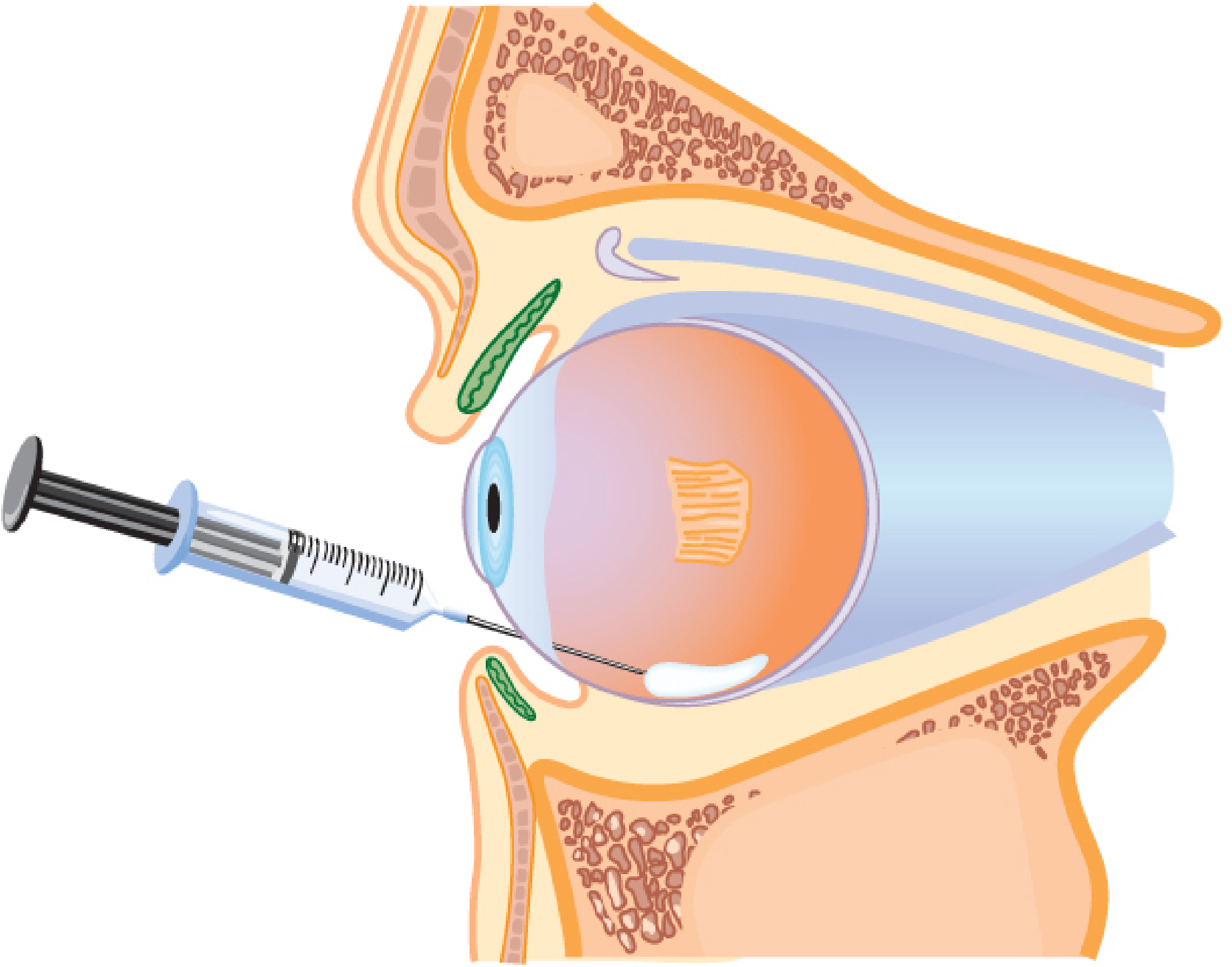
This is currently one of the most popular regional techniques in many parts of the world, overtaking needle-based blocks at many centers. It avoids sharp needle placement deep in the orbit.
Technique
- Topical anesthesia is applied first
- Conjunctiva and Tenon's fascia in the inferonasal quadrant are lifted with forceps
- A small nick is made with blunt-tipped scissors
- A blunt, curved cannula is inserted and guided into the sub-Tenon (episcleral) space past the equator of the globe
- 3-4 mL of local anesthetic is injected
The local anesthetic spreads circularly around the sclera and into extraocular muscle sheaths, producing analgesia and akinesia.
Advantages Over Needle Blocks
- No sharp needle in posterior orbit - lower risk of globe perforation (especially in highly myopic, elongated globes)
- Safer in anticoagulated patients (lower risk of retrobulbar hemorrhage)
- No risk of optic nerve sheath injection
- Rapid onset of analgesia
Newer Modifications (Incisionless Techniques)
- Ultrashort cannulae (6 mm) - reduced insertion depth
- Triport cannula (Allman technique) - no scissors needed
- Conjunctival probe - creates small tunnel without scissors, more consistent aperture, less time
- Metallic lacrimal dilator - less chemosis and postoperative hemorrhage
Complications
Globe perforation (rare), hemorrhage, rectus muscle trauma, postoperative strabismus, orbital cellulitis, optic nerve neuritis, brainstem anesthesia (reported case of death). More complications occur with longer (18-25 mm) rigid metallic cannulae; shorter (12 mm) flexible plastic cannulae are preferred.
- Barash, Cullen, and Stoelting's Clinical Anesthesia, 9e, pp. 4177-4179
4. Peribulbar (Extraconal) Block
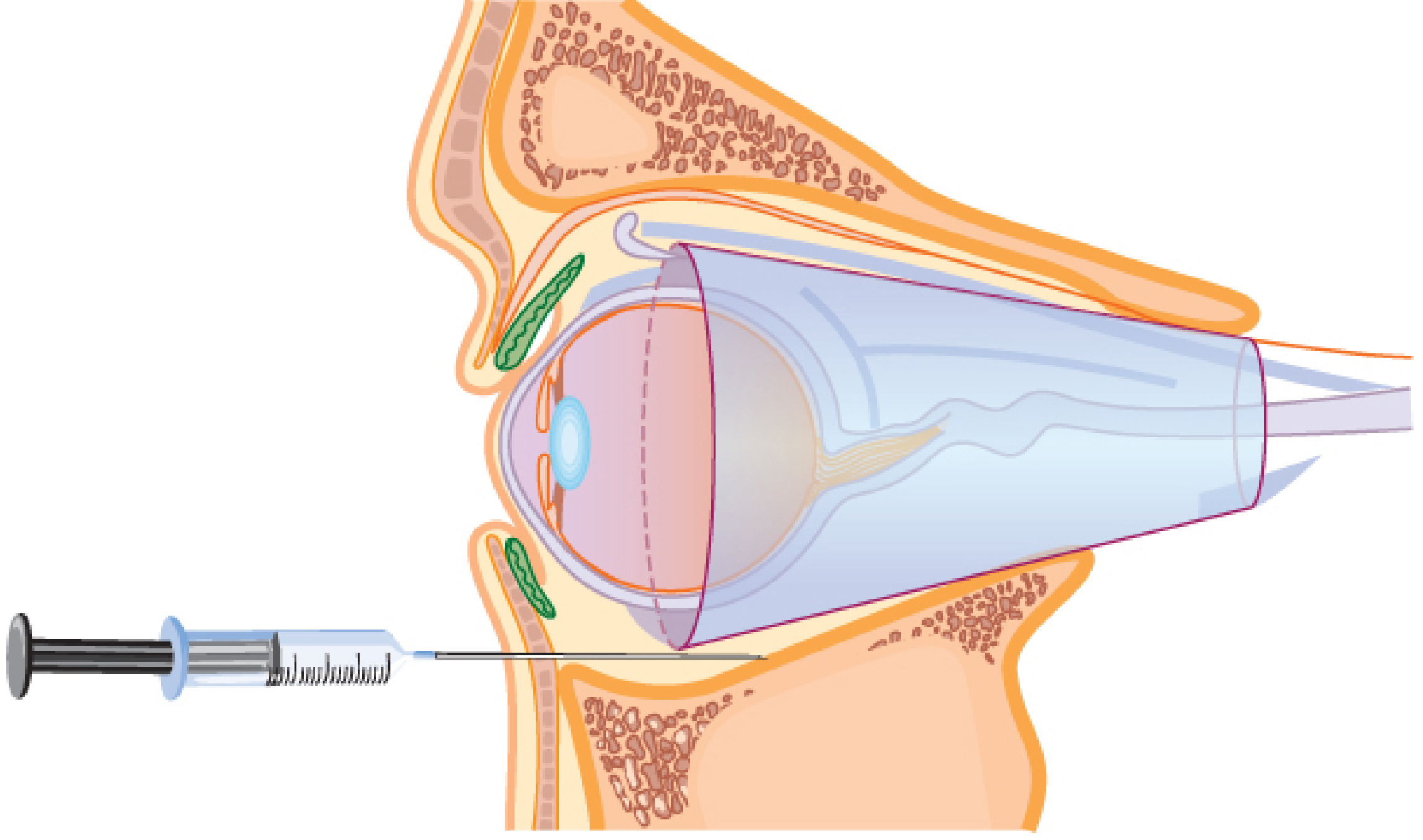
Introduced as a safer alternative to retrobulbar block. The needle is placed outside the muscle cone (extraconal), parallel to the globe, directed toward the greater wing of the sphenoid bone with minimal angulation.
- Local anesthetic diffuses from the extraconal space into the intraconal space (cadaveric dye studies confirm this)
- Theoretically safer because the needle tip stays further from the optic nerve, ophthalmic artery, and other vital structures
- Onset is slightly slower than retrobulbar; requires slightly larger volumes
- Currently preferred over retrobulbar in the United States
- Barash, Cullen, and Stoelting's Clinical Anesthesia, 9e, p. 4169
5. Retrobulbar (Intraconal) Block - Traditional Standard
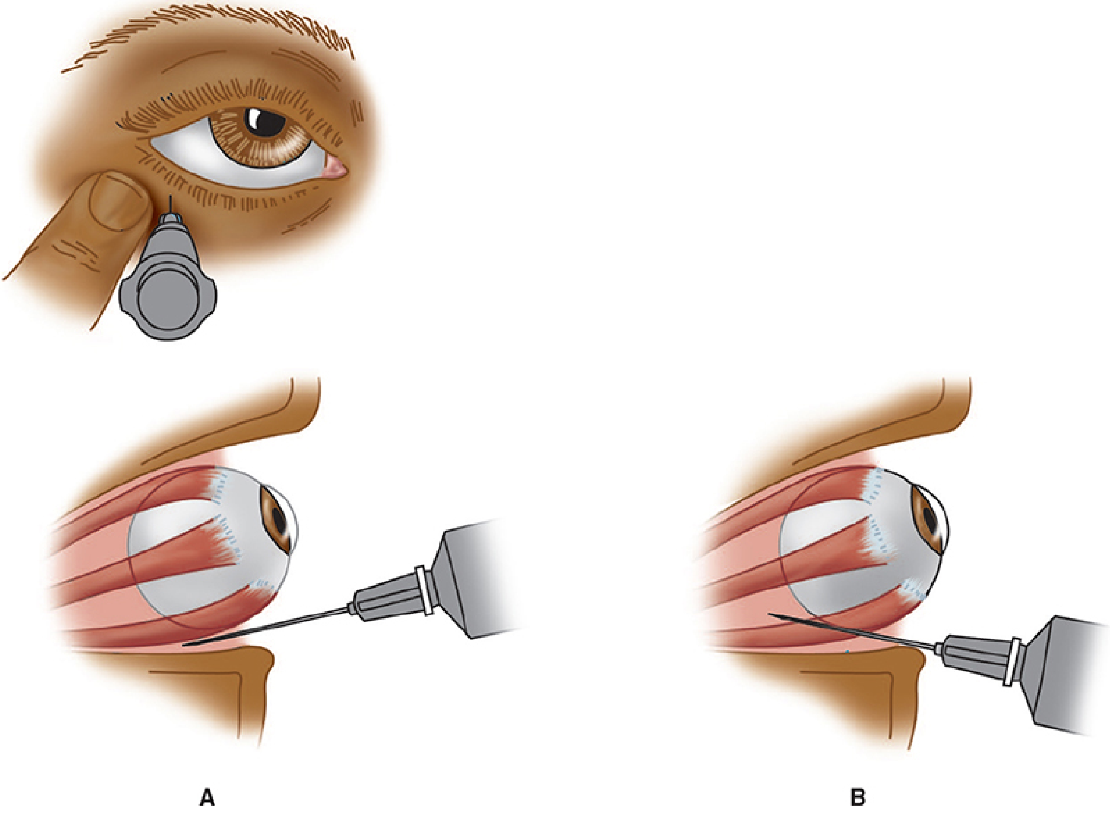
The classic technique, introduced by Atkinson in the early 20th century. A needle is directed through the lower eyelid at the junction of the middle and lateral third of the orbit (0.5 cm medial to the lateral canthus), advanced into the muscle cone toward the orbital apex.
- Patient instructed to look supranasally during needle placement
- 2-5 mL of lidocaine 2% or bupivacaine/ropivacaine 0.75% injected
- Epinephrine may be added to prolong effect and reduce bleeding
- A facial nerve block (van Lint, Atkinson, or O'Brien technique) is required to prevent blinking
- Produces excellent anesthesia and akinesia and abolishes the oculocephalic reflex
Complications (the most serious of all techniques)
| Complication | Notes |
|---|---|
| Retrobulbar hemorrhage | Arterial - precipitous rise in IOP, may need lateral canthotomy |
| Globe perforation | Higher risk in myopic, elongated globes |
| Optic nerve injury | Direct trauma or ischemia |
| Optic nerve sheath injection | Local anesthetic spreads into CSF - postretrobulbar apnea syndrome |
| Intravascular injection | Retrograde flow into ophthalmic artery → instantaneous seizure |
| Brainstem anesthesia | Spread via CSF → loss of consciousness, respiratory arrest |
| Oculocardiac reflex | Bradycardia |
- Morgan and Mikhail's Clinical Anesthesiology, 7e, pp. 1455-1458
Comparison Table
| Feature | Topical | Intracameral | Sub-Tenon | Peribulbar | Retrobulbar |
|---|---|---|---|---|---|
| Akinesia | None | None | Variable/Good | Good | Excellent |
| Analgesia | Surface only | Anterior chamber | Good | Good | Excellent |
| Globe perforation risk | None | None | Very low | Low | Higher |
| Retrobulbar hemorrhage | None | None | Very low | Low | Present |
| CNS spread risk | None | None | Very rare | Low | Present |
| Anticoagulated patients | Safe | Safe | Preferred | Caution | Risky |
| Myopic (long) eyes | Safe | Safe | Preferred | Caution | High risk |
| Typical use | Cataract, glaucoma | Cataract (adjunct) | Most procedures | Most procedures | Declining |
Sedation During Eye Blocks
Deep intraoperative sedation is almost never used (risk of apnea, aspiration, patient movement). A light regimen is standard:
- Small doses of midazolam ± fentanyl/sufentanil intraoperatively
- A brief bolus of propofol or remifentanil (0.1-0.5 mcg/kg) or alfentanil (375-500 mcg) during block placement to provide comfort and amnesia
- The patient must remain responsive, able to follow commands, and maintain their own airway
Current Trends (Summary)
- Topical + intracameral has become the dominant approach for routine cataract surgery worldwide
- Sub-Tenon is favored in the UK/New Zealand and is growing in popularity for cases needing akinesia
- Peribulbar is preferred over retrobulbar in the US for needle-based blocks
- Retrobulbar is declining due to its serious complication profile
- General anesthesia is reserved for children, uncooperative patients, and complex posterior segment surgery (20-30% of all ophthalmic cases)
Sources: Morgan and Mikhail's Clinical Anesthesiology 7e; Barash, Cullen, and Stoelting's Clinical Anesthesia 9e; Miller's Anesthesia 10e
This is a shared conversation. Sign in to Orris to start your own chat.