
I am a 2nd year medical student i want you to make histlogy notes for endocrine also provide evey histlogy slide
pituitary gland histology slide anterior posterior lobe
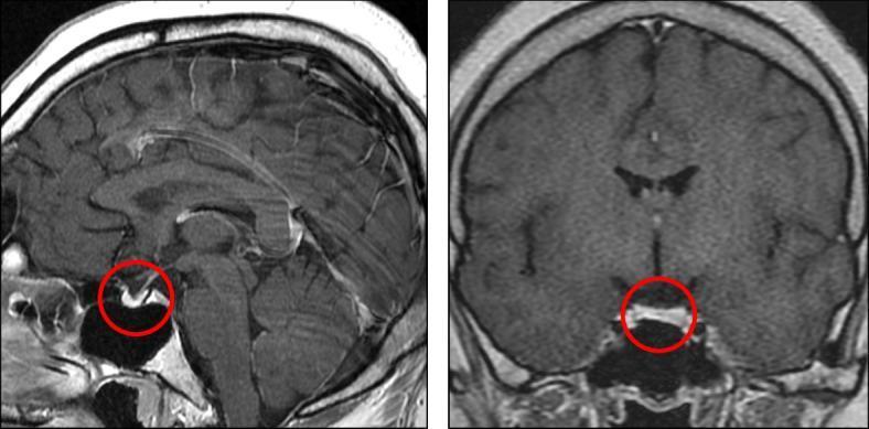
This diagnostic image consists of two panels showing a gadolinium-enhanced brain MRI in sagittal (left) and coronal (right) planes, specifically targeting the sella turcica region. The primary finding, indicated by red circles, is anterior pituitary hypoplasia. The anterior lobe of the pituitary gland appears significantly reduced in volume and height compared to standard anatomical expectations, resulting in a flattened morphology within the sella. In contrast, the posterior pituitary is visible as a hyperintense 'bright spot' on these T1-weighted images, which is a normal finding. The pituitary stalk appears midline and intact without evidence of ectopic posterior pituitary or interruption. This imaging is characteristic of combined pituitary hormone deficiency (CPHD), where underdevelopment of the adenohypophysis correlates with multi-lineage endocrine dysfunction. No other intracranial abnormalities or midline defects are visualized in these views. The scans are relevant for neuroradiology and endocrinology, illustrating the structural correlates of hypopituitarism.
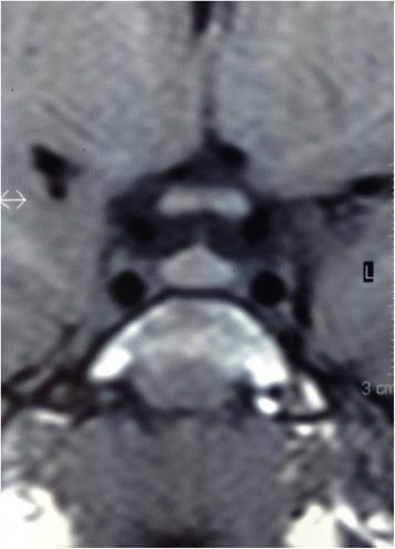
This diagnostic image consists of precontrast T1-weighted Magnetic Resonance Imaging (MRI) views of the sellar region. The visual focus is on the pituitary gland, depicted in both sagittal and coronal orientations. The gland demonstrates a homogeneous internal signal intensity with an enlarged vertical dimension and a prominent convex upper surface, which deviates from the typical flat or concave morphology. Within the sella turcica, the anterior lobe is clearly defined, and the posterior lobe displays its characteristic physiological T1-weighted hyperintensity (the 'pituitary bright spot'). The surrounding neuroanatomical structures, including the optic chiasm above the convex superior border and the adjacent cavernous sinuses, are visible. This imaging is clinically relevant for evaluating pituitary volume and morphology in the context of neuroendocrine disorders or pediatric growth abnormalities.
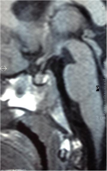
This diagnostic image is a sagittal precontrast T1-weighted MRI focusing on the sellar and suprasellar regions. It depicts the pituitary gland within the sella turcica. The image shows a homogeneous enlargement of the pituitary gland, which measures approximately 7 mm in height. A distinctive feature is the convex upper surface of the gland, which differs from the typically flat or concave surface seen in younger pediatric patients. The posterior lobe of the pituitary gland (neurohypophysis) is clearly identified by its characteristic hyperintense signal (bright spot) on the T1-weighted sequence. The anterior lobe (adenohypophysis) appears normal in signal intensity but contributes to the overall increased volume. Key anatomical landmarks visible include the optic chiasm superior to the gland and the sphenoid sinus inferiorly. This clinical imaging serves to illustrate physiological or pathological pituitary enlargement, often evaluated in the context of rapid growth or endocrine dysfunction.
thyroid gland histology follicles colloid parafollicular cells microscopy
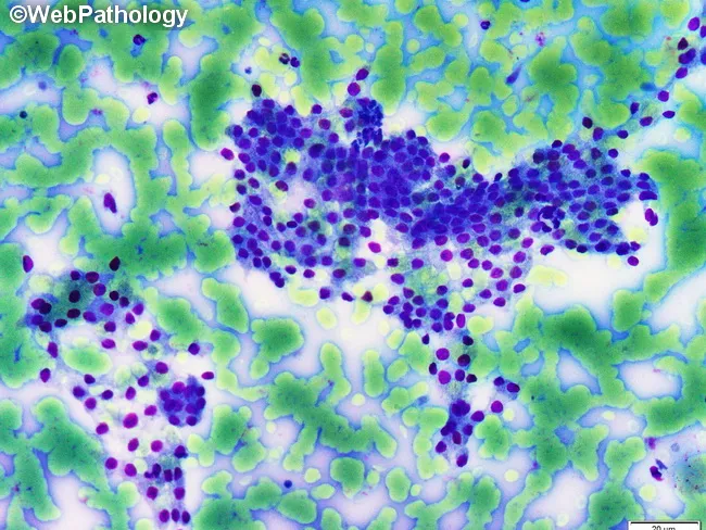
This is a light microscopy histology image of thyroid gland tissue prepared with a Romanowsky-type stain (e.g., Diff-Quik/Wright-Giemsa). The specimen shows normal-appearing follicular epithelium arranged in intact macrofollicles with abundant colloid occupying the luminal spaces. Follicular cells are evenly spaced with uniform, round to oval nuclei and scant to moderate cytoplasm, consistent with well-differentiated thyroid epithelium. The colloid background is pale blue and prominent, reflecting thyroglobulin-containing material released from follicles. The architectural pattern emphasizes large follicle units with a single layer of cuboidal to low-columnar epithelium surrounding expansive colloid pools. No prominent nuclear features of papillary carcinoma are evident, and there is no marked cellular atypia or mitotic activity in the visible fields. This image illustrates classic histology of benign thyroid tissue or colloid-rich goiter, useful for teaching follicular morphologies, thyroid folliculogenesis, and colloid dynamics. Clinically, such samples are evaluated in the context of thyroid nodules and goiter workups, with correlation to serum thyroid function tests and, when applicable, ultrasound or FNA findings. Potential educational uses include cytology-histology correlation, differential diagnosis of thyroid disease, and artifact recognition in Romanowsky-stained preparations. Images are suitable for medical education, pathology review, and database indexing. Correlative data improve student comprehension and diagnostic recall greatly.
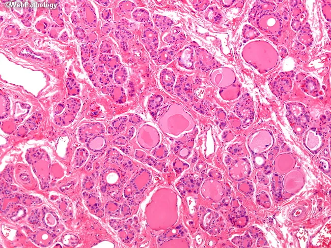
Imaging modality: Light microscopy of thyroid tissue section stained with Hematoxylin and Eosin. Specimen type: thyroid gland biopsy evaluated histologically. Imaging perspective: high-power microscopic view. Anatomical context: thyroid in the neck. Core histology demonstrates diffuse follicular hyperplasia with marked hypercellularity and reduced colloid throughout the gland. Follicles are small, compact, and tightly spaced, with scant colloid and thickened follicular epithelium. The epithelial cells show pronounced cytologic atypia, including bizarre hyperchromatic nuclei, a finding that can simulate neoplasia but is characteristic in dyshormonogenetic goiter due to inborn errors of thyroid hormone synthesis. This pattern is typically diffuse rather than nodular. Clinical significance: these histologic features correlate with congenital or neonatal hypothyroidism in severe cases and with goiter and mild hypothyroidism in milder forms; many patients present by approximately 25 years of age. Diagnostic relevance: supports dyshormonogenesis as an underlying cause of diffuse thyroid enlargement with impaired hormone production. Differential considerations include autoimmune thyroiditis and other causes of diffuse goiter; correlation with thyroid function tests and clinical history is essential. Potential clinical uses: diagnosis of dyshormonetic goiter, gene-level counseling, and guidance of hormone replacement therapy. This histology aids educational cases and research on thyroid hormone synthesis disorders, and informs patient management strategies.
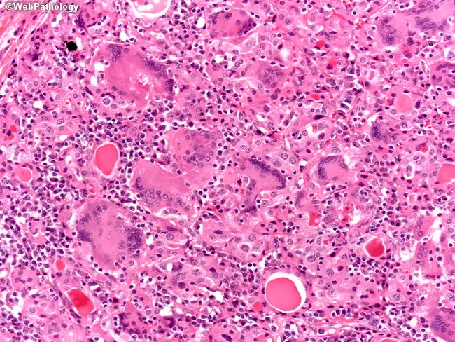
This case utilizes light microscopy of thyroid tissue stained with Hematoxylin and Eosin. The high-magnification image reveals granulomatous thyroiditis characterized by numerous foreign body giant cells and a dense chronic inflammatory infiltrate composed of lymphocytes and plasma cells. Interspersed is residual thyroid parenchyma containing colloid-filled follicles, some diminished or disrupted by inflammation. The tissue architecture shows granulomas centered around disrupted colloid with multinucleated giant cells phagocytosing colloid material, a classic appearance of subacute granulomatous (de Quervain) thyroiditis. There is relative preservation of overall gland structure with focal areas of follicular destruction. These features distinguish subacute thyroiditis from autoimmune thyroiditis (Hashimoto) and bacterial abscesses. Clinically, this pattern correlates with a transient thyrotoxic phase followed by hypothyroidism in many patients; HLA-B35 haplotype association has been reported; etiology often linked to viral trigger; most cases resolve with supportive care. The image supports diagnosis of granulomatous thyroiditis in the appropriate clinical context, guiding management toward observation and symptomatic treatment. This histology is relevant for education in endocrine pathology, differential diagnosis of thyroiditis, and research into post-viral inflammatory thyroid injury. The depiction is representative for pathology teaching slides and radiology correlations in endocrinology. Educational value for residents, fellows, and medical students with accurate histology correlations.
adrenal gland histology cortex medulla zones microscopy
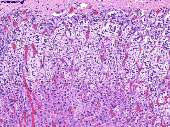
Imaging modality: Light microscopy of hematoxylin and eosin stained adrenal gland cortex section, intermediate magnification. The specimen reveals the three concentric zones of the adrenal cortex arranged from capsule to medulla: the zona glomerulosa immediately beneath the capsule forms small, compact clusters of darker-staining parenchymal cells; the zona fasciculata comprises the majority of cortex with radially oriented cords of pale, lipid-rich cells exhibiting a spongy cytoplasm; the zona reticularis lies nearest the medulla, composed of anastomosing cords of polygonal cells with acidophilic cytoplasm. The cortex shows preserved architecture with slender septa and numerous sinusoidal capillaries evident between cords. The cells demonstrate typical endocrine morphology: small dense nuclei, minimal cytoplasmic granularity in glomerulosa; prominent lipid droplets in fasciculata; reticularis cells with irregular outlines. No overt inflammatory infiltrate, neoplastic glands, or disrupted zonation are observed in this field, consistent with normal histology. Clinically, this zonation underpins steroidogenesis: mineralocorticoids from glomerulosa regulated by angiotensin II and potassium; glucocorticoids from fasciculata, including cortisol; androgen precursors from reticularis. Diagnostic significance lies in confirming trilaminar architecture and zonal differentiation essential for endocrinology education and pathologic assessment of adrenal disorders such as hyperplasia, adenomas, or cortisol-producing tumors. This slide is valuable for education, histology atlases, and research references.
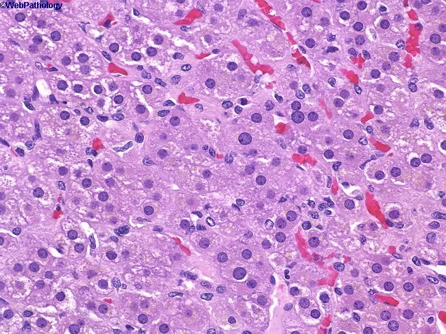
Imaging modality: Light microscopy of adrenal gland tissue, Hematoxylin and Eosin (H&E) stained section, viewed under brightfield illumination at high magnification (approximately 400x). Anatomical location: adrenal cortex with zona reticularis occupying the deepest corticoid layer, immediately superficial to the adrenal medulla, behind zona fasciculata. Visual features: cells arranged in anastomosing cords and small nests; cytoplasm is acidophilic and granular; nuclei are round to vesicular with prominent punctate nucleoli; capillary sinusoids are intermixed, producing a lobular vascular network. The zona reticularis lies between the zona fasciculata and the medulla, forming a reticular, fine meshwork. The cellular morphology indicates steroidogenic chromaffin-adjacent cells with robust endoplasmic reticulum and lipid-poor cytoplasm relative to fasciculata. Notable features include tight cell-to-cell contacts, vascularized stroma, and delineation from the surrounding zones. Pathophysiology/diagnostic significance: Normal zonation of the adrenal cortex is demonstrated; zona reticularis is responsible for glucocorticoid and sex hormone synthesis (androgen precursors), contributing to the endocrine milieu. Clinical relevance: understanding this histology supports differential diagnosis of adrenal cortical neoplasms and endocrine disorders; potential use in educational contexts, research on steroidogenesis, and histopathology training. This image serves as a reference for adrenal cortical anatomy, steroidogenic cell morphology, and the interface with the medulla.
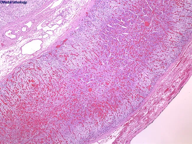
This histology image depicts a low-power hematoxylin-eosin stained cross-section of the mammalian adrenal gland, showing the capsule and the steroidogenic cortex with its three distinct zonal architectures. The adrenal cortex comprises approximately 90% of the gland and is organized into zona glomerulosa (outermost), zona fasciculata (widest middle layer), and zona reticularis (inner zone) adjacent to the medulla. The cortex appears pale and vesiculated due to lipid-rich, foamy cytoplasm within steroid-producing cells, arranged in cords and fascicles separated by sinusoids. At this low magnification, the boundary between capsule and cortex is evident, although a detailed medullary region is not fully resolved. The image demonstrates normal cortical histology without overt neoplasia, hemorrhage, or fibrosis. The adrenal cortex synthesizes glucocorticoids, mineralocorticoids, and androgens under ACTH regulation, with zonation reflecting functional segregation: zona glomerulosa producing aldosterone, zona fasciculata secreting cortisol, and zona reticularis generating dehydroepiandrosterone. Clinically, this pattern is essential for understanding endocrine physiology and adrenal disorders. This composite image serves educational reference for histology, pathology, and medical education, enabling comparison to hyperplasia, adenomas, or pheochromocytoma in other sections. The slide illustrates capsule integrity, distinct cortical zones, and ordinary vascular architecture relevant to teaching. Useful for exams, case discussions, and comparative pathology analyses exercises.
pancreatic islets of Langerhans histology alpha beta cells
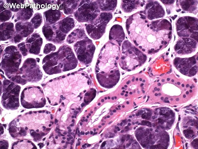
Histology micrograph of human pancreatic tissue analyzed by brightfield light microscopy after Hematoxylin and Eosin staining. The specimen shows classic lobular exocrine pancreas with densely packed acinar cells and intervening islets of Langerhans. Acinar cells display basophilic cytoplasm, basally located round nuclei, and granular eosinophilic zymogen content, contributing to the characteristic purple-blue cellular nests on the slide. The lobular architecture is preserved, with clear separation by connective septa. Within the ducts, cuboidal to low columnar epithelium lines the ductal lumina; small interlobular ducts are visible as white-staining channels. Vascular structures are present, containing erythrocytes. Adipose tissue is variably present at the periphery, consistent with normal pancreatic parenchyma. Notable features include the pale-staining islets that contrast with the intensely staining acinar lobules, providing a focal example of endocrine-exocrine juxtaposition. No overt inflammatory infiltrate, edema, necrosis, or fibrotic changes are evident in this field. Clinically, this image serves as a reference for normal pancreatic histology and as a teaching aid in pathology education. Potential diagnostic uses include benchmarking against pancreatopathic states such as pancreatitis or neoplasia, and facilitating differential diagnosis discussions in exam questions and research reviews. This image supports multiple learning modalities, including atlas reference, slide review, and examination preparation resources.
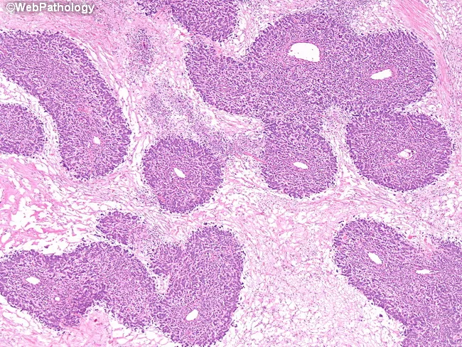
Imaging modality and technique: Brightfield light microscopy of a hematoxylin and eosin stained pancreatic tissue section. The specimen presents pancreatic parenchyma with distinct islets of Langerhans embedded within predominantly acinar lobules. The islets appear as pale, rounded to oval nests that contrast with the surrounding basophilic exocrine cells, producing a characteristic lobulated architecture. Within each islet, endocrine cells display small to medium-sized nuclei with finely dispersed chromatin and scant cytoplasm, arranged in cords and clusters separated by delicate stromal septa. Ductal profiles and occasional small ducts are visible at the periphery, reflecting normal pancreatic histology. The exocrine component is highly cellular, with densely staining acinar cells and visible zymogen-containing granules; connective tissue septa provide lobular delineation. There is no obvious cytologic atypia, mitotic activity, or invasion; no confluent tumor masses are evident. The regional morphology is consistent with healthy pancreatic tissue or benign variation in islet density. This image is valuable for education on endocrine–exocrine pancreatic architecture, comparative pathology, and teaching of islet distribution, size variability, and relationships to exocrine pancreatic tissue. Potential clinical relevance includes assessment of endocrine function and differentiation from islet cell neoplasms in surgical specimens. Interpretations should consider patient history, laboratory data, imaging correlation and context.
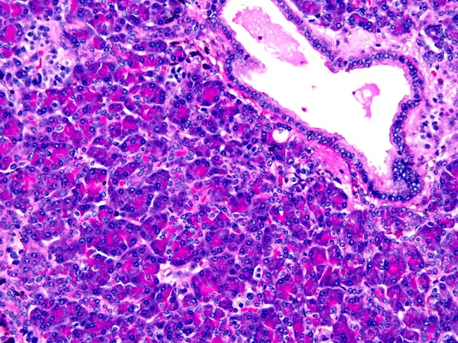
Histology image of ectopic pancreas within the gastric wall. Acinar cells with granular eosinophilic cytoplasm form pancreatic acini adjacent to ductal structures; islets of Langerhans appear as lighter, rounded cell clusters. The specimen demonstrates Heinrich type I ectopia, containing all pancreatic elements (acini, ducts, islets) embedded in gastric tissue, with preserved lobular organization and intervening gastric mucosa. The micrograph is stained with Hematoxylin and Eosin, highlighting basophilic nuclei and eosinophilic cytoplasm; acini display zymogen granules, ductal epithelium lines small ducts, and endocrine islets are dispersed in the exocrine pancreas. The lesion is a congenital anomaly presenting in the stomach, usually incidental but clinically relevant for differential diagnosis of submucosal gastric lesions. Diagnostic significance lies in recognizing heterotopic pancreatic tissue to avoid misdiagnosis as gastric neoplasm or inflammatory process. Potential clinical use cases include educational reference for surgical pathology, gastroenterology differential diagnosis, and radiologic-pathologic correlation. This image illustrates the histomorphology of ectopic pancreas, enabling recognition of all pancreatic components within gastric tissue and supports discussion of Heinrich classification, ectopic pancreas prevalence, and associated clinical scenarios such as pancreatitis or obstruction when symptomatic. The image thereby serves as a reference for teaching, diagnostic reasoning, and histopathological correlation in gastrointestinal pathology and education.
parathyroid gland histology chief cells oxyphil cells
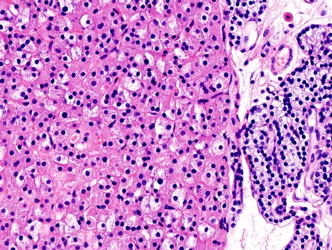
This is a light microscopic histology image of parathyroid tissue stained with hematoxylin and eosin (H&E). The left side shows an oxyphil cell–rich parathyroid adenoma characterized by sheets and cords of large, polygonal cells with abundant eosinophilic cytoplasm and centrally located round to ovoid nuclei. The dense eosinophilic cytoplasm is due to abundant mitochondria, giving the cells a characteristic acidophilic appearance. The stroma is relatively scant, with delicate capillary networks and minimal hematopoietic infiltration. A normal parathyroid gland is visible on the right for comparison, containing smaller, less eosinophilic chief cells arranged in cords with lighter cytoplasm and a richer vascular stroma. The juxtaposition helps distinguish adenomatous parathyroid tissue from normal parenchyma. Clinically, oxyphil cell–predominant adenomas can cause primary hyperparathyroidism, though nonfunctional adenomas exist. Pathology notes include a well-circumscribed lesion lacking significant mitotic activity or atypia, consistent with benign parathyroid adenoma rather than carcinoma. This image is valuable for educational purposes in surgical pathology, endocrine pathology, and histology training, illustrating cellular morphology, dysplastic features, and differential diagnosis with parathyroid hyperplasia. Correlation with serum calcium, parathyroid hormone levels, and radiological localization studies is recommended in clinical workups. It also aids understanding of oxyphilic differentiation and benign behavior in educational settings today.
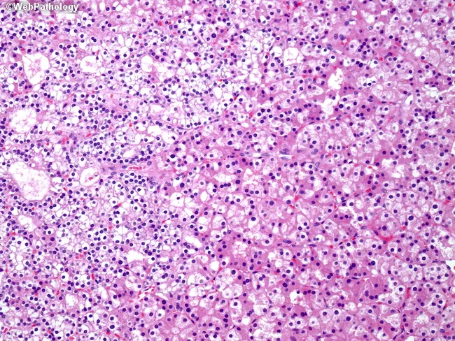
Imaging modality: Light microscopy of Hematoxylin and Eosin stained parathyroid tissue section (parathyroid adenoma). Anatomy: parathyroid gland within the cervical neck, typically near the thyroid; left half features sheets of chief cells; right half shows oxyphil cells; other cell types include water-clear cells and transitional cells. Histology: chief cells with amphophilic to lightly eosinophilic cytoplasm and conspicuous intracellular lipid/fat droplets; oxyphil cells with abundant granular eosinophilic cytoplasm and small pyknotic nuclei; cytoplasmic features reflect numerous mitochondria; cellular arrangement is lobular and highly cellular; vascular stroma is variable; fatty stroma reduced relative to normal parathyroid tissue. Pathology: parathyroid adenoma characterized by clonal proliferation of chief cells with admixture of oxyphil and water-clear cells; cytologic atypia minimal; mitotic activity low; absence of invasive growth in typical cases; indicates benign neoplasm; clinical correlation: hyperparathyroidism due to autonomous parathyroid hormone secretion; diagnostic significance includes correlation with elevated PTH and calcium levels; differential diagnoses include parathyroid hyperplasia, parathyroid carcinoma, and non-neoplastic thyroid tissue; educational use: illustrates cellular heterogeneity and diagnostic features used to differentiate hyperplasia from adenoma by architectural pattern, fat content, and cytoplasmic characteristics; suitable for pathology teaching, exam review, and database annotation. Representative images aid learners in recognizing parathyroid neoplasia and guiding surgical management.
pineal gland histology pinealocytes brain sand corpora arenacea
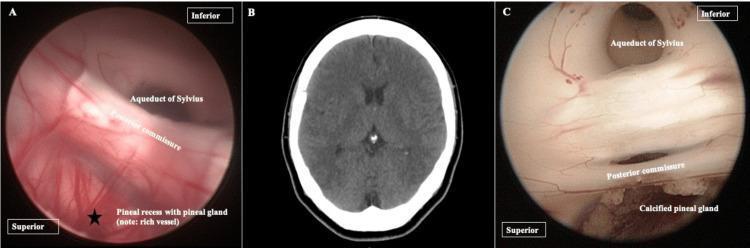
This composite educational graphic illustrates the anatomy and pathology of the pineal gland through a comparison of radiological and neuroendoscopic modalities. Panel A presents a neuroendoscopic view of the posterior third ventricle, highlighting the pineal recess (marked with a star), the posterior commissure, and the Aqueduct of Sylvius. This view emphasizes the rich vascularity of the pineal region. Panel B is a non-contrast axial CT scan of the brain, demonstrating a physiological hyperdense calcification of the pineal gland (circled) located at the midline. Panel C provides a corresponding neuroendoscopic view of a calcified pineal gland (corpora arenacea), showing a distinct 'brain sand' or granular texture below the posterior commissure and Aqueduct of Sylvius. This comparison illustrates how physiological calcifications, commonly seen as hyperdensities on diagnostic CT imaging, appear as physical anatomical features during neurosurgical procedures. The content is suitable for neurosurgical training and advanced anatomical studies focusing on the ventricular system and pineal region pathology.
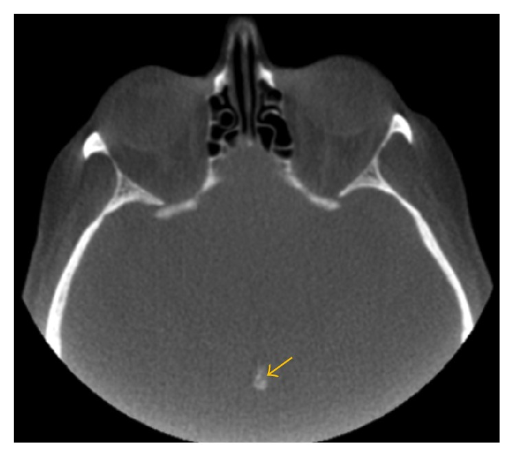
**Imaging Modality:** Non-contrast Computed Tomography (CT) of the head. **Anatomical Region:** Axial section at the level of the midbrain and suprasellar cistern, including the posterior cranial fossa and orbits. **Observed Findings:** There is a distinct, hyperdense, well-circumscribed focus located in the midline of the brain, corresponding to the anatomical position of the pineal gland. This finding represents physiological pineal gland calcification (corpora arenacea). The calcification is centrally positioned with no evidence of mass effect or midline shift. **Characteristic Visual Features:** The lesion exhibits high attenuation (radiodensity) consistent with calcium deposition. An orange arrow highlights the specific site of the calcification. Surrounding brain parenchyma appears grossly homogeneous within the limits of the bone window settings. **Diagnostic Context:** This is a common, typically benign, age-related finding in adult neuroimaging. It serves as a critical radiographic landmark for assessing midline structures and identifying potential pineal region pathology or shift due to intracranial pressure.
anterior pituitary acidophils basophils chromophobes histology
This diagnostic image consists of two panels showing a gadolinium-enhanced brain MRI in sagittal (left) and coronal (right) planes, specifically targeting the sella turcica region. The primary finding, indicated by red circles, is anterior pituitary hypoplasia. The anterior lobe of the pituitary gland appears significantly reduced in volume and height compared to standard anatomical expectations, resulting in a flattened morphology within the sella. In contrast, the posterior pituitary is visible as a hyperintense 'bright spot' on these T1-weighted images, which is a normal finding. The pituitary stalk appears midline and intact without evidence of ectopic posterior pituitary or interruption. This imaging is characteristic of combined pituitary hormone deficiency (CPHD), where underdevelopment of the adenohypophysis correlates with multi-lineage endocrine dysfunction. No other intracranial abnormalities or midline defects are visualized in these views. The scans are relevant for neuroradiology and endocrinology, illustrating the structural correlates of hypopituitarism.
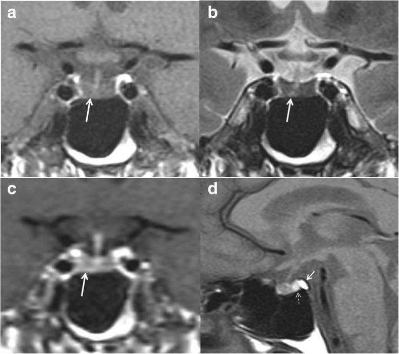
This diagnostic image set consists of four MRI views of the pituitary gland (sella turcica region). Panel (a) shows an unenhanced coronal T1-weighted image where the anterior pituitary displays a low-intermediate signal intensity. Panel (b) is a coronal T2-weighted view exhibiting significantly decreased signal (hypointensity) within the anterior gland. Panel (c) provides an early coronal T1-weighted image following intravenous contrast injection, demonstrating reduced and delayed enhancement of the pituitary parenchyma. Panel (d) shows an unenhanced sagittal T1-weighted view depicting a small (atrophic) anterior pituitary gland. Key landmarks include the normal hyperintense signal of the posterior pituitary (bright spot), the distinct hyperintensity of the fatty marrow in the dorsum sellae, and a normal-appearing pituitary stalk. These findings—diffuse hypointensity on T1 and T2 sequences with reduced gadolinium enhancement—are characteristic of infiltrative disorders such as pituitary amyloidosis, which can lead to panhypopituitarism.
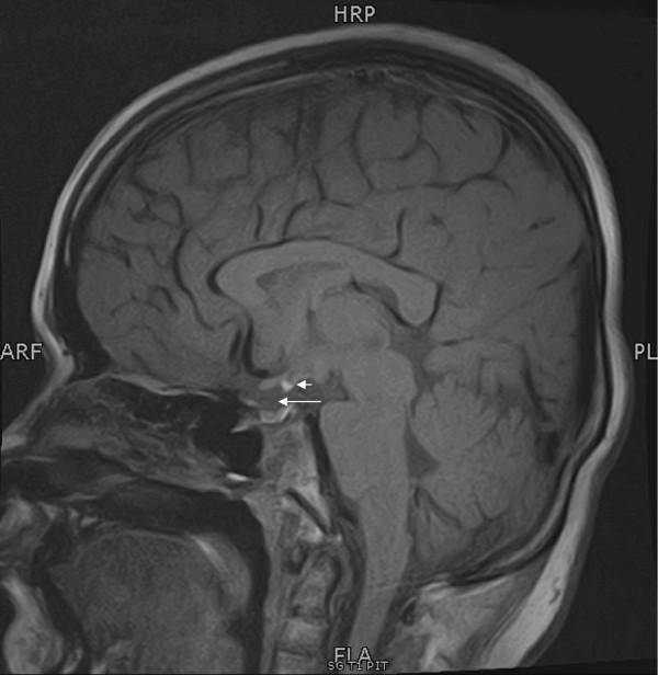
This diagnostic image is a midline sagittal T1-weighted MRI scan of the brain, specifically focusing on the sellar and suprasellar regions. It demonstrates Pituitary Stalk Interruption Syndrome (PSIS). The anterior pituitary gland is visible within the sella turcica, exhibiting normal signal intensity but appearring somewhat small (hypoplastic). A long white arrow indicates the expected location of the pituitary stalk (infundibulum), which is notably absent or non-visualized. A short white arrow identifies an ectopic posterior pituitary, seen as a characteristic T1-hyperintense focus (bright spot) located at the median eminence of the hypothalamus rather than its normal posterior position in the sella. This triad of findings—ectopic posterior pituitary bright spot, absent or attenuated pituitary stalk, and anterior pituitary hypoplasia—is a classic radiological presentation of congenital hypopituitarism. The surrounding neuroanatomical structures, including the corpus callosum, pons, and cerebellum, appear within normal limits.
normal thyroid gland histology follicular cells colloid cuboidal epithelium H&E stain
This is a light microscopy histology image of thyroid gland tissue prepared with a Romanowsky-type stain (e.g., Diff-Quik/Wright-Giemsa). The specimen shows normal-appearing follicular epithelium arranged in intact macrofollicles with abundant colloid occupying the luminal spaces. Follicular cells are evenly spaced with uniform, round to oval nuclei and scant to moderate cytoplasm, consistent with well-differentiated thyroid epithelium. The colloid background is pale blue and prominent, reflecting thyroglobulin-containing material released from follicles. The architectural pattern emphasizes large follicle units with a single layer of cuboidal to low-columnar epithelium surrounding expansive colloid pools. No prominent nuclear features of papillary carcinoma are evident, and there is no marked cellular atypia or mitotic activity in the visible fields. This image illustrates classic histology of benign thyroid tissue or colloid-rich goiter, useful for teaching follicular morphologies, thyroid folliculogenesis, and colloid dynamics. Clinically, such samples are evaluated in the context of thyroid nodules and goiter workups, with correlation to serum thyroid function tests and, when applicable, ultrasound or FNA findings. Potential educational uses include cytology-histology correlation, differential diagnosis of thyroid disease, and artifact recognition in Romanowsky-stained preparations. Images are suitable for medical education, pathology review, and database indexing. Correlative data improve student comprehension and diagnostic recall greatly.
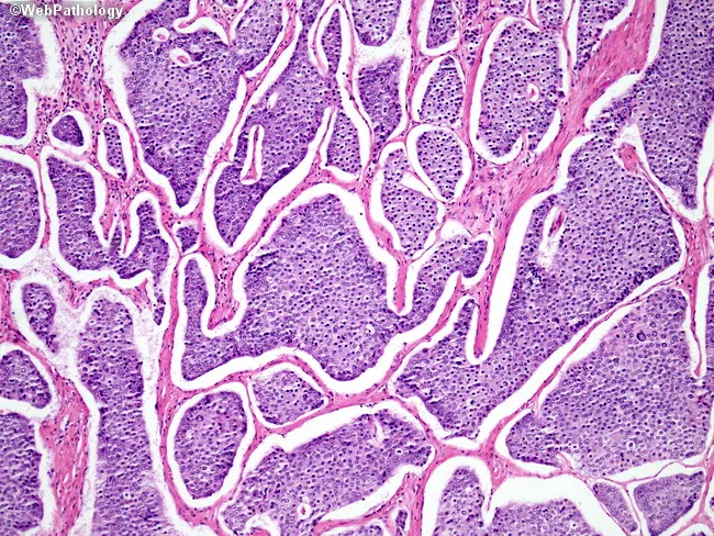
This is a light micrograph of thyroid gland tissue prepared for histopathology and stained with Hematoxylin and Eosin (H&E). The sample reveals multiple spherical to irregular follicles of varying size separated by wispy fibrous septa. The colloid-filled lumens are pink to pale pink, while the surrounding follicular epithelium is a uniform single layer of cuboidal to low-columnar cells with round to oval nuclei and inconspicuous nucleoli. No nuclear grooves, inclusions, or pseudoinclusions characteristic of papillary carcinoma are evident. The stromal background is relatively scant with no dense lymphocytic infiltrate or Hurthle cell metaplasia apparent. The architecture is reminiscent of normal thyroid parenchyma or benign nodular colloid goiter pattern, where follicle size varies and colloid predominates. There are no mitotic figures or cytologic atypia observed. This image demonstrates classic histology of endocrine gland tissue with preserved colloid and regular follicle lining, suitable as reference for normal thyroid architecture in educational, diagnostic, and research contexts. Clinically, such histology supports benign thyroid pathology when correlated with imaging and labs. In malignant differentials, absence of papillary nuclear features or tall columnar cells argues against papillary thyroid carcinoma; however, definitive diagnosis should consider capsule invasion and margin status in surgical specimens.
🔬 Endocrine System — Histology Notes
For 2nd Year Medical Students
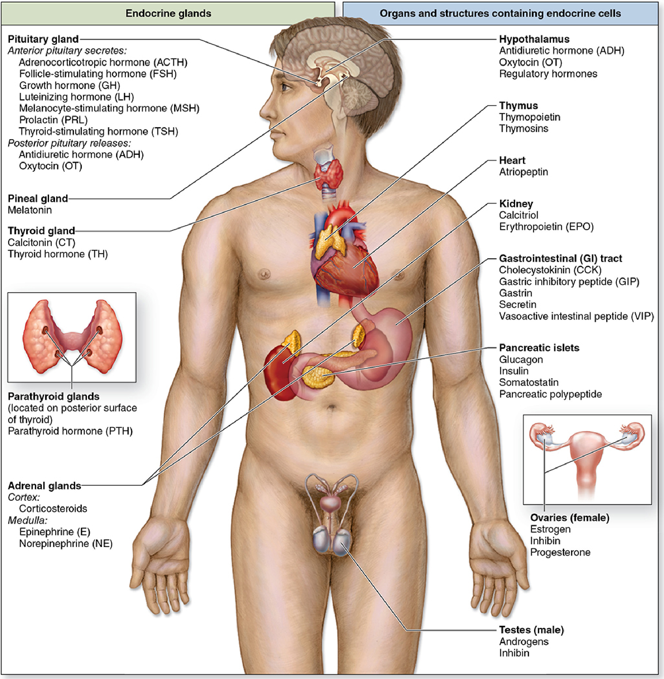
Secretory cells of endocrine glands release hormones directly into the bloodstream - no ducts. Most endocrine cells are epithelial in origin and arranged as cords or clusters around rich capillary networks. - Junqueira's Basic Histology 17e, p.996
1. PITUITARY GLAND (HYPOPHYSIS)
General Facts
- Weight: ~0.5 g; dimensions ~10 × 13 × 6 mm
- Located in the sella turcica of the sphenoid bone
- Dual embryonic origin:
- Adenohypophysis (anterior) - from oral ectoderm (Rathke's pouch)
- Neurohypophysis (posterior) - from neuroectoderm (diencephalon floor)
Parts of the Pituitary
| Part | Origin | Components |
|---|---|---|
| Adenohypophysis | Oral ectoderm | Pars distalis, Pars tuberalis, Pars intermedia |
| Neurohypophysis | Neural tissue | Pars nervosa (posterior lobe), Infundibulum (stalk) |
Adenohypophysis (Anterior Pituitary) - Histology
Cell Types - The Classic Triad
| Cell Type | % of Cells | Staining | Hormones Secreted |
|---|---|---|---|
| Acidophils | ~50% | Pink/red (eosinophilic) | GH (somatotrophs), Prolactin (lactotrophs) |
| Basophils | ~15% | Blue/purple | TSH (thyrotrophs), FSH & LH (gonadotrophs), ACTH (corticotrophs) |
| Chromophobes | ~35% | Pale/unstained | Degranulated or stem cells; minimal secretory activity |
Exam tip: "Acidophils = GH + PRL" (both are protein hormones stored in acidophilic granules). "Basophils make the glycoprotein hormones" (TSH, FSH, LH, ACTH).
Key Histological Features
- Cells arranged in irregular cords and clusters
- Rich sinusoidal fenestrated capillary network between cords
- Herring bodies are NOT found here (they are in the posterior pituitary)
- Portal blood vessels from hypothalamus carry releasing/inhibiting hormones to control secretion
Neurohypophysis (Posterior Pituitary) - Histology
- Does NOT produce hormones - it stores and releases them
- Hormones (ADH/vasopressin and oxytocin) are made in hypothalamic nuclei (supraoptic and paraventricular) and transported down axons
- Pituicytes - glial support cells (the main cellular component); elongated, with irregular nuclei
- Herring bodies - dilated axon terminals filled with secretory vesicles; appear as homogeneous pink blobs on H&E
- Rich capillary network for hormone release
🔑 Key Slide Identification Point:
- Posterior pituitary = pale, fibrous, neuroglia-like tissue with no distinct cell cords
- Anterior pituitary = cellular cords with clear pink/purple staining cells
2. THYROID GLAND
Gross/General
- Two lobes + isthmus, anterior to trachea just below larynx
- Most highly vascularized gland in the body
- Only endocrine gland that stores hormone extracellularly (in colloid)
- Sufficient stored hormone for up to 3 months
🔬 Histology Slide - Thyroid Follicles
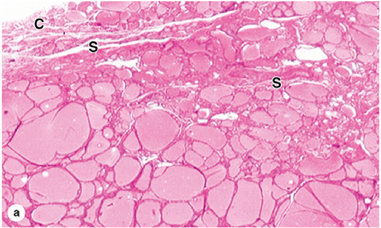
Key Histological Features
Thyroid Follicles
- Spherical units of variable diameter, packed together
- Lined by simple follicular epithelium (thyrocytes)
- Central lumen filled with eosinophilic (pink) colloid containing thyroglobulin
- Separated by sparse reticular connective tissue with fenestrated capillaries
Follicular Cell (Thyrocyte) Morphology - Activity-Dependent
| State | Cell Shape | Nucleus | Colloid |
|---|---|---|---|
| Active (high TSH) | Low columnar | Round, euchromatic | Scalloped edges (resorption lacunae/Sanderson polsters) |
| Inactive/resting | Cuboidal to squamous | Flat | Smooth borders, abundant |
Parafollicular Cells (C-cells)
- Located between follicles or within follicle wall but do not reach the lumen
- Larger than follicular cells; pale cytoplasm
- Produce calcitonin (lowers blood Ca²⁺)
- Neural crest origin (unlike follicular cells which are endodermal)
🔑 Slide Recognition Tips:
- Thyroid = follicles full of pink colloid - the most distinctive feature
- Look for "scalloping" of colloid (resorption lacunae) at the follicular cell border in active glands
- Parafollicular cells = paler, larger cells tucked between follicles
3. PARATHYROID GLANDS
General
- 4 glands, each ~3-8 mm, located on the posterior surface of the thyroid
- Covered by thin connective tissue capsule; thin septa divide into irregular lobules
- Rich capillary network throughout
🔬 Histology Slide - Parathyroid
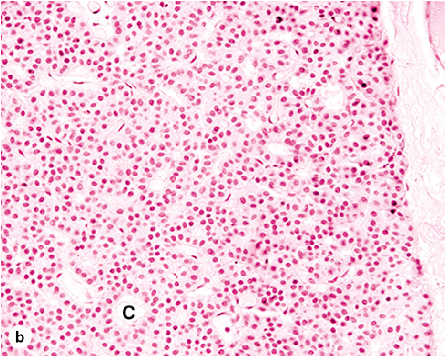
Cell Types
| Cell Type | Size | Cytoplasm | Nucleus | Function |
|---|---|---|---|---|
| Principal (Chief) cells | Small (~8 µm) | Pale, lightly eosinophilic, lipid droplets | Round, central | Secrete PTH (main cell) |
| Oxyphil cells | Large | Intensely eosinophilic (packed mitochondria) | Small, pyknotic | Non-functional (degenerated chief cells); increase with age |
| Water-clear cells | Large | Clear, glycogen-filled | Small | Rare; occasional variant of chief cell |
Key Points
- Chief cells arranged in cords and sheets around capillaries
- PTH raises blood calcium by: stimulating osteoclasts, renal Ca²⁺ reabsorption, and Vitamin D activation
- With age: adipocytes increasingly replace secretory cells (>50% in elderly)
- Oxyphil cells appear after puberty, increase with age
🔑 Slide Recognition:
- Parathyroid = dense cords of small pale cells with round nuclei + richly vascular stroma
- Looks "crowded" compared to thyroid
- Do NOT confuse with thyroid tissue (no follicles, no colloid)
4. ADRENAL GLANDS
Gross/General
- Paired; sit atop kidneys; ~8 g total
- Two distinct regions with different embryonic origins:
- Cortex - from mesoderm → steroid hormones
- Medulla - from neural crest → catecholamines
🔬 Histology Slides - Adrenal Gland
Adrenal Cortex Histology
The Three Zones (from outside in: "GFR")
| Zone | Arrangement | Cell Features | Hormone | Control |
|---|---|---|---|---|
| Zona Glomerulosa (outer) | Small clusters/arches | Small, dark cells; little lipid; no lipid vacuoles | Aldosterone (mineralocorticoid) | Angiotensin II, K⁺ |
| Zona Fasciculata (middle, widest) | Long radial cords (2 cells wide) | Large, pale "spongiocytes" with abundant lipid vacuoles; foamy appearance | Cortisol (glucocorticoid) | ACTH |
| Zona Reticularis (inner) | Anastomosing irregular cords | Smaller, more eosinophilic; lipid-poor; lipofuscin granules | Androgens (DHEA) | ACTH |
Ultrastructure of Cortical Cells (steroid-secreting features)
- Abundant smooth ER (SER) - steroid synthesis enzymes
- Mitochondria with tubular cristae (vs. shelf-like in most cells)
- Many lipid droplets (cholesterol stores for steroidogenesis)
🔑 Slide Recognition Tips for Cortex:
- Zona glomerulosa: small dark clusters just under capsule
- Zona fasciculata: pale/vacuolated ("spongy") cells in straight columns - the thickest zone
- Zona reticularis: darker, net-like cords next to the medulla
Adrenal Medulla Histology
- Large, chromaffin cells (pheochromocytes) - pale pink, polyhedral, arranged in cords/nests
- Chromaffin reaction - turn brown with chromate fixatives (due to oxidized catecholamines)
- Two cell types: epinephrine cells (more common, finer granules) and norepinephrine cells (fewer, larger dense granules)
- Modified postganglionic sympathetic neurons - innervated by preganglionic fibers
- Dual blood supply: arterial (medullary arterioles) + venous (cortical drainage)
- Ganglion cells (true neurons) scattered in the medulla - a unique feature
🔑 Slide Recognition Tips for Medulla:
- Central location
- Pale cells in irregular groups
- Large central vein visible
- Ganglion cells may be seen scattered among chromaffin cells
5. PANCREATIC ISLETS (Islets of Langerhans)
Location
- Scattered throughout the exocrine pancreas
- Approximately 1-2 million islets in the human pancreas
- Islets are paler than surrounding acini on H&E
🔬 Histology Slides - Pancreatic Islets
Islet Cell Types
| Cell Type | % in Islet | Location in Islet | Hormone | Staining |
|---|---|---|---|---|
| B (Beta) cells | ~70% | Central | Insulin | Pale, slightly basophilic |
| A (Alpha) cells | ~20% | Periphery | Glucagon | Slightly more eosinophilic |
| D (Delta) cells | ~5-10% | Periphery | Somatostatin | Hard to distinguish on H&E |
| PP (F) cells | ~5% | Periphery | Pancreatic polypeptide | Requires IHC |
Key Histological Features
- Islet cells arranged in irregular cords and clusters
- Rich fenestrated capillary network (more than exocrine pancreas)
- On H&E, all islet cells look similar - immunohistochemistry (IHC) needed to distinguish types
- Delicate reticular fiber framework
- Surrounded by a thin fibrous capsule-like layer separating them from acini
Blood Supply - "Portal" Flow
- Blood flows from islet center outward - B cells (center) are perfused first, then A and D cells (periphery) are exposed to insulin-rich blood (paracrine regulation)
🔑 Slide Recognition:
- Islets = pale circular nests in a sea of dark purple acinar cells
- Islet cells smaller and paler than acinar cells
- No zymogen granules (unlike acinar cells)
- Centroacinar cells visible at acinar-duct junction nearby
6. PINEAL GLAND (Epiphysis Cerebri)
General
- Small pine cone-shaped organ, ~5-8 × 3-5 mm
- Located in the posterior wall of the 3rd ventricle, attached by a short stalk
- Covered by pia mater; septa divide it into lobules
- Regulates circadian rhythms via melatonin
Histology
Cell Types
| Cell | Features | Function |
|---|---|---|
| Pinealocytes (main cell) | Large, lobulated/irregular nuclei; slightly basophilic; long processes | Synthesize and secrete melatonin |
| Interstitial cells (astrocytes) | Elongated nuclei; glial cell | Support cells, similar to astrocytes |
Key Histological Features
- Pinealocytes have club-shaped cytoplasmic processes ending near capillaries
- Cells arranged in cords and follicle-like groups
- Rich vascular supply
- Corpora arenacea (brain sand/acervuli) - calcified concentric bodies that increase with age
- Composed of calcium/phosphate deposits in an organic matrix
- Common after puberty; visible on X-ray/CT
- Useful as a midline radiological marker
- NOT pathological
🔑 Slide Recognition:
- Small lobules of cells with irregular nuclei
- Calcified concentric corpora arenacea are characteristic (laminated, basophilic deposits)
- Resembles nervous tissue more than a typical endocrine gland
7. SUMMARY TABLE - "The Big Picture"
| Gland | Stain Appearance | Key Cell | Key Hormone | Unique Feature |
|---|---|---|---|---|
| Ant. Pituitary | Cords of acidophils/basophils/chromophobes | Acidophils, basophils | GH, TSH, ACTH, FSH, LH, PRL | Portal blood system |
| Post. Pituitary | Pale, fibrous; Herring bodies | Pituicytes | ADH, Oxytocin (stored, not made here) | Herring bodies |
| Thyroid | Follicles packed with pink colloid | Thyrocytes + C-cells | T3/T4 + Calcitonin | Only gland storing hormone extracellularly |
| Parathyroid | Dense cords of pale chief cells | Chief cells | PTH | Oxyphil cells increase with age |
| Adrenal cortex | 3 zones: GFR (glomerulosa, fasciculata, reticularis) | Spongiocytes (fasciculata) | Aldosterone, Cortisol, Androgens | Smooth ER + tubular mitochondrial cristae |
| Adrenal medulla | Pale chromaffin cells, central vein | Chromaffin cells | Epinephrine, Norepinephrine | Neural crest origin; ganglion cells present |
| Pancreatic islets | Pale nests in dark exocrine tissue | Beta cells (center) | Insulin, Glucagon, Somatostatin | Peripheral A cells, central B cells |
| Pineal | Lobules with lobulated nuclei, brain sand | Pinealocytes | Melatonin | Corpora arenacea (calcification) |
8. HIGH-YIELD EXAM POINTS
- "Which gland stores hormone outside the cell?" - Thyroid (in colloid)
- "Herring bodies are found in...?" - Posterior pituitary (axon terminals storing ADH/oxytocin)
- "Chromaffin cells turn brown with chromate" - Adrenal medulla (basis of pheochromocytoma Dx)
- "Spongiocytes/clear foamy cells" - Zona fasciculata (lipid-rich, cortisol-producing)
- "Neural crest origin" - Adrenal medulla, C-cells of thyroid, chromaffin cells
- "Brain sand increases with age" - Pineal gland (corpora arenacea), useful as midline landmark
- "B cells are central, A cells peripheral in islets" - reflects portal blood flow pattern
- "Oxyphil cells = degenerated chief cells" - Parathyroid; accumulate with age, packed with mitochondria
- "Pars intermedia" - rudimentary in adult humans; vestigial remnant of Rathke's pouch
- "Tubular (not shelf-like) mitochondrial cristae" - Unique feature of all steroid-secreting cells
9. EMBRYOLOGICAL ORIGINS (Exam Favourite)
| Origin | Structure |
|---|---|
| Oral ectoderm (Rathke's pouch) | Adenohypophysis |
| Neuroectoderm (diencephalon) | Neurohypophysis, pineal gland |
| Foregut endoderm | Thyroid (follicular cells), parathyroid, pancreatic islets |
| Neural crest | Adrenal medulla, C-cells (parafollicular) of thyroid |
| Mesoderm | Adrenal cortex, gonads |
Sources: Junqueira's Basic Histology: Text and Atlas, 17th edition | Histology: A Text and Atlas with Correlated Cell and Molecular Biology