
Fat Necrosis - The presentations of fat necrosis are protean and may closely mimic cancer. It may present as a painless palpable mass, skin thickening or retraction, or mammographic densities or calcifications. About half of affected women have a history of breast trauma or surgery. MORPHOLOGY: Acute lesions may be hemorrhagic and contain central areas of liquefactive fat necrosis with neutrophils and macrophages. Over the next few days proliferating fibroblasts and chronic inflammatory cells surround the injured area. Subsequently, giant cells, calcifications, and hemosiderin appear, and eventually the lesion is replaced by scar tissue or is encircled and walled off by fibrous tissue. Ill-defined, firm, gray-white nodules containing small chalky-white foci are seen grossly.
Fat Necrosis of the Breast
Clinical Presentation
- A painless palpable mass
- Skin thickening or retraction (resembling inflammatory or locally advanced carcinoma)
- Mammographic densities or calcifications (mimicking malignant microcalcifications)
Morphology - Chronological Progression
| Phase | Findings |
|---|---|
| Acute | Hemorrhagic lesion; central liquefactive fat necrosis; infiltration by neutrophils and macrophages |
| Early repair (days later) | Proliferating fibroblasts and chronic inflammatory cells surround the injured tissue |
| Later/chronic | Giant cells, calcifications, and hemosiderin deposits appear |
| End stage | Replaced by scar tissue, or walled off by fibrous tissue |
Key Pathological Mechanisms
- Liquefactive necrosis of adipocytes - fat cells lack the structural rigidity to undergo coagulative necrosis; instead they liquefy and release free fatty acids
- Saponification - free fatty acids combine with calcium to form calcium soaps, appearing as chalky-white deposits grossly and as calcifications on mammography
- Foreign body giant cell reaction - macrophages fuse in response to indigestible lipid material
- Hemosiderin deposition - from breakdown of extravasated red blood cells in the hemorrhagic phase
- Fibrosis - the final common pathway, producing the palpable firm mass
Clinical Significance
- Non-neoplastic but easily confused with carcinoma clinically and radiologically
- Biopsy is often required to exclude malignancy
- The calcifications on mammography may appear eggshell-like (oil cyst) or dystrophic/irregular, the latter raising concern for cancer
- No treatment required once malignancy is excluded; the lesion is benign and self-limited
Explain in simple manner
Fat Necrosis - Simply Explained
What is it?
Why does it happen?
- About half the cases have a clear cause - a knock, fall, previous surgery, or biopsy
- The other half - the injury was too minor to remember, or there's no obvious cause
Why is it scary? (Even though it's not cancer)
| Feature | What you notice |
|---|---|
| Palpable lump | A painless hard lump in the breast |
| Skin changes | Skin dimpling or pulling inward |
| Mammogram finding | White spots (calcifications) or dense areas |
What happens inside the breast - step by step?
Fat cells die and the area becomes bloody and liquidy (like pus, but made of fat). The body sends in its first responders - neutrophils (infection-fighting cells) and macrophages (cleanup cells that eat dead tissue).
Fibroblasts (scar-making cells) and chronic inflammatory cells move in to start repairing the area and wall it off.
- Giant cells form (when macrophages can't digest the fat, multiple macrophages fuse together to make a bigger cell - like combining forklifts to move a heavy load)
- Calcium deposits form (dead fat + calcium = chalky white spots - this shows up on mammograms)
- Hemosiderin appears (rusty brown pigment left behind from old blood)
The whole area is replaced by scar tissue or gets walled off by fibrous tissue - like a capsule forming around it.
What does it look like to the naked eye (Gross appearance)?
- Firm, gray-white nodule (hard like a scar)
- Chalky-white spots inside (the calcium deposits - like tiny grains of chalk)
The bottom line
| Point | Simple explanation |
|---|---|
| Cause | Injured/dead fat cells |
| Problem | Looks exactly like cancer |
| Danger | None - it is benign |
| Solution | Biopsy to confirm it's NOT cancer, then reassure the patient |
| Outcome | Heals on its own, leaves a scar |
One-line summary: Fat necrosis is when breast fat dies after injury, triggers a healing reaction that looks frighteningly like cancer on examination and imaging, but is completely harmless once cancer is ruled out by biopsy.
Create a visual diagram showing the stages of fat necrosis
A clean medical educational diagram showing the 4 stages of fat necrosis in the breast, laid out as a horizontal flowchart with arrows between stages. Stage 1 - "Acute Phase" (red/orange): shows fat cells dying, becoming liquefied and hemorrhagic, with neutrophils and macrophages arriving. Label: "Fat cell injury → Liquefactive necrosis, Neutrophils & Macrophages" Stage 2 - "Early Repair (Days later)" (yellow/orange): shows fibroblasts and chronic inflammatory cells surrounding the injured area. Label: "Fibroblasts & Chronic inflammatory cells wall off the area" Stage 3 - "Late Phase (Weeks later)" (yellow/green): shows giant cells forming, calcium chalk-white deposits appearing, and hemosiderin (brown pigment). Label: "Giant cells, Calcifications (chalky white), Hemosiderin deposits" Stage 4 - "End Stage (Months later)" (gray/blue): shows firm scar tissue or fibrous capsule encircling the lesion. Label: "Replaced by Scar tissue or walled off by Fibrous tissue" At the bottom, a clinical note box: "Mimics Breast Cancer - Biopsy required to exclude malignancy" Style: clean medical illustration, white background, bright distinct colors per stage, bold stage labels, simple cell diagrams, arrows connecting stages left to right, professional educational poster style
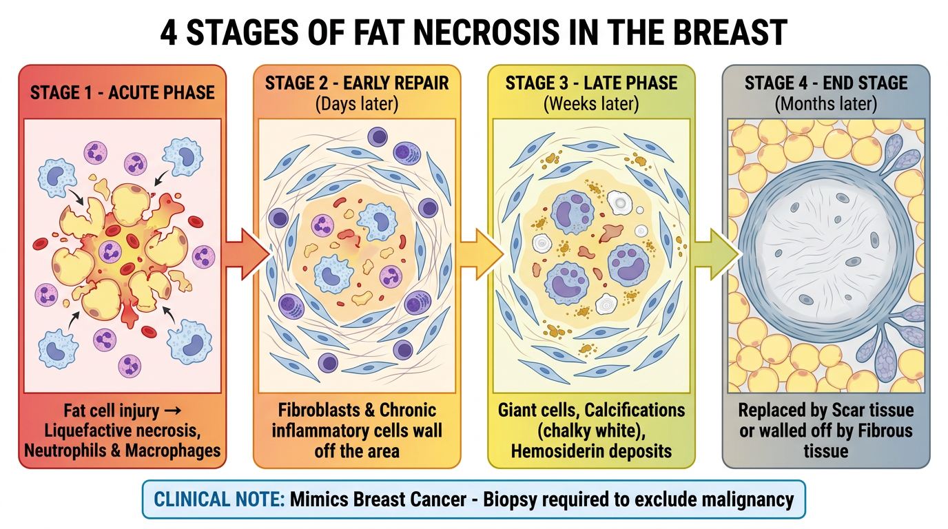
| Stage | Timeframe | Key Events |
|---|---|---|
| Stage 1 - Acute | Hours-Days | Fat cell death, liquefaction, neutrophils & macrophages arrive |
| Stage 2 - Early Repair | Days | Fibroblasts & chronic inflammatory cells wall off the area |
| Stage 3 - Late Phase | Weeks | Giant cells form, chalky calcifications & hemosiderin appear |
| Stage 4 - End Stage | Months | Replaced by scar tissue or encircled by fibrous capsule |