
Share a DM level mcq question with 4 options for a pulmonary medicine resident Topic of mcq is tropical pulmonary eosinophilia
"tropical pulmonary eosinophilia" diagnosis treatment
DM Pulmonary Medicine - MCQ
Answer: (B)
- Why (A) is wrong: TPE can cause a reversible obstructive defect acutely (wheezing, hyperinflation), but the classic late complication is restrictive, not obstructive
- Why (C) is wrong: ABPA is caused by Aspergillus fumigatus sensitization, not filarial antigens; the high IgE here is filarial in origin, and there is no mucoid impaction or central bronchiectasis described
- Why (D) is wrong: DEC is not associated with obliterative bronchiolitis; it is the definitive treatment for TPE
Source: Murray & Nadel's Textbook of Respiratory Medicine, Chapter 58 - Tropical Pulmonary Eosinophilia (lines 2064-2066); Fishman's Pulmonary Diseases and Disorders, Chapter 71

Create a poster .. Keep the content same
A clean medical education poster titled "TPE Management" (Tropical Pulmonary Eosinophilia). The poster has a professional clinical style with a white background and navy/dark blue accents. Content organized clearly: TITLE: "TPE Management" in bold dark blue at top with an underline Section 1 in bold: "D.O.C — Diethylcarbamazine (DEC)" Bullet points below: • 6 mg/kg/day — Orally, in 3 divided doses • Duration: 14–21 days (highlighted in a bordered box) • For a 50 kg adult: Total dose = 300 mg → 100 mg TID (shown with an arrow, both values in rounded banner/ribbon shapes) Clean typography, medical poster layout, no extra decoration, all text clearly readable.
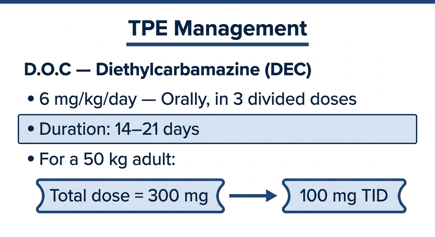
- D.O.C: Diethylcarbamazine (DEC)
- Dose: 6 mg/kg/day orally in 3 divided doses
- Duration: 14-21 days
- For a 50 kg adult: Total 300 mg → 100 mg TID
Can u make the poster more colourful and attractive
A vibrant, colorful medical education poster titled "TPE Management" (Tropical Pulmonary Eosinophilia) for pulmonary medicine residents. Bold, eye-catching design with a bright gradient background (deep teal to purple). TITLE at top: "TPE Management" in large white bold font with a bright yellow underline glow effect, subtitle "Tropical Pulmonary Eosinophilia" in smaller white italic text below title. Content in bright colored cards/sections: 1. A bright coral/orange rounded card labeled "D.O.C" with a star badge, containing white text: "Diethylcarbamazine (DEC)" 2. A bright sky-blue rounded card with a pill icon: "6 mg/kg/day — Orally" in bold white text 3. A lime-green rounded card with a clock icon: "3 Divided Doses" in bold white text 4. A golden-yellow highlighted box with bold border: "Duration: 14 – 21 Days" 5. A pink/magenta section titled "For 50 kg Adult:" with an arrow graphic: - Left bubble in purple: "300 mg/day Total" - Arrow → - Right bubble in bright orange: "100 mg TID" All cards have drop shadows, rounded corners, bright contrasting colors. Fun medical infographic style, professional yet vibrant. White icons next to each point. Clean readable fonts throughout.
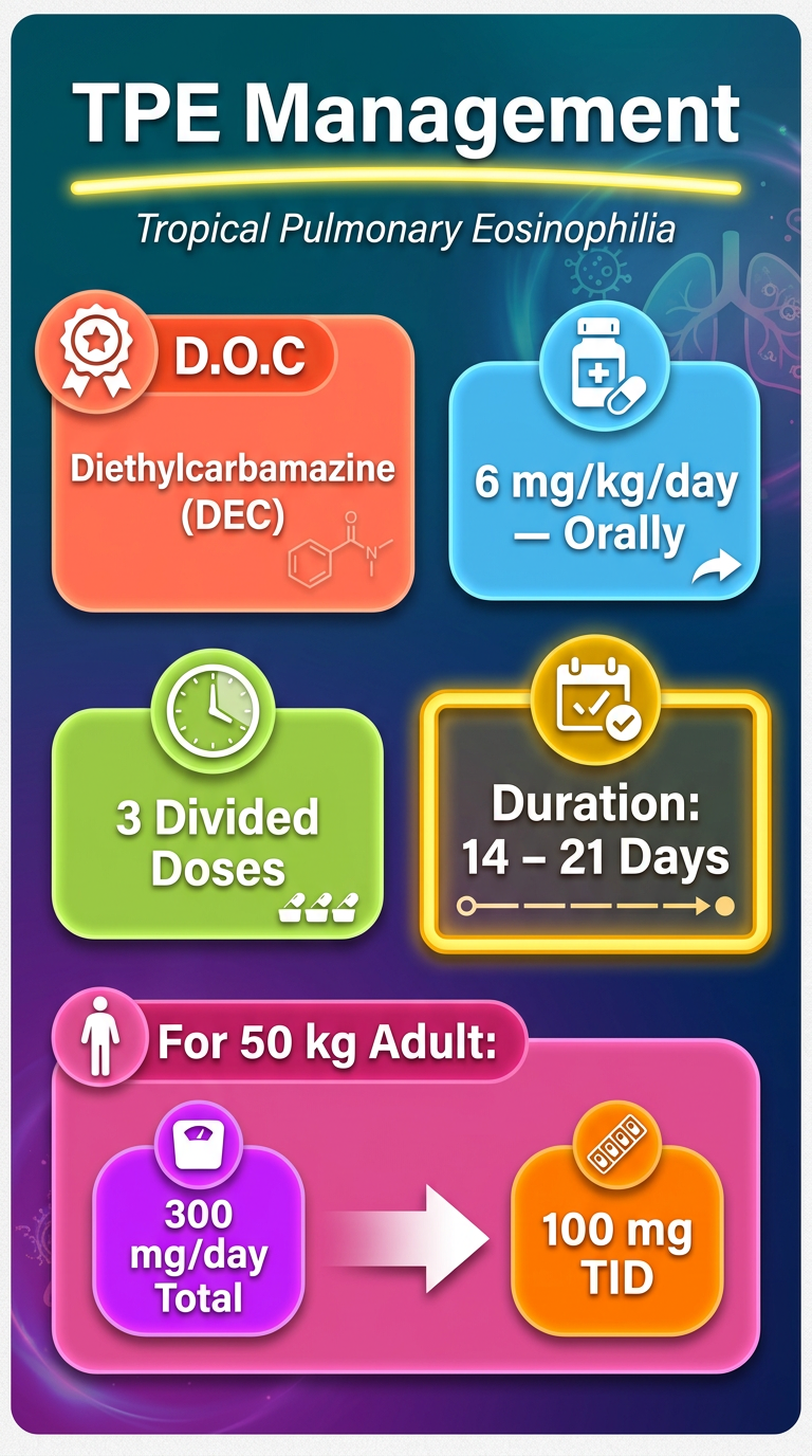
- Gradient background (teal to purple)
- Colour-coded cards for each key point - coral, sky-blue, lime-green, golden, and magenta
- Arrow graphic clearly showing 300 mg/day → 100 mg TID for a 50 kg adult
- Bold, easy-to-read fonts throughout