I now have all the content I need. Let me update the todos and write the comprehensive notes.---
Coronary Anatomy and Physiology - Anaesthesia Notes
Compiled from Miller's Anesthesia 10e, Barash Clinical Anaesthesia 9e, Morgan & Mikhail Clinical Anaesthesiology 7e, Guyton & Hall Medical Physiology, and Sabiston Textbook of Surgery
PART 1: CORONARY ANATOMY
1.1 Origin and Course of the Coronary Arteries
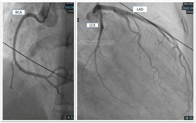
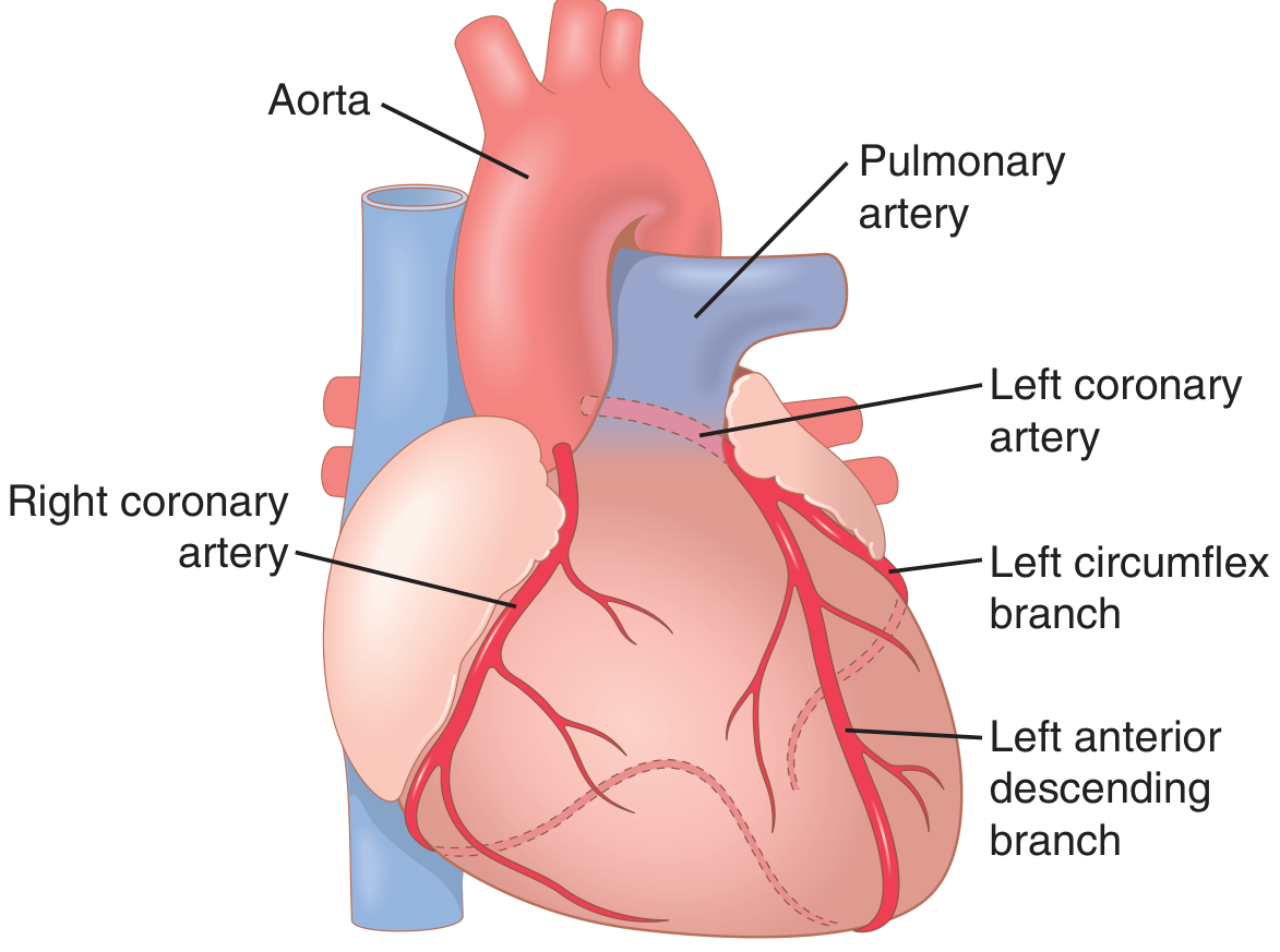
The two main coronary arteries are the first arterial branches of the aorta, each originating from its respective sinus of Valsalva (aortic sinus). The epicardial coronary arteries course along the surface of the heart; smaller arteries then penetrate into the myocardial mass. Only the innermost ~0.1 mm of the endocardial surface can obtain nutrition directly from intracavitary blood - the remainder depends entirely on coronary flow. - Guyton & Hall, Medical Physiology
Figure: Coronary artery anatomy showing the left coronary artery, right coronary artery, left circumflex branch, and left anterior descending branch (Guyton & Hall)
1.2 Left Coronary Artery System
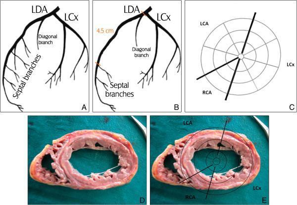
Left Main Coronary Artery (LMCA / LCA)
- Originates from the left coronary sinus of Valsalva
- Courses posterolateral to the main pulmonary trunk for approximately 1 cm
- Varies in length from a few mm to several cm
- Bifurcates into the Left Anterior Descending (LAD) and Left Circumflex (LCx) arteries - Sabiston Textbook of Surgery
Left Anterior Descending Artery (LAD)
- Courses anterolaterally relative to the pulmonary trunk and descends along the anterior interventricular groove
- Terminates at the apex of the left ventricle
- Major branches:
- Diagonal branches - supply the anterolateral aspect of the LV
- Septal perforating branches - supply the anterior 2/3 of the interventricular septum, bundle branches, and Purkinje system
- Supplies: anterior wall of LV (medial half), apex, and anterior 2/3 of the interventricular septum - Barash Clinical Anaesthesia 9e
Left Circumflex Artery (LCx / LCCA)
- Courses along the left atrioventricular groove
- Gives rise to 1-3 obtuse marginal (OM) branches supplying the lateral wall of the LV
- Also supplies anterior and inferior aspects of the lateral wall
- In 45% of patients, the SA node arterial supply arises from the LCx - Miller's Anesthesia 10e
- In 15% of patients (left-dominant system), the LCx gives rise to the posterior descending artery (PDA)
- Coronary blood flow to the LA is derived from branches of the LCx; LA contractility may be compromised when LCx blood flow is impaired - Barash 9e
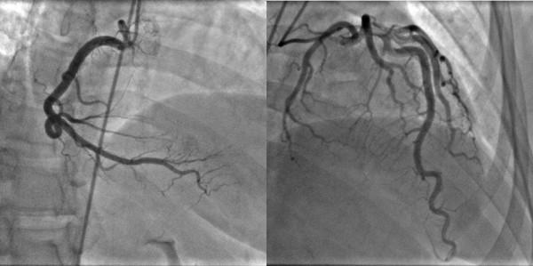
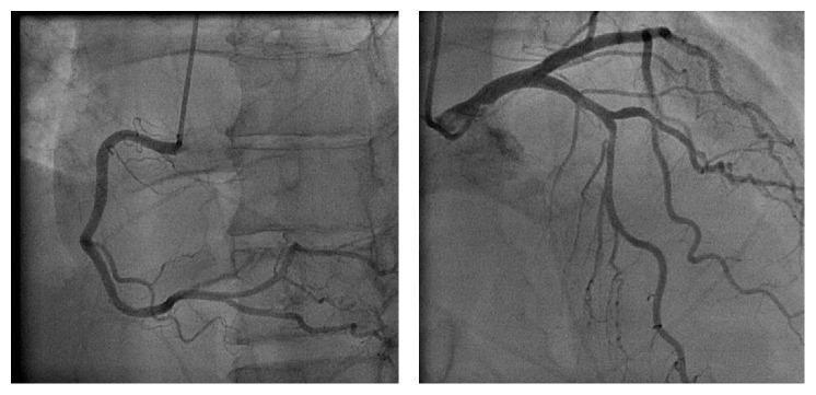
1.3 Right Coronary Artery (RCA)
- Arises from the right coronary sinus of Valsalva; ostium diameter averages 2-3 mm
- Traverses the right atrioventricular groove running rightward and obliquely
- Major branches:
- Acute marginal artery (AMA) - consistent vessel traversing the acute margin of the heart, supplies the lateral aspect of the RV
- Posterior descending artery (PDA / inferior interventricular artery) - in right-dominant patients, supplies the posterior inferior LV and inferior ventricular septum
- Right posterolateral artery
- AV node artery (AVNA) - critical for the conduction system
- In 85% of the population (right-dominant), the RCA gives rise to the PDA - Miller's, Barash, Sabiston
- The RCA and its branches supply most of the RV free wall, but the anterior wall of the RV also receives supply from LAD branches - therefore RV dysfunction can result from either RCA or LAD occlusion - Barash 9e
1.4 Coronary Dominance
| System | Vessel supplying PDA | Frequency |
|---|
| Right-dominant | RCA | 80-85% |
| Left-dominant | LCx | 15-20% |
| Co-dominant | Both | ~5% |
The dominant vessel is defined as the coronary artery that gives rise to the posterior descending artery (PDA), which supplies the inferior interventricular septum. - Sabiston Textbook of Surgery
1.5 Conduction System Blood Supply
| Structure | Primary Supply | Alternative |
|---|
| SA node | RCA (55%) | LCx (45%) |
| AV node | Dominant artery (usually RCA) | - |
| Bundle of His | AV nodal artery | - |
| Right bundle branch | LAD septal perforators | - |
| Left bundle branch | LAD septal perforators | - |
| Purkinje system | LAD septal branches | - |
Clinical pearl: Ischemia in the RCA or LCx territory can delay conduction through the proximal conduction system and cause bradyarrhythmias or heart block. - Barash Clinical Anaesthesia 9e
1.6 Papillary Muscle Supply
| Papillary Muscle | Blood Supply | Clinical Relevance |
|---|
| Anterolateral PM | Dual supply - LAD + LCx | Ischemic dysfunction unusual |
| Posteromedial PM | Usually single - RCA or LCx (2:1 ratio) | Vulnerable to ischemia |
~1/3 of patients have dual supply to the posteromedial papillary muscle (making them less susceptible). Ischemia of the posteromedial PM can cause acute mitral regurgitation. - Barash 9e
1.7 Coronary Venous Drainage
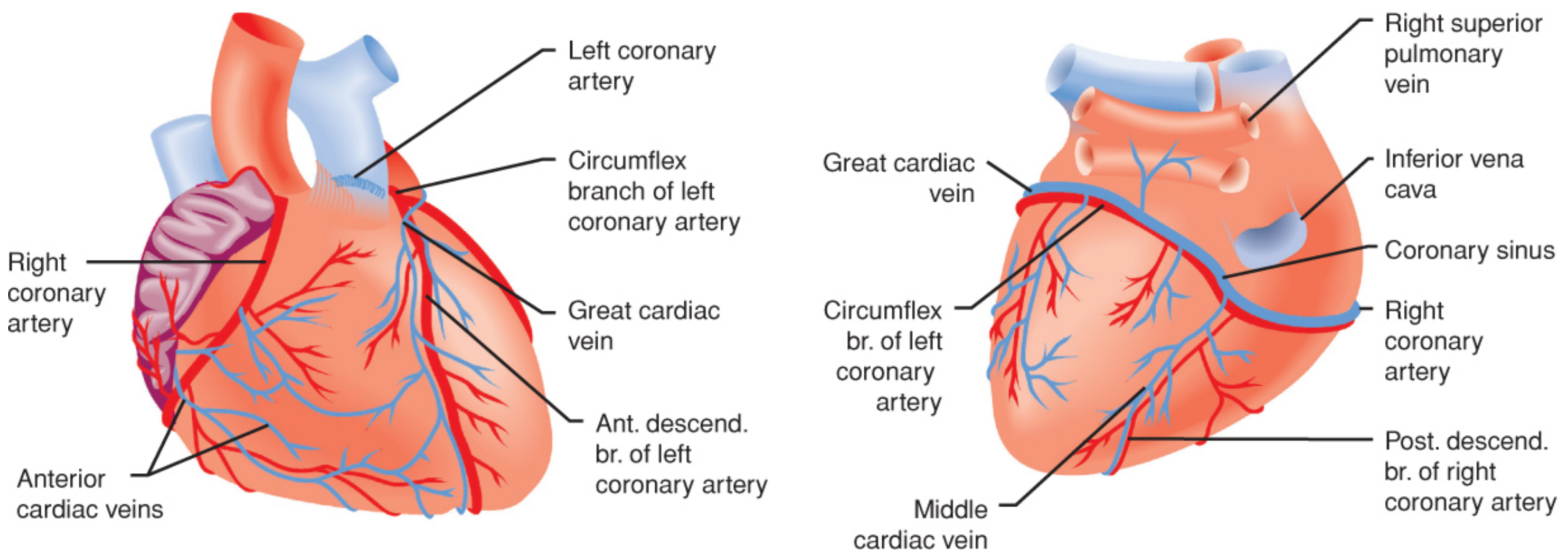
- ~75% of LV venous return drains via the coronary sinus into the right atrium (via the great, middle, and small cardiac veins)
- RV venous drainage - anterior cardiac veins empty directly into the RA
- Thebesian veins - tiny vessels emptying directly into all cardiac chambers (physiological shunt) - Guyton & Hall
Figure: Anterior view (left) shows RCA and LAD. Posterior view (right) shows LCx and PDA. Anterior cardiac veins from RV and coronary sinus (draining primarily the LV) empty into the RA. (Barash Clinical Anaesthesia 9e, Fig. 12-3)
PART 2: CORONARY PHYSIOLOGY
2.1 Baseline Coronary Blood Flow
- Resting coronary blood flow: approximately 225-250 mL/min (~1 mL/min/g) = 4-5% of cardiac output
- During strenuous exercise, cardiac work output may increase 6-9 fold, while coronary blood flow increases 3-4 fold (efficiency increases to compensate) - Guyton & Hall
- Coronary blood flow is governed by: Flow = Coronary Perfusion Pressure / Coronary Vascular Resistance
2.2 Coronary Perfusion Pressure (CPP)
For the Left Ventricle:
CPP (LV) = Aortic Diastolic Pressure - LV End-Diastolic Pressure (LVEDP)
For the Right Ventricle:
CPP (RV) = Aortic Diastolic Pressure - RV End-Diastolic Pressure
- Elevation in LVEDP impedes subendocardial blood flow
- RV, LA, and RA pressures remain lower than aortic pressure throughout the cycle - compressive forces do not impede coronary flow to these chambers - Barash 9e
2.3 Phasic Nature of Coronary Flow - Systole vs. Diastole
This is one of the most important physiological concepts for the anaesthetist:
- 70-80% of LV coronary blood flow occurs during diastole - Miller's Anesthesia 10e
- During systole, cardiac contraction compresses intramyocardial vessels, sharply reducing LV coronary flow - this is the reverse of all other vascular beds
- During diastole, the myocardium relaxes and blood flows freely through the LV capillaries
- RV flow is less affected because RV systolic pressure is far lower than LV systolic pressure
- Subendocardial vulnerability: During systole, the subendocardium is most vulnerable to ischemia. However, preferential flow to the subendocardium during diastole (via metabolically mediated arteriolar dilation) normally preserves uniform transmural perfusion - subepicardial:subendocardial flow ratio is 1:1 - Barash 9e
Anaesthetic relevance of heart rate:
Tachycardia shortens diastole disproportionately - this is a major reason beta-blockers are used as anti-ischemic agents in the perioperative period. Tachycardia can cause subendocardial ischemia when a flow-limiting stenosis or pressure-overload hypertrophy is present. - Miller's Anesthesia 10e
2.4 Coronary Autoregulation
- The myocardium regulates its own blood flow between mean perfusion pressures of 50-120 mmHg - within this range, flow remains relatively constant despite changes in perfusion pressure - Morgan & Mikhail 7e
- Below ~50 mmHg or above ~120 mmHg, flow becomes pressure-dependent (autoregulation lost)
- Small changes in vessel caliber produce large changes in resistance (resistance varies with the 4th power of vessel radius - Poiseuille's law)
- Coronary stenosis causes vessels to dilate maximally distal to the lesion - in this setting, perfusion pressure becomes the primary determinant of flow
2.5 Local Metabolic Regulation (Primary Control)
Local metabolic regulation is the primary controller of coronary blood flow:
- Increased myocardial metabolic activity → local vasodilation → increased coronary flow
- Decreased activity → reduced flow
Key vasodilator mediators released during increased O₂ demand:
- Adenosine (most important) - formed from ATP degradation via AMP → adenosine; potent vasodilator of coronary arterioles
- Other mediators: AMP, potassium ions (K⁺), hydrogen ions (H⁺), CO₂, prostaglandins, nitric oxide (NO)
- Note: pharmacological adenosine blockade does not completely abolish metabolic vasodilation - the other mediators remain active - Guyton & Hall
Flow-metabolism coupling: This intrinsic mechanism is essential because myocardial O₂ extraction is nearly maximal at baseline (~70-75%). The LV has almost no oxygen extraction reserve - it must increase blood flow to meet any increased demand. - Barash 9e
2.6 Nervous Control of Coronary Blood Flow
- Direct effects from autonomic neurotransmitters are generally weak and secondary to indirect metabolic effects
- Sympathetic stimulation - net effect is increased coronary flow because:
- Increased myocardial metabolic demand (dominant effect)
- β₂-receptor activation on smaller intramuscular/subendocardial vessels (vasodilation)
- α₁-receptors on larger epicardial vessels can cause mild vasoconstriction
- Parasympathetic (vagal) effects on coronary vasculature are minor and weakly vasodilatory - Morgan & Mikhail 7e
2.7 Myocardial Oxygen Supply and Demand Balance
This is the central concept for anaesthetic management of patients with CAD.
OXYGEN SUPPLY determinants:
| Factor | Details |
|---|
| Coronary perfusion pressure | Aortic DBP - LVEDP |
| Heart rate / diastolic time | Tachycardia ↓ diastolic filling time = ↓ supply |
| Arterial O₂ content | Haemoglobin concentration + SaO₂ |
| Coronary vessel diameter | Stenosis, vasospasm, vasodilation |
| O₂ release from Hb | Affected by pH, temperature, 2,3-DPG (Bohr effect) |
Note on Hb: A leftward shift of the oxyhemoglobin dissociation curve (caused by alkalosis, hypothermia, or low 2,3-DPG) decreases O₂ release to myocardial tissue. - Miller's 10e
OXYGEN DEMAND determinants:
| Component | Contribution to O₂ demand |
|---|
| Wall tension (pressure work) | 64% (dominant component) |
| Basal metabolic requirements | 20% |
| Volume work | 15% |
| Electrical depolarisation | 1% |
- Wall tension = f(pressure, radius, wall thickness) - Laplace's law: σ = Pr/2h
- Heart rate affects both supply (diastolic time) AND demand (MVO₂)
- LVEDP affects both supply (CPP) AND demand (wall tension via preload)
Key O₂ demand parameters (Double Product): Heart rate × Systolic BP = Pressure-Rate Product - clinical surrogate for MVO₂
2.8 Myocardial O₂ Extraction
- The heart extracts ~65-75% of arterial O₂ at rest (vs. ~25% for most other tissues)
- Coronary sinus O₂ saturation = ~30% (ScvO₂ ~30-35%); O₂ tension only ~20 mmHg
- No meaningful O₂ extraction reserve exists - the only way to increase O₂ delivery is to increase coronary blood flow
- Aerobic metabolism dominates: fatty acids, glucose, and lactate are oxidised in mitochondria. If O₂ supply is restricted, the myocyte depends on anaerobic glycolysis - but this is severely limited, and contraction will cease within 10-15 seconds of acute coronary occlusion without adequate collateral flow - Barash 9e
2.9 Coronary Reserve
- Coronary flow reserve (CFR) = maximal achievable flow / resting flow; normally 4-5x resting flow
- With increasing heart rate, subendocardial flow is maintained by recruiting this reserve
- CFR is exhausted in the presence of:
- Flow-limiting coronary stenosis (>70% diameter reduction)
- Pressure-overload hypertrophy (LVH)
- Tachycardia
- Elevated LVEDP
2.10 Subendocardial vs. Subepicardial Vulnerability
- The subendocardium is most vulnerable to ischaemia because:
- Compressed during systole (high intramyocardial pressure)
- Furthest from epicardial supply vessels
- Highest wall stress (by Laplace's law, inner layers bear greater tension)
- The subendocardial plexus of arteries partially compensates, but this compensation fails under pathological conditions - Guyton & Hall
PART 3: ANAESTHETIC RELEVANCE
3.1 Key Anaesthetic Goals for Myocardial O₂ Balance
| Goal | Intervention |
|---|
| Avoid tachycardia | Beta-blockers, adequate analgesia, avoid light anaesthesia |
| Maintain DBP (CPP) | Vasopressors if hypotensive; avoid excessive vasodilation |
| Avoid ↑ LVEDP | Careful fluid management, avoid myocardial depression |
| Avoid anaemia | Maintain adequate Hb; transfusion threshold guided by clinical context |
| Avoid hypothermia/alkalosis | Prevent leftward O₂Hb curve shift |
3.2 Effects of Anaesthetic Agents on Coronary Circulation
- Volatile agents are generally coronary vasodilators (direct effect); also reduce MVO₂ and offer myocardial preconditioning and protection against reperfusion injury - Morgan & Mikhail 7e
- Net effect on coronary blood flow is variable due to competing effects: direct vasodilation vs. reduced metabolic demand vs. systemic BP reduction
- Volatile agents do not cause significant coronary steal in most patients
3.3 CABG-Specific Anatomy Considerations
From a surgical standpoint, the following lesions are highest risk and require immediate anaesthetic attention:
- Left main coronary artery (LMCA) disease: jeopardises 2/3 of LV
- Proximal LAD disease: jeopardises anterior wall, septum, and apex
- Triple vessel disease: minimal coronary reserve
TEE monitoring for regional wall motion abnormalities (RWMAs) provides earlier detection of ischaemia than ECG ST-segment changes, correlating with specific coronary territories. - Miller's Anesthesia 10e
QUICK REFERENCE SUMMARY TABLE
| Feature | Left Dominant | Right Dominant |
|---|
| Who supplies PDA | LCx | RCA |
| Frequency | 15-20% | 80-85% |
| SA node supply | LCx (45%) or RCA (55%) | - |
| AV node supply | Dominant artery | Usually RCA |
SOURCES
- Miller's Anesthesia, 10th Edition - Chapter 50 (Coronary Anatomy, Determinants of Myocardial O₂ Supply and Demand)
- Barash, Cullen, and Stoelting's Clinical Anaesthesia, 9th Edition - Chapter 12 (Coronary Anatomy and Physiology)
- Morgan and Mikhail's Clinical Anesthesiology, 7th Edition - Chapter 20 (Control of Coronary Blood Flow, Myocardial O₂ Balance)
- Guyton and Hall Textbook of Medical Physiology - Chapter 21 (Physiological Anatomy of the Coronary Blood Supply, Control of Coronary Blood Flow)
- Sabiston Textbook of Surgery - Chapter on Coronary Artery Anatomy and Physiology
Note: Kaplan's Cardiac Anaesthesia is referenced within Barash (Fig. 12-12 caption cites Kaplan JA et al.) but is not available as a standalone volume in this library. The content above incorporates principles directly aligned with Kaplan's approach as reflected in Barash.