
Diseases associated with skin tightening
"skin diseases"[MeSH Terms] AND "scleroderma"
systemic sclerosis sclerodactyly skin tightening hands
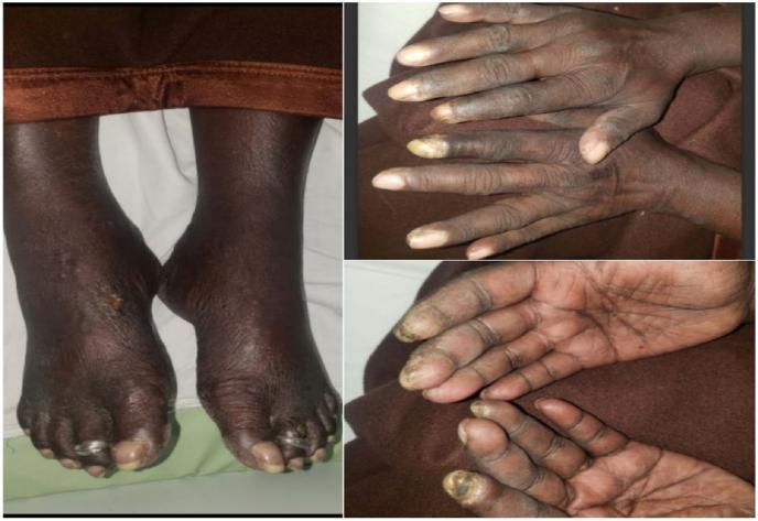
Clinical photograph series of the hands and feet demonstrating dermatologic manifestations of systemic sclerosis (scleroderma). The images show diffuse hyperpigmentation of the extremities with associated sclerodactyly, characterized by taut, thickened skin over the digits. In the dorsal view of the hands, there is notable skin tightening and loss of normal skin creases. The palmar and dorsal views reveal impending digital gangrene, specifically visible as dark, necrotic-appearing lesions at the distal tips of the index fingers bilaterally, indicative of severe Raynaud phenomenon and digital ischemia. The lower extremity photograph shows bilateral diffuse pedal hyperpigmentation and a distinct cutaneous ulcer on the dorsal surface of the right foot. The toes exhibit similar skin tightening and trophic changes. These findings are hallmarks of vascular and fibrotic complications in systemic sclerosis, emphasizing the risk of digital infarction and impaired wound healing in this patient population.
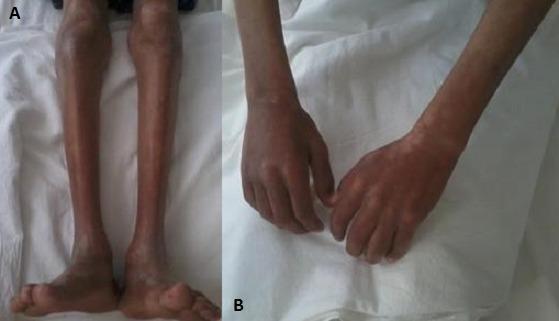
Two-panel clinical photograph demonstrating cutaneous and musculoskeletal manifestations of systemic sclerosis (scleroderma). Image (A) shows the lower limbs with significant cutaneous sclerosis, characterized by skin tightening and thickening. Notable bilateral amyotrophy (muscle wasting) is present, evidenced by a marked reduction in calf circumference and prominence of bony landmarks. Image (B) displays the upper limbs and hands, exhibiting similar skin sclerosis and severe forearm muscle atrophy. The skin in this region also demonstrates salt-and-pepper dyspigmentation, with areas of patchy hyperpigmentation and hypopigmentation. The hands show fixed, slightly flexed posturing consistent with sclerodactyly. These findings are clinically relevant for diagnosing connective tissue diseases and evaluating the extent of skin involvement (modified Rodnan skin score assessment) and secondary muscular complications.
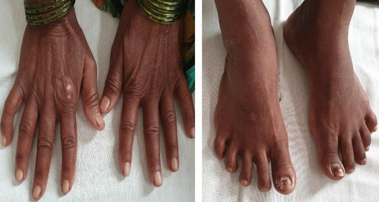
Two clinical photographs demonstrate dermatological manifestations of a systemic connective tissue disease, likely systemic sclerosis. The left panel shows the dorsal aspect of both hands, characterized by significant skin thickening and a taut, bound-down texture. The fingers exhibit tapering and fixed flexion contractures consistent with sclerodactyly. Subtle 'salt and pepper' pigmentary changes (hypopigmentation and hyperpigmentation) are visible on the dorsal surface. The right panel displays the dorsal aspect of the feet, which show similar skin tightening and thickening extending to the toes. Notable findings include xerosis (dryness) and onychodystrophy of the great toenails, suggestive of secondary fungal infection. These visual signs are classic hallmarks of the fibrotic phase of scleroderma, highlighting the loss of skin elasticity and characteristic digital changes used in the clinical diagnosis of CREST syndrome or systemic sclerosis.
1. Systemic sclerosis (scleroderma)
| Subset | Skin pattern | Course |
|---|---|---|
| Limited cutaneous SSc (includes CREST: calcinosis, Raynaud, esophageal dysmotility, sclerodactyly, telangiectasia) | Fingers, forearms, face. Slow onset. | Raynaud phenomenon often comes years before the skin changes. Pulmonary arterial hypertension is late. |
| Diffuse cutaneous SSc | Fingers, extremities, face and trunk. Rapid onset and progression. | Early interstitial lung disease, renal crisis and cardiac involvement. |
2. Scleroderma-like disorders
- Morphea can be plaque, guttate, generalized, deep, pansclerotic or bullous.
- Linear scleroderma, including en coup de sabre and hemifacial atrophy (Parry-Romberg).
- Scleredema, including scleredema diabeticorum (Buschke). It is associated with diabetes, paraproteinemia and post-infectious states, and it typically spares the fingers.
- Scleromyxedema (papular mucinosis). It is associated with a monoclonal gammopathy.
- Diabetic cheiroarthropathy ("stiff hand" or waxy skin of diabetes).
- Chronic graft-versus-host disease (sclerotic type).
- Eosinophilic fasciitis (Shulman disease). It involves the fascia, and it responds better to glucocorticoids than other sclerosing skin disorders (Firestein & Kelley's Rheumatology).
- Overlap syndromes, including mixed connective tissue disease.
- Lichen sclerosus is also listed among SSc mimics in Goldman-Cecil.
- Nephrogenic systemic fibrosis. It is linked to gadolinium exposure in renal failure.
- Eosinophilia-myalgia syndrome. It is linked to contaminated L-tryptophan.
- Vinyl chloride disease, and other chemical or drug-induced sclerosis (for example bleomycin, and some other drugs reported with morpheiform lesions).
- Stiff skin syndrome.
- Pachydermatoperiostosis (primary hypertrophic osteoarthropathy).
- Porphyria cutanea tarda (sclerodermoid changes), listed in Goldman-Cecil.
- Paraneoplastic sclerosis.
3. Clues to tell them apart
- Raynaud phenomenon, nailfold capillary changes, or SSc-specific antibodies (anticentromere, anti-Scl-70, anti-RNA polymerase III) point to true SSc.
- Skin thickening that spares the fingers points to a mimic, such as scleredema, nephrogenic systemic fibrosis or morphea. The ACR/EULAR SSc classification criteria exclude patients with skin thickening that spares the fingers, and also those with a better-explaining scleroderma-like disorder (Goldman-Cecil).
- Peripheral eosinophilia with a "groove sign" and woody induration after exertion suggests eosinophilic fasciitis.
- Monoclonal gammopathy with waxy papules suggests scleromyxedema.
- A history of dialysis or CKD and gadolinium suggests nephrogenic systemic fibrosis.
Evidence update
- Cerebrovascular disease in scleroderma (PMID 37839405).
- Neurological involvement in localized scleroderma (PMID 42812294).
- Regenerative treatments for facial involvement in scleroderma (PMID 38976297).