
Answer the following questions as per bds 3rd year( india) oriented for university exam pattern of oral pathology with histological diagram ( to drawable with h and e pencil) : Define , classify fibroosseous lesions. Clinical features , histologic features, radiologic features of fibrous dysplaisa
fibrous dysplasia jaw histology Chinese letters pattern
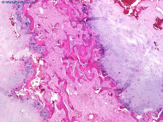
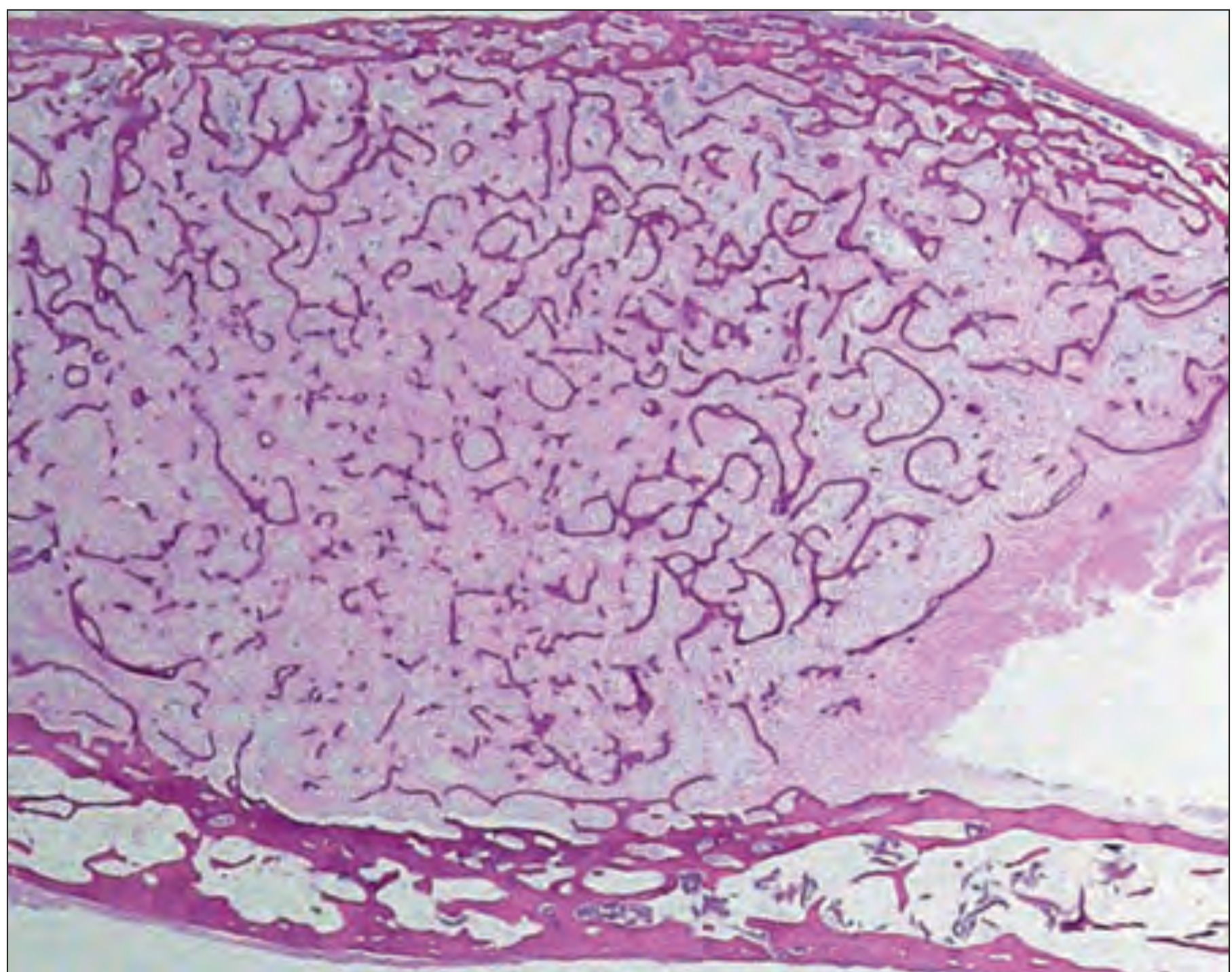
Histology of fibrous dysplasia. This hematoxylin and eosin stained bone biopsy section reveals a fibroblastic stroma populated by spindle-shaped cells and irregular, immature woven bone trabeculae. The trabeculae are variably sized, often curved or irregular in shape, resembling a Chinese character pattern, and they lack a consistent osteoblastic rim. The surrounding stroma is fibrous and moderately cellular, with a bland appearance and sparse mitotic activity. In this case, nodules of hyaline cartilage are present on either side of the osseous component, illustrating chondroid differentiation within the lesion. The chondroid component can be strikingly prominent in some fibrous dysplasia cases, contributing to a mixed fibro-osseous phenotype. Overall, the combination of woven bone within fibrous stroma and cartilaginous nodules supports a benign fibrous-osseous process rather than a malignant tumor; however, the cartilaginous elements may raise differential considerations such as chondromatous differentiation or low-grade chondrosarcoma if atypia were evident. Clinically, these features correspond to a slow-growing, benign lesion of bone that may cause deformity or structural weakness depending on location. This histologic pattern is classic for fibrous dysplasia and highlights heterogeneity within the lesion, including possible cartilage-rich variants and variable radiographic correlation. These features aid surgical planning and prognosis.
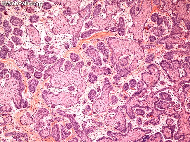
Histology: Light microscopy of a jaw lesion shows classic ameloblastoma morphology on hematoxylin and eosin stained tissue. The neoplasm is composed of multiple epithelial islands and cords embedded in a fibrous connective tissue stroma. Follicular pattern predominates, with nests of odontogenic epithelium displaying peripheral palisading of columnar cells and reversal of polarity, nuclei oriented away from the basement membrane. Central cells resemble stellate reticulum with loosely arranged, angular morphology. Some islands exhibit a loosely arranged, anastomosing arrangement compatible with plexiform architecture. Overall cellularity is moderate, with minimal pleomorphism and no overt mitotic activity in the field, consistent with benign but locally aggressive behavior. Basal layer cells are darkly basophilic with prominent nucleoli, while surrounding stroma is densely fibrous and occasionally myxoid, producing a subtle desmoplastic impression in portions of the section. The tumor borders appear infiltrative into adjacent connective tissue, reflecting its known propensity for local invasion despite benign cytology. There is an absence of keratinization, dysplasia, or metastatic features. The image highlights the diagnostic hallmarks: enamel organ–like islands, reverse polarity, stellate reticulum–like center, and a fibrous stroma. These histologic features underline confirmation of diagnosis and guide surgical planning in odontogenic tumors and jaw neoplasms. For educational and diagnostic purposes.
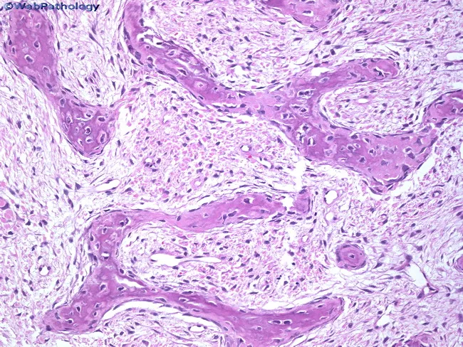
Histology of fibrous dysplasia showing a fibroblastic stroma with irregular immature woven bone trabeculae. The bony trabeculae are narrow, curvilinear or irregularly shaped and arranged haphazardly within a dense fibrous matrix. Trabeculae are immature and inadequately mineralized, lacking osteoblastic rimming, reflecting a maturation defect with failure to form lamellar bone. Reversal lines may be present within the woven bone, a feature that can resemble Paget’s disease in some fields but without the extensive osteolysis or mosaic lamellar pattern. A chondroid component is seen in about 10% of cases, sometimes strikingly prominent, contributing to a mixed fibrous–chondroid pattern. Areas of cystic change can occur, containing lipid-laden macrophages and occasional multinucleated giant cells. Overall, the lesion demonstrates fibrous tissue interlacing with irregular osseous trabeculae, with relatively bland fibroblast populations and no significant cytologic atypia. These histologic features indicate a benign, developmental bone lesion driven by abnormal osteogenesis and fibroplasia. Clinically, the pattern may range from monostotic to polyostotic disease, often presenting in childhood or adolescence with painless swelling or deformity. The diagnostic significance lies in recognizing the maturation defect of bone and distinguishing it from other fibro-osseous lesions. Correlation with radiographs and clinical findings is essential for accurate diagnosis and management planning.
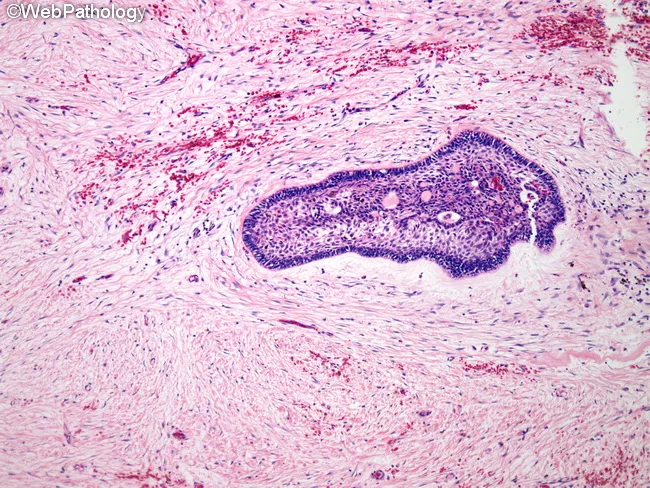
Histopathology of ameloblastoma, desmoplastic variant, on a Hematoxylin and Eosin stained slide from jaw bone tissue. The lesion shows dense, sclerotic stroma with compressed bone trabeculae and scattered islands and thin cords of odontogenic epithelium embedded in the fibrous matrix. Epithelial components consist of cuboidal to columnar cells, with partial reverse polarity, and areas reminiscent of stellate reticulum are minimal or absent. Peripheral palisading is often not evident in this variant. The stroma demonstrates marked desmoplasia with abundant type I collagen and sparse inflammatory infiltrate. Keratinization is uncommon, and mitotic activity is typically low. The tumor is locally infiltrative despite its benign designation, and the desmoplastic pattern can mimic fibrous or desmoplastic fibromatous lesions radiographically. Diagnostic significance rests on identifying odontogenic epithelium within a densely fibrous milieu, consistent with desmoplastic ameloblastoma, a distinct histologic subtype of ameloblastoma. Differential considerations include conventional ameloblastoma subtypes (follicular, plexiform), fibrous dysplasia, odontogenic fibroma, and fibromatosis. Clinically, lesions tend to occur in the anterior jaws and may present as swelling. Surgical management requires complete excision due to recurrence risk. This image is educational for pathology training, histology atlases, and radiology–histology correlation studies. Interpretation should integrate clinical context, radiographs, and immunohistochemical confirmation when available, guiding management.
fibrous dysplasia ground glass appearance radiograph jaw panoramic
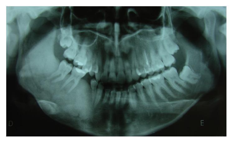
This diagnostic image is a panoramic dental radiograph (orthopantomogram) of a human mandible and maxilla. The primary finding is a large, poorly defined radiopaque lesion located in the right hemimandible. The lesion exhibits a classic 'ground glass' appearance, characterized by a fine, granular, and uniform increase in bone density that blends into the surrounding healthy bone. This finding is highly suggestive of fibrous dysplasia, specifically the monostotic form given the localized presentation. The lesion involves the mandibular body and ramus, causing expansion and altering the normal trabecular pattern. Dental examination reveals the absence of teeth 4.5 and 4.8, consistent with historical surgical extraction. The contralateral (left) mandible appears normal in bone density and structure. This radiograph serves as an educational example of fibro-osseous lesions of the jaws, emphasizing the importance of recognizing specific radiographic textures like 'ground glass' for differential diagnosis between fibrous dysplasia, ossifying fibroma, and chronic osteomyelitis.
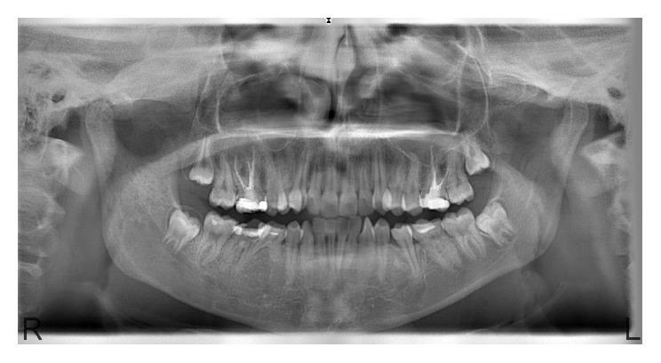
This diagnostic image is a panoramic radiograph (orthopantomogram) of the maxilla and mandible, illustrating radiographic signs characteristic of polyostotic craniofacial fibrous dysplasia. The image reveals multiple, ill-defined lesions with a classic 'ground-glass' appearance. Key affected anatomical regions include the right mandibular corpus and ramus, the mandibular symphysis, and the right posterior maxillary region. These lesions exhibit increased radiopacity and a hazy, homogeneous texture that obscures the normal trabecular bone pattern. The transition zones between the pathological lesions and the adjacent normal bone are indistinct, indicating a gradual, infiltrative process rather than a well-circumscribed border. In the right maxilla, there is evidence of slight bony expansion. The radiographic findings are essential for medical and dental students to identify the presentation of fibro-osseous lesions, emphasizing the diagnostic importance of bone texture changes (ground-glass vs. trabecular) and the loss of clear anatomical demarcation in craniofacial pathology.
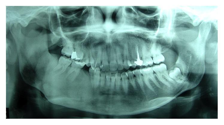
This panoramic dental radiograph (orthopantomogram) displays a significant pathological alteration in the right hemimandible. The primary finding is a large, poorly defined radiopaque lesion with a characteristic 'ground glass' appearance, which has replaced the normal trabecular bone pattern. This appearance is highly suggestive of fibrous dysplasia. The lesion extends through the mandibular body on the right side, causing an apparent increase in bone volume and density. Dental findings include evidence of previous restorative and endodontic treatments, visible as radiopaque fillings and a root canal post. There is also evidence of tooth extractions in the posterior right mandible, indicated by missing dentition in the affected area. The maxillary and contralateral mandibular structures appear relatively normal, providing a comparative baseline for the density of the pathology. This diagnostic image is used to monitor the progression or recurrence of fibro-osseous lesions in a clinical dental or maxillofacial context.
A labeled pencil-style histological diagram of fibrous dysplasia suitable for BDS exam drawing. The diagram shows: (1) a background of moderately cellular fibrous stroma made of spindle-shaped fibroblasts; (2) irregular, curved/curvilinear trabeculae of woven bone scattered throughout resembling Chinese letters or alphabets; (3) absence of osteoblastic rimming around the bone trabeculae; (4) occasional osteoclasts; (5) areas of cystic degeneration with foamy macrophages in one corner. All structures clearly labeled with arrows. Black and white pencil sketch style with clean labels. Title at top: "Histology of Fibrous Dysplasia (H&E)"
FIBROOSSEOUS LESIONS OF THE JAWS
(University Exam Pattern - BDS 3rd Year, India)
DEFINITION
Key phrase for exam: "Normal bone is replaced by fibrous connective tissue within which varying amounts of mineralized material are deposited."
CLASSIFICATION OF FIBROOSSEOUS LESIONS
WHO Classification (Neville / Shafer - commonly used in Indian BDS curriculum)
- Monostotic fibrous dysplasia
- Polyostotic fibrous dysplasia
- McCune-Albright Syndrome
- Mazabraud Syndrome
- Craniofacial fibrous dysplasia (some authors classify separately)
- Periapical (periapical cemental dysplasia)
- Focal cemento-osseous dysplasia
- Florid cemento-osseous dysplasia
- Conventional ossifying fibroma
- Juvenile ossifying fibroma
- Juvenile trabecular ossifying fibroma
- Juvenile psammomatoid ossifying fibroma
FIBROUS DYSPLASIA
ETIOLOGY / PATHOGENESIS
- Produces constitutive activation of Gs protein
- Leads to increased intracellular cyclic AMP (cAMP)
- This alters Wnt signaling, promotes cellular proliferation and inhibits osteoblast differentiation
- Results in replacement of normal bone by fibrous tissue containing poorly mineralized woven bone
- Very early embryogenesis → McCune-Albright syndrome
- Later in an osteoblast precursor → monostotic lesion
CLINICAL FEATURES
Types
| Feature | Monostotic FD | Polyostotic FD | McCune-Albright |
|---|---|---|---|
| Bones involved | Single bone | Multiple bones | Multiple bones |
| Frequency | ~80% of cases | ~20% of cases | Rare |
| Skin pigmentation | Absent | May be present | Cafe-au-lait spots (coast of Maine border - irregular) |
| Endocrinopathy | Absent | Absent | Precocious puberty (girls), hyperthyroidism |
| Jaw involvement | Common | Common | Common |
General Clinical Points
- Age of onset: During skeletal development (childhood/adolescence); 25% are not diagnosed until after age 30
- Sex: Slight female predilection in craniofacial involvement
- Site: Maxilla > Mandible (in jaws). Maxilla most commonly involved craniofacial bone
- Symptoms:
- Painless, slow, progressive swelling of the jaw
- Facial asymmetry
- Displacement/tilting of teeth
- Malocclusion
- Nasal obstruction (when maxilla involved)
- Proptosis (if orbital walls involved)
- Leontiasis ossea - "lion face" appearance (severe, extensive involvement)
- Behavior: Lesion tends to stabilize or stop growing after growth plate closure (puberty). Polyostotic may continue into adulthood.
- Complications:
- Pathological fracture (especially long bones; rare in jaw)
- Rare malignant transformation to sarcoma (usually osteosarcoma or fibrosarcoma) - more common in polyostotic, especially post-irradiation
- Shepherd's crook deformity of femur (in polyostotic, long bone involvement)
RADIOLOGICAL FEATURES
-
"Ground-glass" appearance - Most characteristic finding
- Fine granular haziness, like frosted glass
- Due to mineralized woven bone spicules within fibrous tissue
- Obliteration of normal trabecular pattern
-
Ill-defined borders - lesion merges imperceptibly with surrounding normal bone (no sclerotic rim, no clear cortication) - this is a KEY differentiator from ossifying fibroma
-
Expansion of cortex - buccal and lingual cortical plates expand; cortical thinning present
-
"Long lesion in a long bone" pattern in appendicular skeleton; in jaw - involves a large portion of the bone
-
May show zones of:
- Radiolucency (early/purely lytic stage)
- Mixed radiolucency-radiopacity (ground-glass)
- Radiopacity (mature/late stage - "orange peel" or "fingerprint" pattern on CT)
-
Loss of lamina dura around involved teeth
-
Displacement of teeth - without root resorption usually
-
CT scan: "Orange peel" or "fingerprint" bony texture; expansion of medullary cavity; no periosteal reaction
- Early: Radiolucent
- Intermediate: Ground-glass (most characteristic)
- Late: More radiopaque / mixed
HISTOLOGICAL FEATURES
Key Microscopic Features (H&E)
-
Fibrous stroma - moderately cellular, composed of bland spindle-shaped fibroblasts (no atypia, low mitotic activity)
-
Irregular trabeculae of woven (immature) bone - the single most important feature:
- Curvilinear, curved, irregular shapes
- Haphazardly arranged
- Classically described as resembling "Chinese letters / Chinese writing / alphabets" - this is the hallmark phrase
-
Absence of osteoblastic rimming - the bony trabeculae LACK a rim of osteoblasts around them (this differentiates FD from ossifying fibroma, which has prominent osteoblastic rimming)
-
Immature woven bone - fails to mature into lamellar bone; reflects a maturation defect
-
Occasional osteoclast-type giant cells may be present
-
Cystic degeneration - areas of cystic change containing:
- Hemorrhage
- Foamy macrophages (lipid-laden)
- Blood-filled spaces
-
Chondroid nodules - seen in ~10% of cases (cartilaginous differentiation)
-
No capsule
HISTOLOGICAL DIAGRAM (Drawable with H&E Pencil)
Photomicrograph from Robbins Pathology (H&E, low power):
High power H&E:
DIAGRAM TO DRAW IN EXAM (H&E Pencil Diagram)
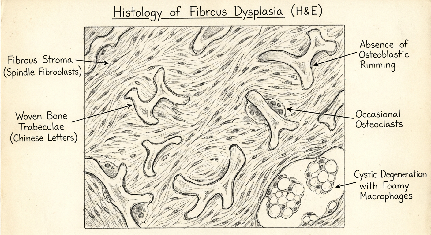
HOW TO DRAW - FIBROUS DYSPLASIA (H&E):
Step 1: Draw a background of wavy spindle-shaped cells = FIBROUS STROMA
Step 2: Draw irregular, curved "C", "S", "U"-shaped pink trabeculae = WOVEN BONE
(arrange haphazardly - like Chinese letters)
Step 3: Do NOT draw osteoblasts lining the bone = NO OSTEOBLASTIC RIMMING
Step 4: Add 1-2 multinucleated cells near bone = OSTEOCLASTS (occasional)
Step 5: In one corner, draw a clear space with foamy cells = CYSTIC DEGENERATION
LABELS TO ADD (with arrows):
→ Fibrous stroma (spindle fibroblasts)
→ Woven bone trabeculae (curvilinear / "Chinese letter" pattern)
→ No osteoblastic rimming (point to edge of bone)
→ Osteoclast-type giant cells (occasional)
→ Cystic degeneration with foamy macrophages
DIFFERENTIAL DIAGNOSIS (Table)
| Feature | Fibrous Dysplasia | Ossifying Fibroma |
|---|---|---|
| Border | Ill-defined, merges with bone | Well-defined, often corticated |
| Osteoblastic rimming | ABSENT | PRESENT |
| Encapsulation | Not encapsulated | Encapsulated |
| Recurrence after curettage | Common | Less common (can be shelled out) |
| Expansion | Yes | Yes |
| Radiograph | Ground-glass, ill-defined | Well-defined radiolucent/mixed lesion |
TREATMENT
- Wait and watch - if asymptomatic, especially if growing age
- Conservative surgical contouring/recontouring - after lesion stabilizes (post-puberty), for cosmetic deformity
- Curettage - recurrence is common in younger patients
- Bisphosphonates (e.g., pamidronate) - for polyostotic disease to reduce bone pain
- Radiation therapy is CONTRAINDICATED - increases risk of malignant transformation to sarcoma
- Regular follow-up is mandatory due to recurrence and rare malignant transformation risk
SUMMARY TABLE FOR QUICK REVISION
| Parameter | Fibrous Dysplasia |
|---|---|
| Definition | Replacement of normal bone by cellular fibrous tissue containing woven bone trabeculae |
| Gene mutation | GNAS1 (somatic, gain-of-function) |
| Most common jaw bone | Maxilla |
| Key clinical feature | Painless, progressive bony expansion; facial asymmetry |
| Characteristic radiograph | Ground-glass appearance, ill-defined borders |
| Characteristic histology | Curvilinear woven bone trabeculae ("Chinese letters") WITHOUT osteoblastic rimming, in fibrous stroma |
| Treatment | Conservative recontouring after stabilization |
| Contraindication | Radiation therapy |
| Important syndrome | McCune-Albright (polyostotic FD + cafe-au-lait + precocious puberty) |
Exam Tip: The two most important phrases to memorize are:
- Histology - "Curvilinear trabeculae of woven bone resembling Chinese letters/alphabets WITHOUT osteoblastic rimming, in a cellular fibrous stroma"
- Radiology - "Ground-glass appearance with ill-defined borders that merge imperceptibly with surrounding normal bone"
Here’s another question and answer it the same way: Classify epithelial and non epithelial tumor of oral cavity. Etiopath and h/f and grading of oscc
oral squamous cell carcinoma histology well differentiated keratin pearl H&E
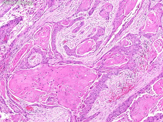
High-magnification histopathology image of oral mucosa showing keratinizing, well-differentiated squamous cell carcinoma with classic keratin pearl formation. The neoplasm displays nests and cords of malignant squamous cells invading the fibrous lamina propria, with abundant eosinophilic cytoplasm and intercellular bridges. Keratinization is evident both centrally within tumor nests and at the periphery, producing concentric keratin pearls of pale eosinophilic keratin. Tumor cells are relatively well differentiated, with recognizable squamous maturation and occasional mitotic figures. The adjacent mucosa demonstrates diverse epithelial alterations, including squamous metaplasia (keratinizing and nonkeratinizing variants), dysplasia, and areas consistent with squamous cell carcinoma in situ, reflecting field cancerization around the invasive focus. Inflammatory infiltrate and edema are present in the superficial lamina propria, while deeper stroma shows variable desmoplastic response. The surface epithelium may show thinning or ulceration over the lesion. No clear vascular invasion is evident at this magnification, though deeper planes could reveal invasion into connective tissue and perineural spread. This image provides valuable diagnostic features for distinguishing keratinizing SCC from nonkeratinizing variants and other mucosal neoplasms, and highlights the relevance of adjacent dysplasia/CIS as indicators of tumor field effect and oncologic margins. It serves as an educational reference for pathology, histology, and clinical tumor biology.
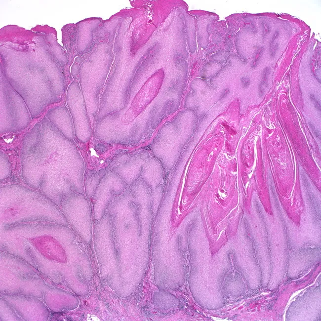
Histology: Light microscopy of an Hematoxylin and Eosin (H&E) stained biopsy from oral mucosa. The tissue shows stratified squamous epithelium over fibrous stroma with an exophytic, verruciform surface. The neoplasm is well differentiated and displays bulbous, finger-like downgrowths with broad, pushing borders into the underlying connective tissue. Tumor cells exhibit minimal cytologic atypia, abundant keratinization, intercellular bridges, and occasional keratin pearls. Invasion is present but shallow and non-destructive, consistent with a verrucous carcinoma phenotype. The architecture emphasizes exophytic growth with prominent surface maturation and blunt-to-rounded rete pegs that markedly extend into stroma while preserving cell polarity. The lesion tends to be locally aggressive yet with low metastatic potential. These features differentiate it from conventional keratinizing squamous cell carcinoma, which shows more infiltrative margins and higher mitotic activity. Clinically, verrucous carcinoma manifests as a large, cauliflower-like exophytic mass on mucosal surfaces (oral cavity, oropharynx), often causing local destruction without distant spread. Management typically involves wide local excision with clear margins; radiotherapy is generally avoided due to risk of anaplastic transformation. This image illustrates classic histopathologic hallmarks and provides diagnostic and educational value for surgical pathology, oncology, and oral medicine. Correlates with clinicopathologic staging and informs prognosis and treatment planning in multidisciplinary tumor boards and educational contexts.
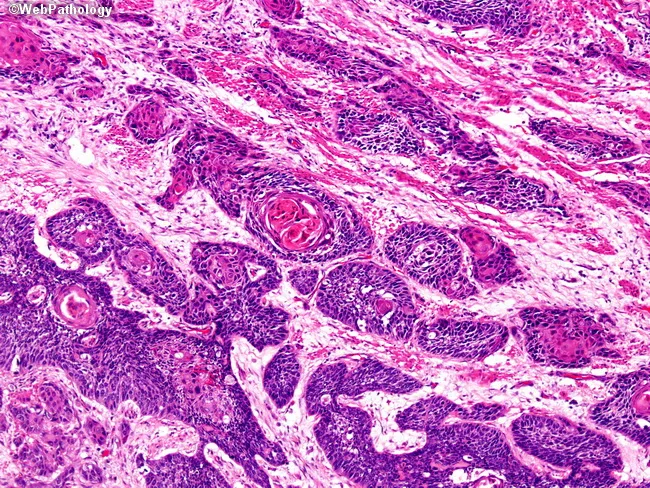
This histopathology image depicts esophageal squamous cell carcinoma, using brightfield light microscopy on an H&E-stained tissue section. The lesion is located in the mid-to-lower esophagus, consistent with common anatomical distribution of esophageal squamous cell carcinoma in males over 50. The tumor shows nests and cords of malignant squamous cells with sharp borders, abundant eosinophilic cytoplasm, intercellular bridges, and keratin pearl formation, reflecting a well-differentiated phenotype. The surrounding stroma exhibits desmoplastic reaction with inflammatory infiltrates. Individual tumor cells display relatively mild atypia given the differentiation, though mitotic figures may be present at higher magnification. Invasion into the submucosa and muscularis propria is evident in places, indicating advanced local disease despite the well-differentiated histology. These histologic features align with classic radiographic and clinical patterns: progressive dysphagia, weight loss, chest pain in some cases, and late presentation. Risk factors include tobacco use, alcohol consumption, prior caustic ingestion (lye), achalasia, Plummer-Vinson syndrome, and prior radiation. Pathologic assessment informs prognosis and staging, guiding therapeutic decisions such as neoadjuvant therapy, esophagectomy, or definitive chemoradiation. This image serves educational and research purposes for pathology, oncology, and gastroenterology to study squamous differentiation, keratinization, and local invasion patterns in esophageal cancer. Correlates with staging and multidisciplinary treatment planning today.
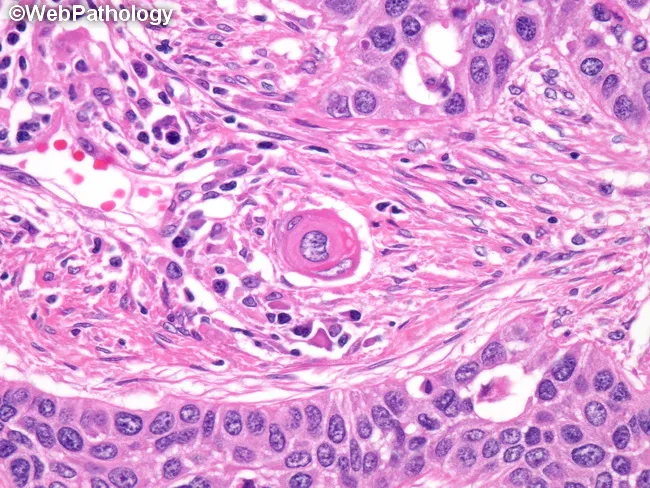
This is a brightfield histopathology image of an invasive, well-differentiated squamous cell carcinoma in epidermal/mucosal squamous epithelium. The H&E stained section demonstrates malignant squamous cells in nests and cords penetrating the dermis with desmoplastic stromal reaction. Characteristic features include abundant eosinophilic cytoplasm, keratinization in the form of keratin pearls, and pronounced intercellular bridges between adjacent tumor cells. Some tumor clusters show concentric keratinization around a central keratin pearl; intracellular mucin may be present in scattered malignant cells, though not a dominant feature in well-differentiated tumors. The cells exhibit minimal pleomorphism and relatively preserved nuclear-to-cytoplasmic ratio compared with poorly differentiated SCC. The presence of keratin pearls and intercellular bridges supports squamous differentiation and a diagnosis of squamous cell carcinoma. In non-keratinizing or poorly differentiated variants, immunohistochemistry for squamous markers (p40, p63, CK5/6) and absence of TTF-1 would aid differential; mucin positivity would favor adenosquamous or mucinous differentiation. Clinically, this finding is relevant for tumor staging and treatment planning, including surgical excision with clear margins and consideration of adjuvant therapy depending on invasion depth and perineural or lymphovascular invasion. This image is suitable for educational reference and image-based pathology teaching, and for validating histologic criteria of keratinizing SCC.
poorly differentiated squamous cell carcinoma oral cavity histology H&E
Histology: Light microscopy of an Hematoxylin and Eosin (H&E) stained biopsy from oral mucosa. The tissue shows stratified squamous epithelium over fibrous stroma with an exophytic, verruciform surface. The neoplasm is well differentiated and displays bulbous, finger-like downgrowths with broad, pushing borders into the underlying connective tissue. Tumor cells exhibit minimal cytologic atypia, abundant keratinization, intercellular bridges, and occasional keratin pearls. Invasion is present but shallow and non-destructive, consistent with a verrucous carcinoma phenotype. The architecture emphasizes exophytic growth with prominent surface maturation and blunt-to-rounded rete pegs that markedly extend into stroma while preserving cell polarity. The lesion tends to be locally aggressive yet with low metastatic potential. These features differentiate it from conventional keratinizing squamous cell carcinoma, which shows more infiltrative margins and higher mitotic activity. Clinically, verrucous carcinoma manifests as a large, cauliflower-like exophytic mass on mucosal surfaces (oral cavity, oropharynx), often causing local destruction without distant spread. Management typically involves wide local excision with clear margins; radiotherapy is generally avoided due to risk of anaplastic transformation. This image illustrates classic histopathologic hallmarks and provides diagnostic and educational value for surgical pathology, oncology, and oral medicine. Correlates with clinicopathologic staging and informs prognosis and treatment planning in multidisciplinary tumor boards and educational contexts.
![Histopathology image of prostatic tissue stained with Hematoxylin and Eosin (H&E) at high magnification. The specimen demonstrates two divergent neoplastic components: poorly formed glands with intraluminal mucin representing glandular/prostatic adenocarcinoma, and an adjacent region of moderately differentiated squamous cell carcinoma (visible in the upper right). The glandular glands show architectural distortion, nuclear atypia, prominent nucleoli, and mitotic activity within a mucin-rich luminal compartment. The intraluminal mucin is variably distended and characteristically aligns with dysplastic glandular epithelium. The squamous component displays nests of keratinizing polygonal cells, intercellular bridges, and moderate cytologic atypia consistent with squamous differentiation. Immunohistochemistry (PSA and prostatic acid phosphatase [PAP]) shows strong, diffuse positivity in the glandular areas, confirming prostatic origin, while the squamous component is negative for PSA and PAP, supporting a divergent differentiation lineage. These findings indicate an adenosquamous (mixed) carcinoma of the prostate or a prostatic adenocarcinoma with squamous differentiation. Clinically, this histology implies heterogeneity with potential implications for prognosis and treatment, as squamous differentiation can be less responsive to androgen deprivation therapy. Differential diagnosis includes non-prostatic mucin-producing adenocarcinomas, metastasis, or sarcomatoid carcinoma; correlation with clinical history and additional markers is recommended for definitive classification.](https://cdn.orris.care/cdss_images/Pathology_1760054677215_733e1638-123d-4401-a20b-3ec7b56a0fcf.jpg)
Histopathology image of prostatic tissue stained with Hematoxylin and Eosin (H&E) at high magnification. The specimen demonstrates two divergent neoplastic components: poorly formed glands with intraluminal mucin representing glandular/prostatic adenocarcinoma, and an adjacent region of moderately differentiated squamous cell carcinoma (visible in the upper right). The glandular glands show architectural distortion, nuclear atypia, prominent nucleoli, and mitotic activity within a mucin-rich luminal compartment. The intraluminal mucin is variably distended and characteristically aligns with dysplastic glandular epithelium. The squamous component displays nests of keratinizing polygonal cells, intercellular bridges, and moderate cytologic atypia consistent with squamous differentiation. Immunohistochemistry (PSA and prostatic acid phosphatase [PAP]) shows strong, diffuse positivity in the glandular areas, confirming prostatic origin, while the squamous component is negative for PSA and PAP, supporting a divergent differentiation lineage. These findings indicate an adenosquamous (mixed) carcinoma of the prostate or a prostatic adenocarcinoma with squamous differentiation. Clinically, this histology implies heterogeneity with potential implications for prognosis and treatment, as squamous differentiation can be less responsive to androgen deprivation therapy. Differential diagnosis includes non-prostatic mucin-producing adenocarcinomas, metastasis, or sarcomatoid carcinoma; correlation with clinical history and additional markers is recommended for definitive classification.
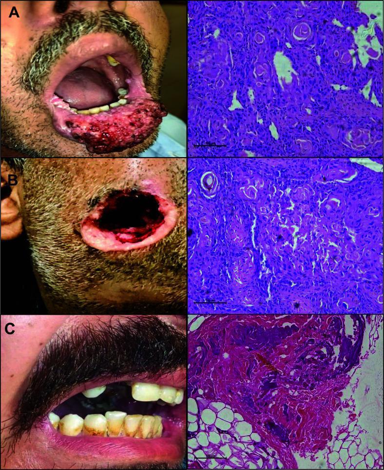
A medical comparison chart illustrating Oral Squamous Cell Carcinoma (OSCC) through clinical photographs (left column) and corresponding histopathological H&E stained slides (right column), categorized by tumor differentiation grades. Panel A demonstrates well-differentiated OSCC, showing an exophytic, irregular, and erythematous mass on the lower lip; the microscopy reveals nests of squamous cells with distinct keratin pearls. Panel B shows moderately differentiated OSCC with an extensive, deep ulcerative lesion of the chin/perioral region and histological features including disorganized cellular arrangements and fewer keratinization signs. Panel C represents poorly differentiated OSCC, showing severe dental decay and gingival recession clinically; the histopathology displays highly pleomorphic cells with a loss of typical squamous architecture, increased cellular dispersion, and invasion into underlying adipose tissue (visible as clear adipocytes). This visual resource is designed for dental and oncological education to correlate gross clinical presentations of oral malignancy with microscopic grading based on cellular morphology and tissue organization.
oral squamous cell carcinoma clinical appearance ulcer tongue floor of mouth
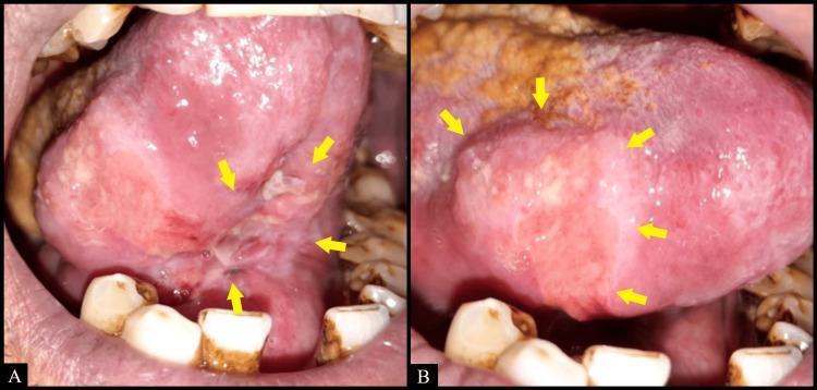
Two-panel intraoral clinical photograph (A and B) demonstrating a large, irregularly shaped ulcer located on the tongue and floor of the mouth. Panel A shows the ulcer extending from the ventral surface of the tongue to the floor of the mouth, while Panel B highlights its involvement along the right lateral border of the tongue. Yellow arrows delineate the margins of the lesion, which features edematous, raised, and slightly indurated borders. The ulcerated bed exhibits a heterogeneous appearance with mixed areas of erythematous (reddish) and leukoplakic (whitish) tissue, along with surface irregularities and sloughing. The surrounding oral mucosa appears erythematous, and there is evidence of yellowish-white coating on the dorsal tongue, suggestive of secondary candidiasis. The clinical presentation is highly relevant for the differential diagnosis of oral squamous cell carcinoma, deep fungal infections (such as histoplasmosis), or chronic traumatic ulcers. The educational focus is on identifying morphology and anatomical distribution of chronic oral ulcerative lesions in clinical dentistry and pathology.
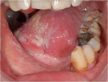
This clinical intraoral photograph displays the oral cavity 12 months following resection of an oral squamous cell carcinoma (OSCC) of the right floor of the mouth. The image highlights the postoperative and post-radiation appearance of the tongue and reconstructed floor of the mouth. The ventral and lateral surfaces of the tongue show areas of mucosal thinning, erythema, and white patches consistent with chronic post-radiation changes or scarring. The floor of the mouth exhibits altered tissue architecture and color, with generalized pallor indicative of fibrotic changes from surgical reconstruction and adjuvant therapy. The mandibular dentition is visible, showing significant dental attrition, decay, and amalgam restorations, particularly in the lower left quadrant. The surrounding gingiva and buccal mucosa appear intact but show signs of mild recession and chronic inflammatory changes. This visual serves to demonstrate long-term tissue remodeling and the clinical status of the oral cavity following radical resection, mandibulectomy, and fibular transplant reconstruction for advanced oral cancer.
A labeled pencil-style histological diagram for BDS exam of well-differentiated oral squamous cell carcinoma (H&E). The diagram shows: (1) surface oral epithelium with ulceration; (2) invasive islands and cords of squamous epithelial cells breaking through the basement membrane into connective tissue; (3) prominent keratin pearls (concentric whorls of pink keratin) centrally within tumor nests; (4) intercellular bridges (prickles) between tumor cells; (5) nuclear pleomorphism and hyperchromatism; (6) individual cell keratinization; (7) desmoplastic fibrous stroma around tumor nests; (8) inflammatory infiltrate (lymphocytes and plasma cells) at tumor-stroma interface; (9) mitotic figures. Black and white pencil sketch labeled with arrows. Title: "Well-Differentiated OSCC (H&E)" Clean exam diagram style.
TUMORS OF THE ORAL CAVITY + ORAL SQUAMOUS CELL CARCINOMA (OSCC)
BDS 3rd Year - Oral Pathology - University Exam Pattern
CLASSIFICATION OF TUMORS OF THE ORAL CAVITY
I. EPITHELIAL TUMORS
A. BENIGN EPITHELIAL TUMORS
- Squamous papilloma
- Keratoacanthoma
- Verruca vulgaris (common wart)
- Condyloma acuminatum
- Focal epithelial hyperplasia (Heck's disease)
- Sebaceous adenoma / Fordyce's granules (sebaceous origin)
B. MALIGNANT EPITHELIAL TUMORS (CARCINOMAS)
- Squamous Cell Carcinoma (SCC) - most common (>90% of all oral malignancies)
- Well differentiated
- Moderately differentiated
- Poorly differentiated
- Verrucous Carcinoma (Ackermann's tumor)
- Basaloid Squamous Cell Carcinoma
- Spindle Cell Carcinoma (Sarcomatoid carcinoma)
- Adenoid Squamous Cell Carcinoma
- Lymphoepithelial Carcinoma
- Basal Cell Carcinoma (rare intraorally; common on lip)
C. SALIVARY GLAND EPITHELIAL TUMORS
- Pleomorphic adenoma (most common salivary tumor)
- Monomorphic adenoma (Warthin's tumor, etc.)
- Oncocytoma
- Mucoepidermoid carcinoma (most common malignant salivary tumor)
- Adenoid cystic carcinoma
- Acinic cell carcinoma
- Adenocarcinoma NOS
- Carcinoma ex pleomorphic adenoma
II. NON-EPITHELIAL TUMORS
A. BENIGN NON-EPITHELIAL TUMORS
- Fibroma (irritation fibroma - most common)
- Lipoma
- Myxoma
- Hemangioma (capillary, cavernous)
- Lymphangioma
- Rhabdomyoma
- Leiomyoma
- Granular cell tumor (Abrikossoff's tumor - origin disputed)
- Neurofibroma
- Schwannoma (Neurilemmoma)
- Traumatic neuroma
- Osteoma
- Chondroma
- Torus palatinus / torus mandibularis (reactive bony growths)
- Ameloblastoma
- Odontoma
- Odontogenic myxoma
- Cementoblastoma
B. MALIGNANT NON-EPITHELIAL TUMORS (SARCOMAS)
- Fibrosarcoma
- Liposarcoma
- Kaposi's sarcoma
- Angiosarcoma
- Rhabdomyosarcoma (most common soft tissue sarcoma of H&N in children)
- Leiomyosarcoma
- Osteosarcoma (most common primary bone malignancy of jaw)
- Chondrosarcoma
- Malignant melanoma
- Non-Hodgkin lymphoma (most common lymphoma of oral cavity)
- Hodgkin lymphoma
- Multiple myeloma / Plasmacytoma
ORAL SQUAMOUS CELL CARCINOMA (OSCC)
DEFINITION
ETIOPATHOGENESIS
A. ETIOLOGICAL FACTORS
1. Tobacco
- Most important risk factor for OSCC in India
- Smoked tobacco: cigarettes, bidis, cigars, pipes, hookah
- Smokeless tobacco (most significant in Indian population): gutka, pan masala, khaini, mawa, snuff, zarda
- Carcinogens: polycyclic aromatic hydrocarbons (PAH), nitrosamines (NNK, NNN), benzopyrene
- Act as initiators and promoters
2. Alcohol
- Independent risk factor; synergistic effect with tobacco (multiplicative, not additive)
- Mechanism: direct solvent action on mucosa, impairs DNA repair, acts as carcinogen solvent, increases mucosal permeability
3. Betel nut (Areca nut) / Betel quid
- Unique to South/Southeast Asia
- Areca nut contains arecoline - promotes fibrosis and epithelial changes
- Leads to Oral Submucous Fibrosis (OSMF) - a recognized precancerous condition (~7% malignant transformation)
4. Human Papillomavirus (HPV)
- HPV 16 and HPV 18 are high-risk oncogenic strains
- More important in oropharyngeal SCC (base of tongue, tonsil)
- Mechanism: viral oncoproteins E6 (degrades p53) and E7 (inactivates Rb protein)
5. Chronic sun exposure / UV radiation
- Risk factor for lower lip OSCC (actinic cheilitis → SCC)
- UV-B is most carcinogenic
6. Chronic irritation
- Ill-fitting dentures, sharp tooth edges, chronic ulcers
7. Diet / Nutrition
- Iron deficiency anemia (Plummer-Vinson / Paterson-Kelly syndrome) - risk for post-cricoid and tongue carcinoma
- Deficiency of vitamins A, C, E, riboflavin
8. Immunosuppression
- Organ transplant patients, HIV/AIDS
9. Syphilis (historically linked to tongue carcinoma)
10. Oncogenic viruses: EBV (rare oral association)
B. PATHOGENESIS (MOLECULAR BASIS)
Normal Epithelium → Hyperplasia → Dysplasia (mild→moderate→severe)
→ Carcinoma in situ → Invasive Carcinoma
-
p53 tumor suppressor gene mutation (chromosome 17p) - most common genetic alteration in OSCC
- Tobacco carcinogens cause G:C → T:A transversions at p53 hotspots
- Results in loss of apoptosis, uncontrolled proliferation
-
Loss of chromosome 9p - loss of CDKN2A (p16/INK4a) - early event
- p16 normally inhibits CDK4/6 which phosphorylates Rb
- Loss → uncontrolled cell cycle progression through G1/S checkpoint
-
Rb (Retinoblastoma) gene inactivation - cell cycle deregulation
-
EGFR (Epidermal Growth Factor Receptor) overexpression - found in ~80-90% OSCC
- Drives proliferation through RAS-MAPK and PI3K-AKT pathways
-
Loss of chromosome 3p, 13q - additional tumor suppressor losses
-
Field cancerization (Slaughter's concept, 1953):
- Entire field of mucosa exposed to carcinogens undergoes genetic damage
- Explains multifocal tumors, second primary tumors, and high local recurrence rates
-
HPV pathway: E6 → p53 degradation; E7 → Rb inactivation → p16 overexpression paradoxically (used as HPV surrogate marker)
-
Telomerase activation - immortalization of tumor cells
-
Angiogenesis (VEGF upregulation) - tumor neovascularity
HISTOLOGICAL FEATURES OF OSCC
GENERAL HISTOLOGICAL CRITERIA FOR MALIGNANCY
| Feature | Description |
|---|---|
| Loss of normal architecture | Disorganized growth, loss of stratification |
| Nuclear pleomorphism | Variation in nuclear size and shape |
| Hyperchromatism | Dark-staining nuclei due to increased DNA |
| Increased N:C ratio | Nucleus enlarged relative to cytoplasm |
| Abnormal mitoses | Tripolar, star-shaped, atypical mitotic figures |
| Loss of polarity | Cells disoriented, no orderly maturation |
| Invasion | Tumor nests breaking through basement membrane |
| Desmoplasia | Reactive fibrous stroma around tumor nests |
SPECIFIC HISTOLOGICAL FEATURES OF OSCC
-
Surface changes: ulceration of overlying epithelium; or intact surface with underlying invasion
-
Invasive tumor islands: nests, cords, and sheets of malignant squamous epithelial cells invading the connective tissue, breaking through the basement membrane
-
Keratin pearl formation (epithelial pearl / cell nest):
- Concentric whorls of keratinized squamous cells
- Central amorphous pink keratin surrounded by layers of flattened cells
- Pathognomonic of squamous differentiation
-
Individual cell keratinization: single cells with abundant pink cytoplasm within tumor nests
-
Intercellular bridges (prickles): desmosomes between adjacent squamous cells - evidence of squamous differentiation
-
Nuclear pleomorphism and hyperchromatism
-
Increased mitotic figures, including atypical/abnormal mitoses (tripolar, multipolar)
-
Desmoplastic stroma: dense fibrous connective tissue reaction surrounding tumor nests
-
Inflammatory infiltrate: lymphocytes, plasma cells, and occasionally eosinophils at the tumor-stroma interface (host response)
-
Perineural invasion and lymphovascular invasion (markers of aggressive behavior)
GRADING OF OSCC (BRODERS' GRADING SYSTEM)
BRODERS' ORIGINAL 4-GRADE SYSTEM
| Grade | Name | % Undifferentiated cells |
|---|---|---|
| Grade I | Well differentiated | <25% undifferentiated |
| Grade II | Moderately differentiated | 25-50% undifferentiated |
| Grade III | Poorly differentiated | 50-75% undifferentiated |
| Grade IV | Anaplastic | >75% undifferentiated |
HISTOLOGICAL GRADING TABLE (as used in Indian BDS exams)
| Feature | Grade I (Well Differentiated) | Grade II (Moderately Differentiated) | Grade III (Poorly Differentiated) |
|---|---|---|---|
| Keratin pearl | Abundant, well-formed | Few, poorly formed | Absent |
| Intercellular bridges | Prominent | Occasional | Absent or rare |
| Individual cell keratinization | Present | Present | Absent |
| Nuclear pleomorphism | Mild | Moderate | Severe |
| Mitotic figures | Few, typical | Moderate, some atypical | Many, atypical (tripolar, multipolar) |
| Cell differentiation | Closely resembles normal squamous epithelium | Partial resemblance | No resemblance to squamous epithelium |
| Prognosis | Relatively better | Intermediate | Poorest |
| N:C ratio | Near normal | Increased | Markedly increased |
| Hyperchromatism | Mild | Moderate | Severe |
CLINICAL PHOTOGRAPHS + H&E GRADING CORRELATION
PHOTOMICROGRAPH - Well-Differentiated OSCC (H&E)
H&E DIAGRAM FOR EXAM (Drawable)
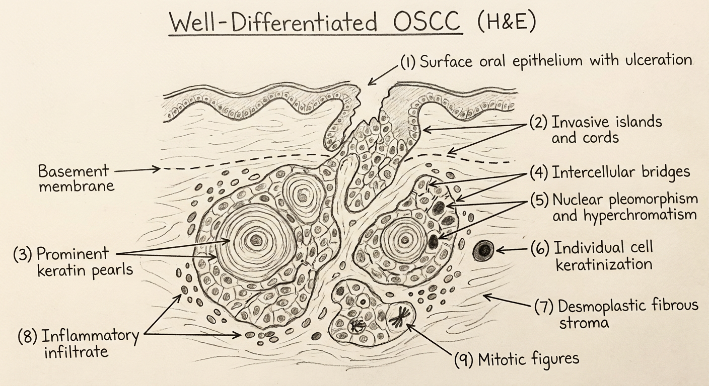
HOW TO DRAW OSCC (H&E) IN EXAM:
Step 1: Draw surface epithelium - show ulceration / break in surface epithelium
Step 2: Show basement membrane disruption (critical!)
Step 3: Draw irregular nests/islands of cells invading downward into fibrous stroma
Step 4: Inside tumor nests - draw concentric whorls = KERATIN PEARLS (pink, concentric rings)
Step 5: Between cells - draw small bridges = INTERCELLULAR BRIDGES (prickles)
Step 6: Nuclear details - enlarged, hyperchromatic nuclei, prominent nucleoli
Step 7: Around nests - pink dense fibrous tissue = DESMOPLASTIC STROMA
Step 8: At periphery of nests - small dark cells = INFLAMMATORY INFILTRATE
Step 9: Show 1-2 ATYPICAL MITOTIC FIGURES (starred/tripolar shapes)
LABELS (with arrows):
→ Ulcerated surface epithelium
→ Disrupted basement membrane
→ Invasive tumor nests / islands
→ Keratin pearl (cell nest / epithelial pearl)
→ Individual cell keratinization
→ Intercellular bridges (prickles)
→ Nuclear pleomorphism and hyperchromatism
→ Atypical mitotic figures
→ Desmoplastic fibrous stroma
→ Inflammatory infiltrate (lymphocytes, plasma cells)
CLINICAL IMAGE - OSCC of Tongue / Floor of Mouth
ADDITIONAL GRADING SYSTEMS (for completeness)
Anneroth et al. / Bryne's Malignancy Grading System (1987, modified 1992)
| Parameter | Score 1 | Score 2 | Score 3 | Score 4 |
|---|---|---|---|---|
| Degree of keratinization | Highly keratinized | Moderately | Minimal | No keratinization |
| Nuclear polymorphism | Little | Moderate | Abundant | Extreme |
| Mitoses per HPF | 0-1 | 2-3 | 4-5 | >5 |
| Pattern of invasion | Pushing borders | Solid cords | Small groups | Individual cells |
| Stage of invasion | In situ | Lamina propria | Muscle/bone | Perineural/vascular |
| Lymphoplasmacytic infiltration | Marked | Moderate | Slight | Absent |
SUMMARY TABLE FOR QUICK REVISION
| Parameter | OSCC |
|---|---|
| Most common oral malignancy | >90% of all oral cancers |
| Most common site | Tongue (lateral border) > Floor of mouth > Lower lip |
| Most common site in India | Buccal mucosa (due to tobacco habit) |
| Most important risk factor | Tobacco (smoked and smokeless) |
| Synergistic risk | Tobacco + Alcohol |
| Key molecular alteration | p53 mutation (most common); loss of 9p (earliest) |
| Concept of multifocal risk | Field cancerization (Slaughter, 1953) |
| Grading system | Broders' system (Grade I-III) |
| Best prognosis grade | Grade I (well differentiated) |
| Key histological feature | Keratin pearls + intercellular bridges + invasive nests |
| Most important prognostic factor | TNM stage (lymph node status) |
Exam Tips:
- Grading is NOT staging - grading = histologic differentiation (Broders); staging = clinical extent (TNM)
- Best grade to identify in diagram = Grade I - "keratin pearls are the single most important feature to draw"
- Broders' classification - always remember the 4-grade system, then mention the 3-grade simplification used in practice
- Field cancerization - Slaughter (1953) - always a 2-mark question
- E6 destroys p53; E7 inactivates Rb - for HPV-associated mechanism questions
Enumerate viral lesions of oral cavity. Detail about hsv and oral manifestation of hiv infection
herpes simplex stomatitis oral vesicles ulcers gingivostomatitis clinical
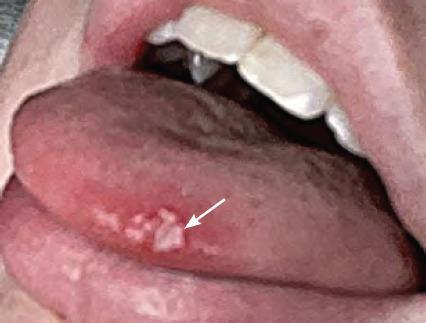
A clinical photograph of the oral cavity focusing on the lateral and dorsal aspect of the tongue, illustrating features of primary herpetic gingivostomatitis. The image demonstrates a cluster of multiple small, discrete, and coalescing whitish-yellow vesicles and ulcers located on the lateral border of the tongue. These lesions are slightly raised with irregular margins. A white arrow highlights a prominent ulcerated area within the cluster. The surrounding mucosa is markedly erythematous and inflamed, providing a sharp contrast to the lighter-colored lesions. This visual represents the acute phase of a viral infection (Herpes Simplex Virus Type 1), focusing on the presentation of painful intraoral vesicles that rupture to form fibrin-covered ulcers. It is a key educational example for dental and medical students in recognizing oral manifestations of primary viral stomatitis and differentiating it from other ulcerative conditions like aphthous ulcers.
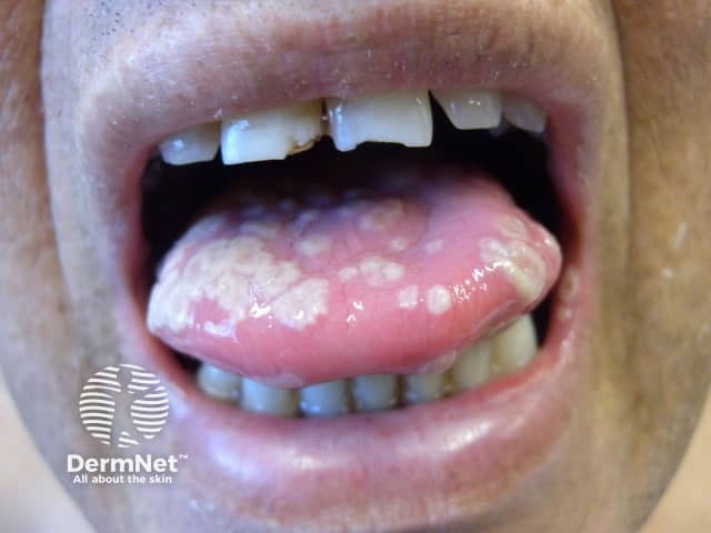
Clinical photograph of the oral cavity documenting mucosal vesicles and erosions associated with suspected herpes simplex infection. The image shows diffuse mucosal involvement of the tongue with white to yellowish plaques, and scattered vesicular lesions along the gingiva and buccal mucosa with mild surrounding erythema. The tongue surface is swollen, with a smooth, shiny appearance; plaques are patchy, and some vesicles have ruptured, leaving shallow erosions covered by pseudomembranous debris. Lips and labial mucosa may show fissuring and crusting. Notable absence of radiographic features. This morphological pattern is typical of primary herpes simplex virus type 1 infection, more common in pediatric patients and in primary exposure. Clinically, vesicles progress to painful ulcers with erythematous halos; fever, irritability, and reduced oral intake are common. Differential diagnoses include candidiasis, aphthous stomatitis, erythema multiforme, and recurrent herpetic lesions. Diagnostic significance: recognition of classic mucocutaneous HSV pattern supports prompt antiviral therapy (acyclovir) and can shorten illness; confirmatory testing from a lesion (PCR, viral culture, or antigen assays) may be employed. Clinically correlate with recent exposure to orolabial HSV, gingivostomatitis symptoms, and systemic signs. This image is educational for recognizing orofacial HSV lesions, guiding differential diagnosis, and illustrating progression from vesicles to ulcers in dermatology and infectious disease training.
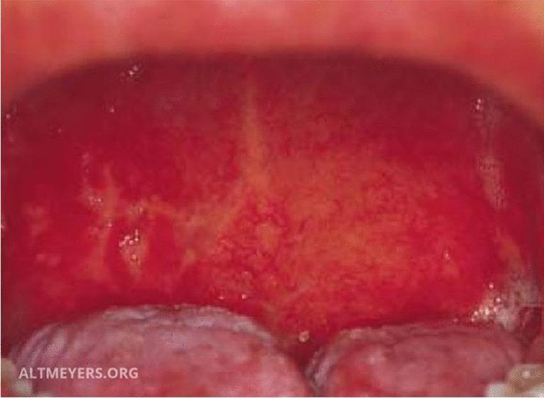
This clinical photograph displays a detailed intraoral view of the posterior oral cavity and oropharynx, demonstrating classic signs of Herpes simplex virus (HSV) stomatitis. The oral mucosa exhibits diffuse, intense erythema (redness) across the soft palate and pharyngeal wall. Superimposed on this inflamed background are multiple irregular, yellowish-white exudative patches and small fibrin-covered erosions, which represent ruptured vesicles characteristic of herpetic infection. The lesions are unevenly distributed and show a tendency toward confluence in certain areas. In the lower foreground, segments of the dorsal tongue are visible; these appear relatively spared, presenting a pale pink color and normal surface texture without obvious ulceration. This image serves as a clinical reference for primary herpetic gingivostomatitis or secondary HSV stomatitis, highlighting the transition from viral vesicles to painful mucosal erosions and the associated generalized inflammatory response in the epithelial tissues.
oral hairy leukoplakia HIV tongue Epstein Barr virus
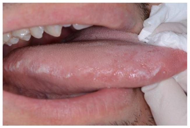
This clinical photograph displays a close-up intraoral view of the lateral margin of the tongue. The primary clinical finding is oral hairy leukoplakia, characterized by multiple adherent, whitish-gray plaques. These lesions exhibit a distinct corrugated, vertically-oriented, or 'hairy' surface texture and are localized along the lateral tongue border. The lesions are non-scrapable and irregular in shape. Surrounding oral structures, including the visible maxillary teeth and lower lip, appear within normal limits without evidence of generalized inflammation. In an educational context, this manifestation is a classic marker of Epstein-Barr virus (EBV) replication within the oral epithelium, frequently occurring in immunocompromised individuals, such as those with HIV infection. The image serves as a diagnostic reference for identifying opportunistic oral viral infections and highlights the importance of oral examination in systemic disease monitoring.
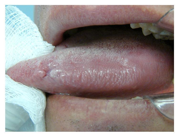
This clinical photograph displays a close-up, intraoral view of a human tongue, specifically focusing on the lateral border. The image depicts oral hairy leukoplakia (OHL), characterized by a non-removable, whitish, slightly elevated plaque with a corrugated or 'hairy' surface texture. The primary lesion is situated along the lateral margin, exhibiting vertical striations that are characteristic of this condition. Surrounding the main lesion are smaller, subtle areas of similar whitish discoloration against the normal pink, papillated mucosa of the tongue. The tongue is partially retracted with a gauze square for better visualization of the lateral aspect. This presentation is highly suggestive of Epstein-Barr virus (EBV) infection, often occurring in immunocompromised individuals, such as those with HIV/AIDS. The clinical focus illustrates the classic morphology and anatomical site preference (lateral tongue) for OHL, serving as an important diagnostic sign in oral pathology and infectious disease.
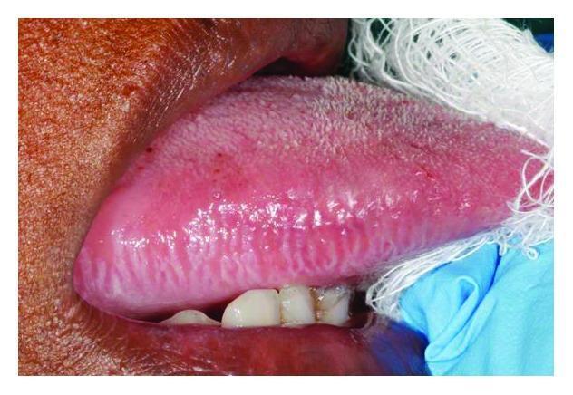
This clinical intraoral photograph displays a localized white lesion on the lateral border of the tongue, characteristic of oral hairy leukoplakia. The lesion is primarily hyperkeratotic and exhibits a distinct corrugated or 'hairy' surface texture with prominent vertical ridges. It is situated along the right lateral margin, showing a clear demarcation from the surrounding normal, pinkish-red tongue mucosa. The image provides clinical context for evaluating opportunistic infections in immunocompromised patients, specifically those with HIV/AIDS. The presence of these vertically oriented white plaques is strongly associated with Epstein-Barr virus (EBV) replication within the oral epithelium. Educational focus includes recognizing the morphology, anatomical site predilection, and clinical significance of this benign but diagnostic marker of immune suppression.
herpes simplex histology Tzanck cells ballooning degeneration intranuclear inclusion
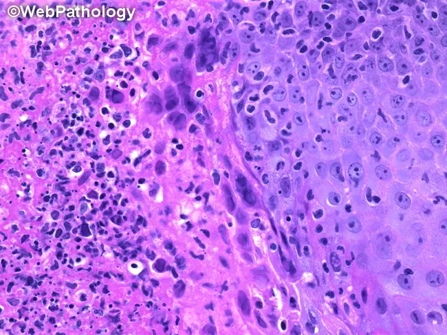
Light microscopy of a hematoxylin and eosin stained mucocutaneous biopsy demonstrates histopathologic features of herpes simplex virus infection. The specimen comprises stratified squamous epithelium from a genital or oral lesion with mucosal involvement. At high magnification, epithelial cells show ballooning degeneration, multinucleation, and prominent nuclear molding. Intranuclear eosinophilic inclusions (Cowdry type A) occupy the karyoplasm with marginated chromatin and variable nuclear clearing. These changes are frequently accompanied by vesiculation, keratinocyte crowding, and focal necrosis at the lesion edge. An inflammatory infiltrate dominated by lymphocytes and neutrophils resides in a dense subepithelial zone. The combination of multinucleated keratinocytes, eosinophilic intranuclear inclusions, and nuclear enlargement is highly suggestive of active HSV infection and helps differentiate from varicella-zoster virus or cytomegalovirus, which may show different inclusion bodies or owl’s eye nuclei, respectively. Clinically, HSV-2 is commonly genital; HSV-1 is typically orolabial, but overlap occurs. Histology supports clinical differential and, when combined with PCR or immunohistochemistry, confirms HSV typing. This image is clinically relevant for dermatology, infectious disease, pathology education, and research on mucocutaneous viral pathology and antiviral therapy evaluation. The image serves as a teaching reference for recognizing classic HSV cytopathic effects on routine histology and correlates with molecular typing and lesion localization.
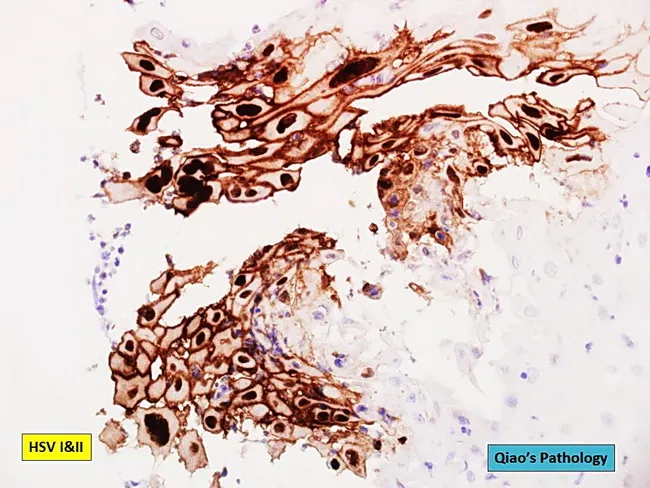
Histology with immunohistochemistry for herpes simplex virus (HSV-1/HSV-2) applied to an esophageal ulcer biopsy. The specimen shows mucosal ulceration with loss of epithelial integrity and dense inflammatory infiltrate in the underlying lamina propria. Numerous infected squamous epithelial cells exhibit classic cytopathic effects, including multinucleation, nuclear enlargement and prominent eosinophilic intranuclear inclusions (Cowdry type A). The HSV immunostain labels the infected cells with a brown chromogen, delineating scattered and clustered positive cells throughout the lesion. Adjacent noninfected mucosa demonstrates preserved architecture with basal cell hyperplasia and regenerative changes. In some areas, ballooning degeneration and acantholysis contribute to the desquamation of epithelial cells. The stain is specific for HSV-1/HSV-2 and supports a diagnosis of herpes esophagitis, especially when clinical context includes immunosuppression or underlying mucosal damage. Background inflammatory cells include lymphocytes and plasma cells; occasional neutrophils are present in ulcer craters. The morphological pattern helps distinguish from cytomegalovirus infection, which typically shows large cells with prominent owl's eye inclusions, and from candidal infection, which reveals fungal organisms on special stains. Thus HSV immunostaining provides definitive confirmation and guides antiviral therapy. Correlation with patient symptoms, serology, and endoscopic findings improves diagnostic confidence and informs antiviral management, isolation precautions, and monitoring for complications such as bleeding or perforation.
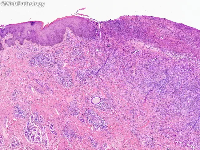
Penile foreskin and glans mucosal biopsy obtained during evaluation of confluent genital ulcers in an immunocompromised patient with chronic lymphocytic leukemia. Hematoxylin and eosin stained tissue shows epidermal ulceration with underlying dermal inflammatory infiltrate and necrosis. The epidermis often displays ballooning degeneration of keratinocytes and occasional multinucleated giant cells, reflecting herpes simplex virus cytopathic effect. Intranuclear eosinophilic Cowdry type A inclusions may be present within atypical keratinocytes, though at this low magnification they can be difficult to appreciate. Dermal perivascular inflammation, edema, and abundant neutrophils are common in the ulcer bases, with focal lymphoplasmacytic infiltrates. The lesion is confluent and shallow, with subepithelial pseudomembrane and necrotic debris. In immunocompromised hosts the histology may be more extensive with epidermal necrosis and persistent ulceration. These morphological features, together with compatible clinical history of recurrent painful genital ulcers, support a diagnosis of genital herpes simplex infection caused by herpes simplex virus type 1 or type 2. Differential diagnosis includes syphilitic chancres, chancroid, candidal balanitis, and non-infectious etiologies; culture, PCR for HSV DNA, or direct fluorescent antibody testing can confirm the diagnosis. Appropriate antiviral therapy is indicated and infection control measures advised to prevent transmission.
A labeled pencil-style histological diagram for BDS exam of herpes simplex virus (HSV) oral infection (H&E). Shows: (1) stratified squamous epithelium with intraepithelial vesicle formed by ballooning degeneration of cells; (2) ballooned/swollen epithelial cells with pale cytoplasm; (3) multinucleated giant cells (Tzanck cells) with nuclear molding and marginated chromatin; (4) Cowdry type A intranuclear eosinophilic inclusions in nuclei; (5) acantholysis - separation of cells; (6) roof of vesicle and floor labeled; (7) dense inflammatory infiltrate below vesicle in lamina propria (lymphocytes, neutrophils); (8) necrosis of cells. Black and white pencil sketch, clean labels and arrows. Title: "Histology of HSV Oral Infection (H&E)"
VIRAL LESIONS OF THE ORAL CAVITY + HSV + ORAL MANIFESTATIONS OF HIV
BDS 3rd Year - Oral Pathology - University Exam Pattern
ENUMERATION OF VIRAL LESIONS OF THE ORAL CAVITY
A. HERPESVIRUS GROUP (Herpesviridae)
- Herpes Simplex Virus (HSV-1, HSV-2)
- Primary herpetic gingivostomatitis
- Recurrent herpes labialis (cold sore / fever blister)
- Recurrent intraoral herpes
- Varicella Zoster Virus (VZV / HHV-3)
- Chickenpox (primary)
- Herpes zoster (shingles - reactivation)
- Epstein-Barr Virus (EBV / HHV-4)
- Infectious mononucleosis (kissing disease)
- Oral hairy leukoplakia (in immunocompromised)
- Burkitt's lymphoma (association)
- Cytomegalovirus (CMV / HHV-5)
- CMV ulcers (in HIV/immunocompromised)
- Human Herpesvirus 8 (HHV-8)
- Kaposi's sarcoma
B. PAPOVAVIRUS GROUP (Papillomaviridae)
- Human Papillomavirus (HPV)
- Squamous papilloma (HPV 6, 11)
- Verruca vulgaris / common wart (HPV 2, 4)
- Condyloma acuminatum / venereal wart (HPV 6, 11)
- Focal epithelial hyperplasia / Heck's disease (HPV 13, 32)
- HPV-associated oropharyngeal SCC (HPV 16, 18)
C. PARAMYXOVIRUS GROUP
- Mumps virus - parotitis (viral sialadenitis)
- Measles (Rubeola) virus - Koplik's spots on buccal mucosa
D. COXSACKIEVIRUS (Enterovirus)
- Herpangina (Coxsackievirus A)
- Hand, Foot and Mouth Disease (Coxsackievirus A16, Enterovirus 71)
E. HIV / RETROVIRUS
- HIV / AIDS - multiple oral manifestations (see below)
F. HEPATITIS VIRUSES
- Hepatitis C - association with oral lichen planus
G. OTHER
- Molluscum contagiosum (Poxvirus) - rare perioral lesions
HERPES SIMPLEX VIRUS (HSV) - DETAILED
THE VIRUS
- Family: Herpesviridae
- Type: HSV-1 (orolabial, above waist) - most relevant for oral pathology
- HSV-2 (genital, below waist) - occasionally causes oral lesions
- Large, enveloped, double-stranded DNA virus
- Key property: Latency - virus persists in sensory ganglia (trigeminal ganglion for HSV-1) after primary infection and reactivates periodically
PATHOGENESIS OF HSV INFECTION
PRIMARY INFECTION
↓
Virus contacts oral mucosa → enters epithelial cells
↓
Replication → cell lysis → intraepithelial vesicle formation
↓
Virus travels retrograde along sensory nerve axons
↓
LATENCY in Trigeminal (Gasserian) Ganglion
↓
REACTIVATION TRIGGERS: fever, stress, sunlight, trauma,
menstruation, immunosuppression, GI disturbances
↓
Anterograde travel → RECURRENT LESION at original site
A. PRIMARY HERPETIC GINGIVOSTOMATITIS
Definition
Epidemiology
- Most common in children 6 months to 5 years (though can occur in young adults)
- Spread by direct contact with infected saliva or lesions
- Many primary infections are subclinical (asymptomatic seroconversion)
Clinical Features
- Fever (may be high, 38-40°C), malaise, irritability
- Loss of appetite
- Headache, myalgia
- Regional lymphadenopathy (cervical nodes - tender, enlarged)
- Diffuse, fiery red gingival erythema (marginal and attached gingiva, characteristic)
- Multiple small vesicles (1-3 mm) that quickly rupture to form:
- Shallow, painful ulcers with yellowish-grey fibrinous pseudomembrane and erythematous halo
- Ulcers affect any oral mucosal surface - gingiva, palate, tongue, buccal mucosa, lips
- Lesions may coalesce to form large irregular ulcers
- Intense oral pain - child refuses to eat or drink (risk of dehydration)
- Halitosis
- Excessive salivation
Clinical Photographs:
B. RECURRENT HERPES LABIALIS (Cold Sore / Fever Blister)
Definition
Clinical Features
- Occurs in same individual repeatedly (recurrence)
- Common site: vermilion border of lip (herpes labialis); occasionally anterior hard palate and attached gingiva (intraorally)
- Prodrome (hours before lesion): burning, tingling, itching, or pain at the site
- Lesion progression:
- Erythema at site
- Small grouped vesicles (cluster of 2-3 mm)
- Vesicles rupture → shallow ulcers with crust (scab) on lip skin
- Healing in 7-10 days (no scarring)
- Less severe than primary; no systemic symptoms usually
Differences: Primary vs. Recurrent HSV
| Feature | Primary Herpetic Gingivostomatitis | Recurrent Herpes Labialis |
|---|---|---|
| Age | Children (infants - 5 yr) | Any age (adults) |
| Immunity status | Non-immune (first contact) | Pre-immune (reactivation) |
| Site | Widespread - any oral mucosa + gingiva | Localized - lips / attached gingiva / hard palate |
| Severity | Severe, systemic symptoms | Mild, localized |
| Gingival involvement | Diffuse, bright red gingivitis | Only attached gingiva |
| Lymphadenopathy | Present (cervical) | Absent or minimal |
| Duration | 10-14 days | 7-10 days |
| Fever | Yes (high grade) | Absent |
| Trigger | First exposure | Stress, UV, fever, trauma |
| Latent site | Trigeminal ganglion | Same - trigeminal ganglion |
HISTOLOGICAL FEATURES OF HSV
Microscopic Findings (H&E):
-
Intraepithelial vesicle - fluid-filled space within the epithelium formed by:
- Ballooning degeneration - epithelial cells swell, become rounded with pale/clear cytoplasm (hallmark)
- Acantholysis - loss of intercellular connections → cells float free in vesicle
-
Multinucleated giant cells (Tzanck cells):
- Multiple nuclei within one cell
- Nuclei show molding (fitting together like puzzle pieces)
- Margination of chromatin (chromatin pushed to periphery of nucleus)
- Produced by viral-induced cell fusion
-
Cowdry Type A intranuclear inclusions (most diagnostic):
- Eosinophilic (pink) intranuclear inclusion bodies
- Surrounded by a clear halo
- Chromatin marginated to nuclear membrane
- Reflects viral DNA replication in nucleus
-
Reticular degeneration - early stage, cells separate at intercellular junctions
-
Inflammatory infiltrate in lamina propria: lymphocytes, neutrophils
-
Necrosis of affected epithelial cells
Tzanck Smear (Cytology):
- Scraping from base of fresh vesicle, stained with Giemsa or H&E
- Shows multinucleated giant cells - quick diagnostic test
H&E Histology Photomicrograph:
DIAGRAM TO DRAW IN EXAM (H&E Pencil Diagram)
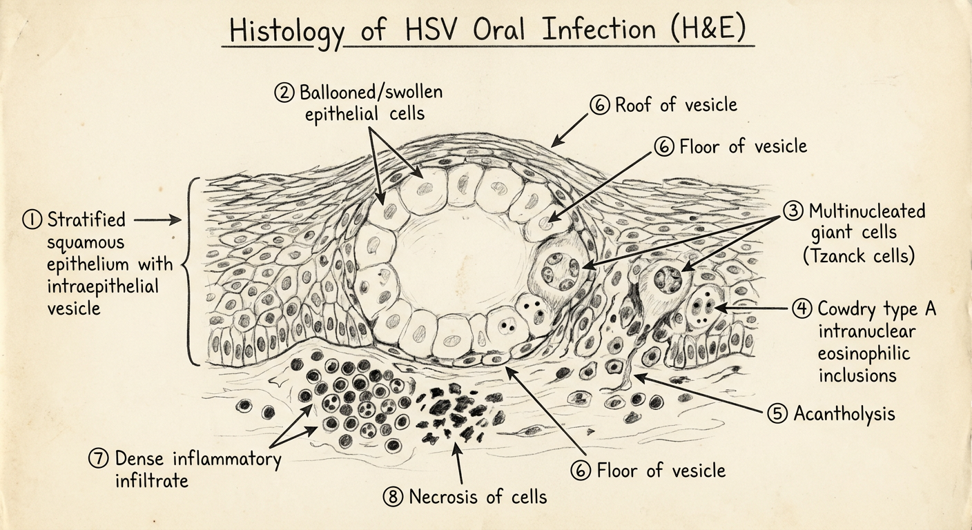
HOW TO DRAW HSV HISTOLOGY (H&E):
Step 1: Draw stratified squamous epithelium with a clear space = INTRAEPITHELIAL VESICLE
Step 2: At edges of vesicle - large round swollen cells = BALLOONING DEGENERATION
Step 3: Floating cells in vesicle fluid = ACANTHOLYTIC CELLS
Step 4: Large cell with 3-4 nuclei pressed together = MULTINUCLEATED GIANT CELL (Tzanck cell)
Step 5: Inside nuclei - draw pink dot with clear halo = COWDRY TYPE A INCLUSION
Step 6: Nuclei show chromatin pushed to edges = MARGINATED CHROMATIN
Step 7: Below vesicle in connective tissue - dark cells = INFLAMMATORY INFILTRATE
(lymphocytes + neutrophils)
Step 8: Draw some necrotic/ghost cells = NECROSIS
LABELS (with arrows):
→ Intraepithelial vesicle
→ Ballooning degeneration
→ Acantholysis / acantholytic cells
→ Multinucleated giant cell (Tzanck cell)
→ Cowdry Type A intranuclear inclusion (eosinophilic)
→ Marginated chromatin / nuclear molding
→ Inflammatory infiltrate in lamina propria
→ Necrosis of epithelial cells
TREATMENT OF HSV
- Acyclovir (antiviral of choice) - topical for recurrent labialis; systemic (200-400 mg 5x/day x 7-10 days) for primary
- Valacyclovir (prodrug of acyclovir) - better bioavailability
- Supportive: analgesics, antipyretics, fluids, good oral hygiene
- Topical acyclovir cream for herpes labialis if applied in prodromal stage
ORAL MANIFESTATIONS OF HIV INFECTION
INTRODUCTION
- May be the first sign of HIV infection
- Indicate the degree of immune suppression (inversely correlated with CD4 count)
- May indicate progression to AIDS (CD4 <200 cells/μL)
CLASSIFICATION OF ORAL MANIFESTATIONS OF HIV
- Candidiasis (pseudomembranous, erythematous, angular cheilitis)
- Hairy leukoplakia (EBV-associated)
- HIV-associated periodontal disease
- Linear gingival erythema (LGE)
- Necrotizing ulcerative gingivitis (NUG)
- Necrotizing ulcerative periodontitis (NUP)
- Kaposi's sarcoma
DETAILED ORAL LESIONS
1. ORAL CANDIDIASIS (Most common oral manifestation)
-
Pseudomembranous candidiasis (Thrush) - most common
- Creamy white, curd-like removable plaques
- Any site: tongue, palate, buccal mucosa
- Scraped off leaving red, raw, bleeding surface
- Distinguishes from hairy leukoplakia (OHL cannot be scraped off)
-
Erythematous candidiasis
- Red, atrophic patches; often on palate or dorsal tongue
- May present before pseudomembranous form
-
Angular cheilitis (perlèche)
- Cracking, erythema, soreness at corners of mouth
- Often mixed Candida + Staphylococcus
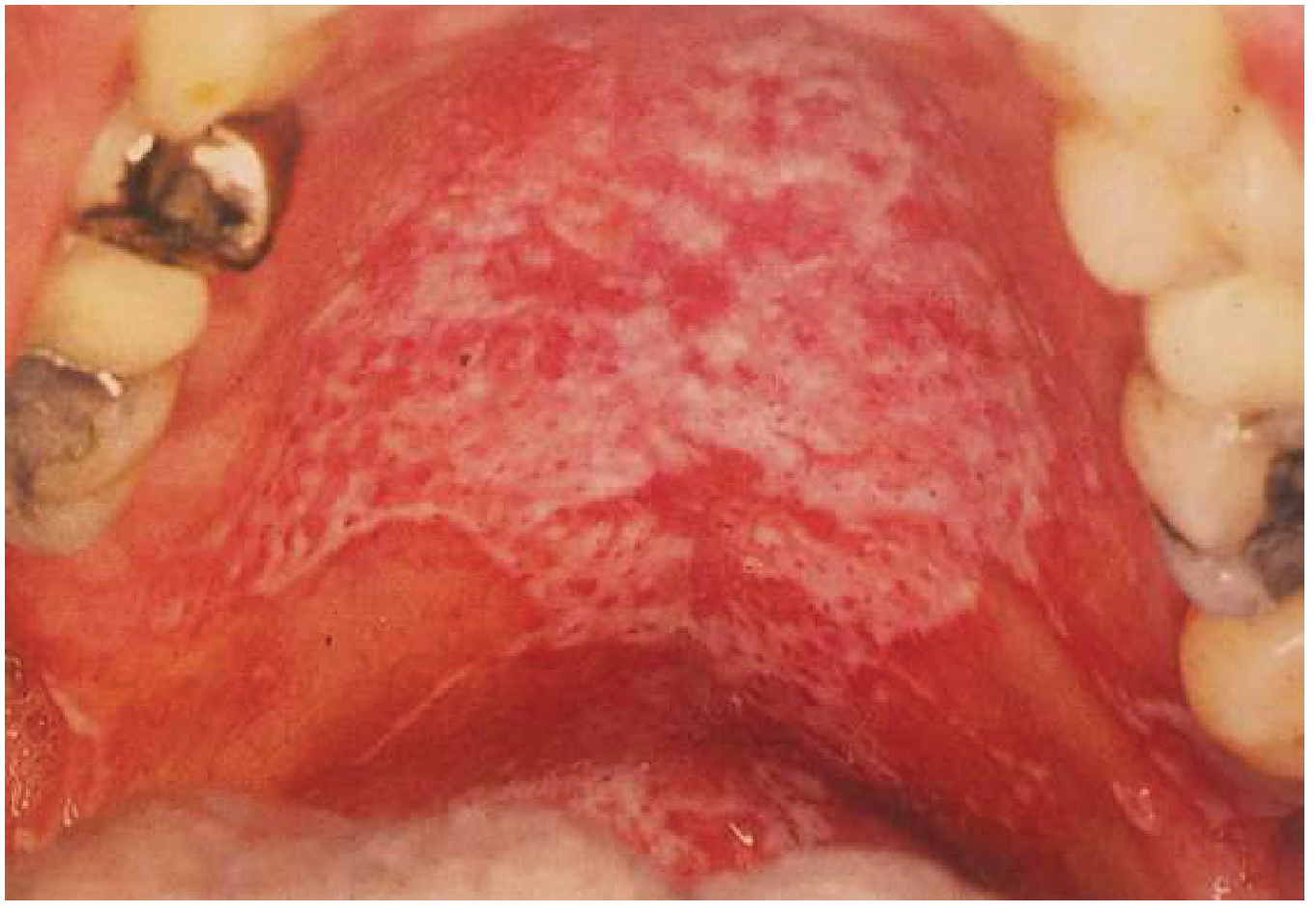
2. ORAL HAIRY LEUKOPLAKIA (OHL)
- Caused by: Epstein-Barr Virus (EBV) replication in epithelial cells
- Pathognomonic of HIV (virtually diagnostic)
- Not seen in healthy individuals; indicates significant immunosuppression
- Site: Lateral borders of tongue (bilateral); occasionally ventral tongue
- Appearance: White, corrugated ("hairy"), vertically oriented ridges or folds
- Cannot be scraped off (unlike candidiasis) - key differentiating point
- Usually asymptomatic
- CD4 count usually <300 cells/μL when it appears
- Hyperparakeratosis with surface "hair-like" projections
- Acanthosis (epithelial thickening)
- Koilocytes - balloon cells in the upper spinous layer (viral cytopathic effect)
- No inflammation in epithelium
- Koilocytes resemble HPV effect but EBV-specific
3. HIV-ASSOCIATED PERIODONTAL DISEASE
- Fiery red band (2-3 mm) along free gingival margin
- Disproportionate to amount of plaque present
- Bleeds spontaneously or on probing
- Does not respond to routine scaling
- Gram-negative and Candida organisms involved
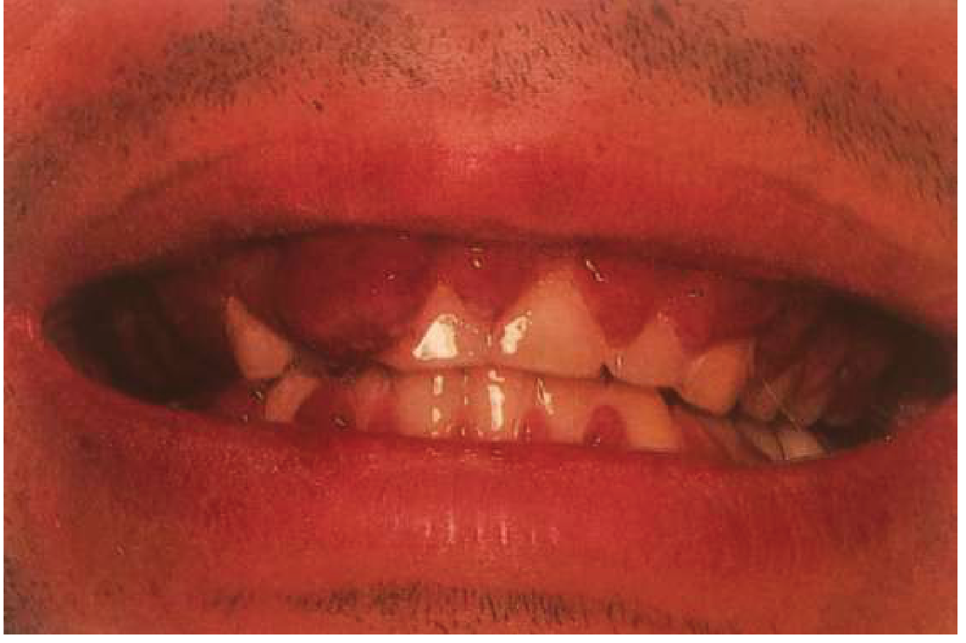
- Punched-out, crater-like destruction of interdental papillae
- Necrosis of gingival tissue
- Pseudomembrane formation
- Severe pain and fetor oris (halitosis)
- Extension of NUG into supporting bone
- Rapid, severe bone loss (several mm within days/weeks)
- Exposure of bone, spontaneous bleeding
- Occurs with CD4 <200 cells/μL
- Can progress to Necrotizing stomatitis (full-thickness necrosis)
4. KAPOSI'S SARCOMA (KS)
- Caused by HHV-8 (Human Herpesvirus 8)
- AIDS-defining malignancy
- Oral involvement occurs in ~50% of AIDS-KS cases
- Site: Hard palate (most common) > gingiva > tongue
- Appearance: Flat or slightly raised, red/purple/brown to violaceous (purple) macule or nodule
- Early lesions - flat, red-purple patches
- Later - raised nodular, may ulcerate, bleed
- Non-tender (unless secondarily infected)
- Diagnosis: biopsy required
- Proliferating spindle-shaped endothelial cells
- Slit-like vascular channels filled with RBCs
- Extravasated RBCs in stroma
- Hemosiderin deposits
- Inflammatory cells (plasma cells, lymphocytes)
5. ATYPICAL / MAJOR APHTHOUS ULCERS
- Large, deep, extremely painful ulcers (>1 cm)
- Prolonged healing (weeks to months)
- Site: soft palate, oropharynx, tongue
- Occurs with severe immunosuppression (CD4 <100)
- Refractory to usual treatment
- May require systemic steroids or thalidomide
6. SALIVARY GLAND DISEASE
- Xerostomia (dry mouth): due to antiretroviral drugs or lymphocytic sialadenitis
- Bilateral parotid enlargement: due to diffuse infiltrative lymphocytosis syndrome (DILS) - CD8 cells infiltrate glands
7. VIRAL INFECTIONS (in HIV patients)
- Recurrent herpes labialis - more frequent, severe, prolonged
- CMV ulcers - large, deep, non-healing ulcers (CMV coinfects 90% of HIV patients)
- HPV lesions - condyloma acuminatum, squamous papillomas increase in frequency
- Molluscum contagiosum - perioral, large lesions
8. NON-HODGKIN LYMPHOMA
- AIDS-defining malignancy
- Site: palate, gingiva, tongue, posterior oral cavity
- Presents as painless, rapidly enlarging mass
- B-cell lymphoma most common
- Poor prognosis
SUMMARY TABLE - CD4 COUNT AND ORAL LESIONS
| CD4 Count | Oral Lesions Expected |
|---|---|
| <500 cells/μL | Oral candidiasis, hairy leukoplakia, LGE |
| <200 cells/μL | NUG, NUP, major aphthous ulcers, KS |
| <100 cells/μL | CMV ulcers, extensive candidiasis, NHL |
QUICK REVISION TABLE - KEY DIFFERENTIATORS
| Feature | OHL | Oral Candidiasis (Thrush) |
|---|---|---|
| Cause | EBV (HHV-4) | Candida albicans (fungus) |
| Site | Lateral tongue (bilateral) | Any mucosa - tongue, palate |
| Surface | Corrugated, hairy, white | Creamy white plaques |
| Scrapability | Cannot be scraped off | Scraped off (leaves raw surface) |
| Specific to HIV | Yes (pathognomonic) | No (can occur in non-HIV) |
| Treatment | Acyclovir (resolves) | Antifungals (nystatin, fluconazole) |
| Histology | Koilocytes, EBV | Pseudohyphae, spores on PAS |
Exam Tips:
- HSV histology - 3 key words: Ballooning degeneration + Tzanck cells (multinucleated giant cells) + Cowdry Type A intranuclear inclusions
- OHL vs Candidiasis - OHL cannot be scraped; Candidiasis can be scraped - always comes in short answer
- EC-Clearinghouse Group I = strongly associated with HIV (candidiasis, OHL, KS, HIV periodontitis)
- Kaposi's sarcoma - HHV-8, hard palate, violaceous, AIDS-defining
- Latent site of HSV-1 = Trigeminal (Gasserian) ganglion - always mentioned in 2-mark questions
- Recurrent herpes intraorally occurs only on keratinized mucosa (hard palate, attached gingiva) - distinguishes it from aphthous ulcers (on non-keratinized mucosa)
Developmental disturbances in formation of dentin
dentinogenesis imperfecta teeth opalescent clinical radiograph shell teeth
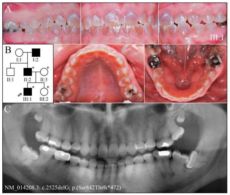
Educational composite image illustrating the clinical and radiographic features of Dentinogenesis Imperfecta Type II (DGI-II). Panel A presents multiple intraoral clinical photographs of a 19-year-old patient (proband III:1), showing a permanent dentition with characteristic amber-brown opalescent discoloration. There is evidence of enamel chipping on the buccal surfaces of the maxillary central incisors, while overall occlusal attrition appears mild and the wear facets remain smooth. Panel B shows a three-generation pedigree indicating an autosomal dominant inheritance pattern of the condition within the family. Panel C displays a panoramic radiograph (panorex) revealing significant internal dental anomalies: generalized obliteration of pulp chambers and root canals, bulbous posterior crowns with cervical constriction, and abnormally short roots on the anterior teeth. Radiographically, the enamel thickness appears within normal limits despite the underlying dentin defect. Several radiopaque dental restorations (crowns) are visible. This visual summary correlates clinical dental phenotypes with radiographic findings and genetic lineage in hereditary dentin disorders.
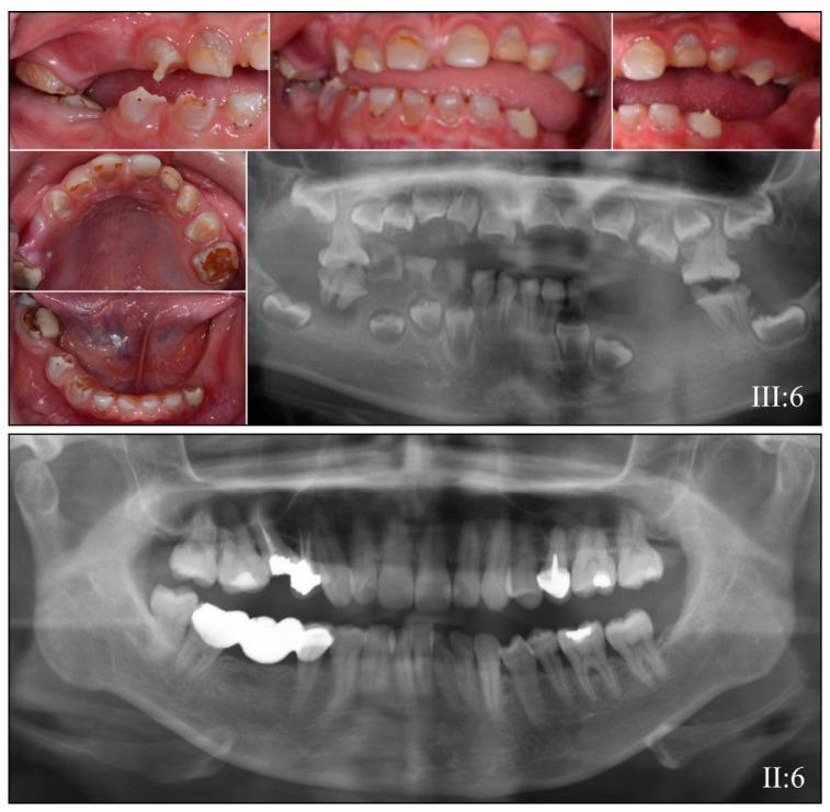
This composite educational image illustrates the clinical and radiographic features of Dentinogenesis Imperfecta (DGI) and Dentin Dysplasia (DD) in a pediatric patient compared to an unaffected adult. The upper section consists of clinical intraoral photographs of an 8-year-old in the mixed dentition stage. The teeth demonstrate characteristic amber-brown opalescent discoloration, bulbous crowns, and severe occlusal attrition leading to enamel loss and dentin exposure. Accompanying these photos is a panoramic radiograph (labeled III:6) showing diagnostic hallmarks including short, tapered roots and partial to complete obliteration of pulp chambers and root canals in both primary and permanent teeth. In contrast, the bottom section displays a panoramic radiograph of an unaffected adult (labeled II:6), showing normal dental morphology with well-defined pulp chambers, standard root lengths, and evidence of routine dental restorations. The comparison highlights the significant structural and developmental abnormalities associated with DSPP gene mutations, emphasizing the diagnostic importance of radiographic pulp obliteration and clinical crown discoloration in hereditary dentin defects.
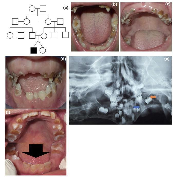
This composite image illustrates clinical and radiographic features of Dentinogenesis Imperfecta (DI) in a pediatric patient. Panel (a) shows a pedigree indicating a familial inheritance pattern. Panels (b, c, d, f) are intraoral clinical photographs showing both primary and permanent dentition. The teeth exhibit characteristic amber-to-opalescent discoloration, significant enamel attrition, and bulbous crown morphology. Severe destruction of the clinical crowns is visible, particularly in the deciduous teeth. Panel (e) is a panoramic radiograph demonstrating hallmark diagnostic features of DI, including bulbous molar crowns with cervical constriction, thin or shortened roots (blue arrow), and enlarged pulp chambers (orange arrow), indicating a failure of normal dentin deposition. The radiographic appearance also shows a lack of clear demarcation between the enamel and dentin layers. This collection of images serves as a clinical reference for the diagnosis of hereditary dentin defects, specifically DI Type II, commonly associated with DSPP gene mutations.
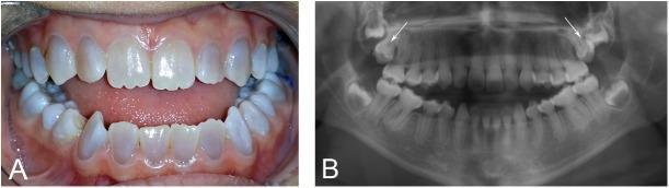
This composite educational image illustrates the dental manifestations of dentinogenesis imperfecta (DGI) in a patient with osteogenesis imperfecta (OI). Panel A is a clinical photograph showing the permanent dentition. Key features include the characteristic opalescent, grey-blue or amber discoloration of the teeth. A significant skeletal and dental anomaly is visible as a Class III malocclusion with a prominent anterior open bite, where the maxillary and mandibular incisors fail to meet. Panel B is an orthopantomogram (panoramic radiograph) demonstrating pathognomonic radiographic signs of DGI. The teeth exhibit bulbous crowns with marked cervical constriction (tulip-shaped) and progressive obliteration of the pulp chambers and root canals due to excessive dentin deposition. Additionally, white arrows indicate the retention and vertical impaction of both maxillary permanent second molars. These findings are clinically relevant for understanding the syndromic dental complications associated with COL1A1/COL1A2 mutations in patients with osteogenesis imperfecta.
dentin dysplasia type 1 rootless teeth radiograph
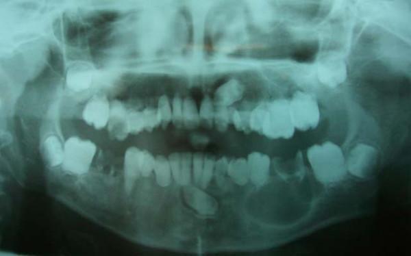
A panoramic dental radiograph (orthopantomogram) of a pediatric patient illustrating hallmarks of Dentin Dysplasia Type I (DD-I). The imaging shows a generalized lack of root development across the primary and permanent dentition, with teeth exhibiting either extremely short, blunted roots or a complete absence of root structure. Multiple teeth present with large radiolucent areas consistent with extensive dental caries. Notably, there is a large, well-defined, round unilocular radiolucency associated with the apex of the mandibular left first permanent molar, suggestive of a radicular cyst or apical inflammatory lesion. Furthermore, the left maxillary and mandibular canines are horizontally impacted within the alveolar bone. The overall presentation highlights the characteristic 'rootless teeth' appearance and associated secondary pathologies such as premature mobility, periapical lesions, and tooth impaction common in hereditary dentin disorders.
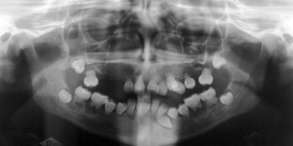
A panoramic radiograph (orthopantomogram) of the maxilla and mandible demonstrating pathognomonic features of Dentin Dysplasia Type I. The primary diagnostic finding is a striking absence of root development (radicular dysplasia) across both the primary and permanent dentition, with teeth appearing as 'rootless' crowns. While the visible dental crowns maintain relatively normal morphology, pulp chambers are obliterated or significantly reduced. Multiple periapical radiolucencies are visible, consistent with spontaneous periapical cysts or inflammatory lesions occurring in the absence of obvious caries. The image further shows several impacted and retained teeth (including the third molars and lower left canine) and congenitally missing teeth in the maxillary quadrants. Structurally, the maxilla and mandible exhibit vertical and sagittal underdevelopment, contributing to an abnormal alveolar ridge profile. This diagnostic image illustrates a rare hereditary dentin defect relevant to pediatric dentistry, oral pathology, and maxillofacial surgery.
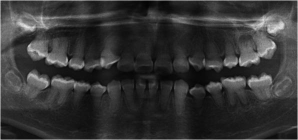
This panoramic dental radiograph (orthopantomogram) of a 12-year-old female demonstrates the classic radiographic features of Dentin Dysplasia Type I (DD-1), also known as radicular dentin dysplasia. The most striking abnormality is the near-total obliteration or significant reduction of pulp chambers and root canals across the entire dentition, appearing as solid radiopaque structures where radiolucent pulp spaces would normally exist. The roots appear atypically shortened, blunted, or 'crescent-shaped' in several segments. Additionally, multiple periapical radiolucencies are visible surrounding the apices of teeth 15, 12, 26, 36, 32, 31, and 46. These lesions are particularly significant as they occur in the absence of dental caries or visible restorations, a pathognomonic finding for DD-1 where abnormal dentin structure leads to spontaneous pulpal necrosis and apical periodontitis. The image serves as a key diagnostic reference for identifying hereditary dentin defects and differentiating them from common inflammatory pathologies.
dentinogenesis imperfecta histology atubular dentin interglobular dentin microscopy
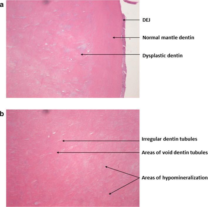
This composite of two light microscopy images (H&E stain) illustrates the histopathology of dentin in Dentinogenesis Imperfecta Type II (DI-II). Image (a) focuses on the dentino-enamel junction (DEJ), which exhibits an abnormally smooth, even appearance lacking typical scalloping. While the immediately adjacent mantle dentin appears relatively normal, the deeper layer consists of dysplastic dentin characterized by incomplete mineralization and pale-staining interglobular areas. Image (b) provides a high-magnification view of the circumpulpal dentin, demonstrating significant structural irregularities. Key features include enlarged and irregular dentin tubules with frequent branching, alongside areas where tubules are entirely void. Notable regions of hypomineralization are present throughout the dentin matrix. These findings characterize the hereditary dentin defect associated with DSPP gene mutations, explaining the clinically observed fragility and detachment of enamel from the underlying compromised dentin.
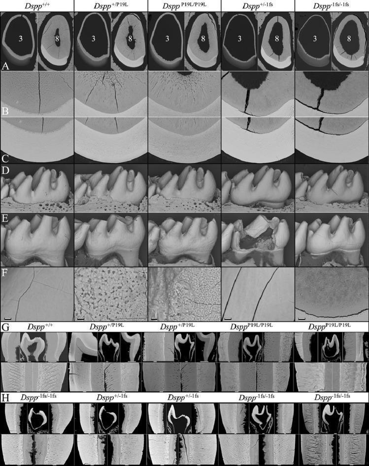
This composite diagnostic image displays backscattered scanning electron microscopy (bSEM) views of mouse tooth development, serving as a model for human dentinogenesis imperfecta and enamel defects related to DSPP (Dentin Sialophosphoprotein) mutations. The figure compares wild-type (Dspp+/+) specimens with various mutant genotypes: Dspp+/P19L, DsppP19L/P19L, Dspp+/-1fs, and Dspp-1fs/-1fs. (A) Cross-sections of 7-week-old mandibular incisors at levels 3 and 8 show a progressively thinner dentin layer and enlarged pulp chamber in mutant mice, most severe in the -1fs genotypes. (B, C) High-magnification views (400x) of incisor dentin and enamel; notable is the complete absence of dentinal tubules in the Dspp-1fs mice and more conspicuous enamel rod outlines in DsppP19L mice, indicating hypomineralization. (D, E) 3D surface reconstructions of D14 and 7-week molars demonstrate normal initial morphology but subsequent accelerated attrition, enamel surface roughness in DsppP19L, and catastrophic crown failures in Dspp-1fs mice. (F-H) Microscopic analysis of molar surfaces and cross-sections highlights uneven mineralization, loss of rod/interrod architecture in P19L mutants, and extremely thin dentin lacking tubules in -1fs mutants.
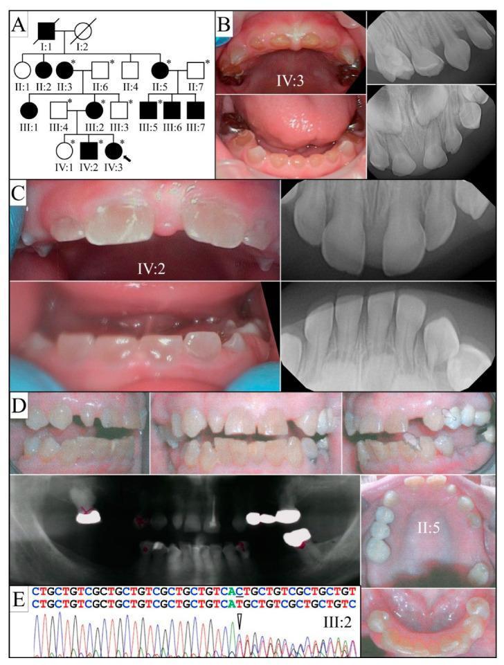
This composite educational graphic details the clinical and genetic presentation of Dentinogenesis Imperfecta Type II (DGI-II) within a single family. Panel A shows a four-generation pedigree indicating an autosomal dominant inheritance pattern. Panels B, C, and D provide clinical photographs and corresponding diagnostic radiographs (panorex and periapical views) of different family members. Clinical photographs demonstrate hallmark features including characteristic amber-brown or opalescent discoloration, significant enamel fracturing (shedding), and extensive occlusal attrition resulting in exposed dentin. Radiographic findings show bulbous crowns, cervical constriction, and the progressive obliteration of pulp chambers and root canals, which are diagnostic indicators of the condition. Panel E displays a sequencing chromatogram identifying a heterozygous frameshift mutation in the DSPP gene (c.3461delG), providing a molecular basis for the observed phenotype. This material illustrates the correlation between genetic mutations, radiographic markers of dentin dysplasia, and the resulting clinical dental pathology.
A labeled pencil-style diagram for BDS exam showing cross-section of a tooth with Dentinogenesis Imperfecta features. The diagram shows: (1) normal enamel crown but with chipped/fractured enamel edges; (2) abnormal dentin - irregular, atubular with large interglobular spaces; (3) obliterated or very small pulp chamber; (4) constriction at CEJ (cervical constriction); (5) short blunt roots; (6) comparison with normal tooth showing normal pulp, normal dentin tubules, normal root length. Labels with arrows. Second smaller diagram showing histology: atubular dentin, interglobular dentin, irregular dentin tubules, obliterated pulp. Title: "Dentinogenesis Imperfecta - Tooth Cross-section and Histology". Black and white pencil sketch BDS exam style.
DEVELOPMENTAL DISTURBANCES IN FORMATION OF DENTIN
BDS 3rd Year - Oral Pathology - University Exam Pattern
CLASSIFICATION
I. HEREDITARY / GENETIC DISTURBANCES
- Type I - DI with Osteogenesis Imperfecta
- Type II - Hereditary Opalescent Dentin
- Type III - Brandywine Type (Shell teeth)
- Type I - Radicular Dentin Dysplasia (Rootless teeth)
- Type II - Coronal Dentin Dysplasia
II. SYSTEMIC / ENVIRONMENTAL DISTURBANCES
- Interglobular dentin (hypocalcification / rickets / vitamin D deficiency)
- Tomes' granular layer abnormalities
- Dead tracts (response to stimuli)
- Sclerotic / Transparent dentin (aging, attrition, caries)
- Reparative / Tertiary (Irregular secondary) dentin (response to injury)
III. LOCALIZED CONDITIONS
- Pulp stones (Denticles) - true and false
- Diffuse calcifications
A. DENTINOGENESIS IMPERFECTA (DI)
Synonyms: Hereditary Opalescent Dentin / Capdepont Teeth
DEFINITION
GENETICS
- Autosomal dominant inheritance (AD)
- Gene involved: DSPP gene (Dentin Sialophosphoprotein gene) on chromosome 4q21
- DSPP encodes dentin sialoprotein (DSP) and dentin phosphoprotein (DPP) - both critical for dentin mineralization
- Exception: DI Type I is caused by COL1A1 / COL1A2 mutations (same as osteogenesis imperfecta)
SHIELDS' CLASSIFICATION (1973) - Most Used in Indian BDS
| Feature | Type I (DI with OI) | Type II (Hereditary Opalescent Dentin) | Type III (Brandywine Type) |
|---|---|---|---|
| Association | Osteogenesis imperfecta | Isolated (no OI) | Isolated, rare (Maryland/Delaware) |
| Gene | COL1A1 / COL1A2 | DSPP | DSPP |
| Primary teeth | Severely affected | Severely affected | Severely affected |
| Permanent teeth | Less affected | Equally affected | Mildly affected |
| Pulp chambers | Obliterated | Obliterated | Large / "shell teeth" |
| Special feature | Blue sclerae, fragile bones | Most common type | Multiple pulp exposures |
DENTINOGENESIS IMPERFECTA TYPE II (Most Commonly Asked)
Clinical Features
-
Color:
- Characteristic opalescent (translucent) discoloration - most important feature
- Color ranges: amber/yellow-brown to grey-blue or purple
- Translucency gives a "opalescent" hue in light - like mother of pearl
- Both primary and permanent teeth affected
-
Enamel chipping / fracture:
- Enamel is structurally normal but chips off readily
- Due to lack of normal scalloping at DEJ → poor enamel-dentin bonding
- Exposed dentin undergoes rapid wear (attrition)
-
Attrition:
- Severe, rapid wearing down of exposed dentin
- Teeth may wear down to gingival level
-
Crown shape: Normal crown form initially
-
Both dentitions affected (primary more severely)
-
No systemic association (differentiates from Type I)
Clinical Photograph - Dentinogenesis Imperfecta Type II:
Radiological Features
- Bulbous / bell-shaped crowns with cervical constriction (thistle-tube / tulip appearance)
- Obliteration of pulp chambers and root canals (most important radiographic sign) - due to continued, excessive dentin deposition
- Short, blunted, thin roots (higher fracture risk)
- Normal enamel thickness
- No periapical pathology (pulp is sealed off)
- "Shell teeth" in Type III: Very thin dentin shell with large pulp chambers (opposite to Type II)
Histological Features of DGI
-
Abnormal DEJ (Dentino-enamel junction):
- Normal DEJ has scalloping/interdigitation
- In DGI: DEJ is smooth, flat - no normal scalloping
- This weakens enamel-dentin bond → enamel chips off
-
Mantle dentin (outermost layer):
- Often relatively normal in appearance
- This explains why enamel is histologically normal
-
Circumpulpal dentin (bulk of dentin):
- Irregular, abnormal dentin tubules - fewer, irregular in course, branching
- Areas completely devoid of tubules (atubular dentin)
- Interglobular dentin - zones of incompletely mineralized matrix (pale areas)
- Hypomineralization of dentin matrix
- Overall: disorganized, poorly calcified dentin
-
Pulp obliteration:
- Continued deposition of irregular dentin → pulp chamber filled
- Pulp tissue eventually replaced by dentin
H&E Histology of DGI:
Diagram for DGI:
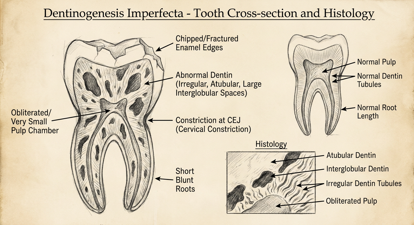
HOW TO DRAW DGI DIAGRAM IN EXAM:
Normal tooth (left side) vs DGI tooth (right side):
→ Normal: Normal pulp chamber, normal root length, normal DEJ scalloping
→ DGI: Obliterated pulp, short blunt roots, cervical constriction, bulbous crown
Histology inset:
→ Smooth DEJ (no scalloping)
→ Normal thin mantle dentin
→ Dysplastic deeper dentin - irregular, sparse tubules
→ Interglobular dentin (pale unmineralized zones)
→ Atubular areas
LABELS:
→ Bulbous crown
→ Cervical constriction
→ Obliterated pulp chamber
→ Short, blunt root
→ Smooth DEJ (no scalloping)
→ Normal mantle dentin
→ Atubular / dysplastic dentin
→ Interglobular dentin
DGI - Comparison of All 3 Types
| Feature | Type I | Type II | Type III |
|---|---|---|---|
| Synonym | DI with OI | Hereditary opalescent dentin | Brandywine type |
| Systemic | OI (blue sclerae, fractures) | None | None |
| Color | Opalescent | Opalescent (yellow-brown, grey-blue) | Similar |
| Pulp | Obliterated | Obliterated | Large / shell teeth |
| Primary teeth | Severely affected | Severely affected | Mildly affected |
| Inheritance | AD (COL1A1/COL1A2) | AD (DSPP) | AD (DSPP) |
B. DENTIN DYSPLASIA (DD)
DEFINITION
DENTIN DYSPLASIA TYPE I (Radicular DD / "Rootless Teeth")
Clinical Features
- Teeth appear clinically normal in color (or slightly amber)
- Premature exfoliation / mobility - due to extremely short roots
- Tendency for spontaneous periapical abscess without apparent caries
- Affects both dentitions
Radiological Features (Most Diagnostic)
- "Rootless teeth" - extremely short, truncated, or completely absent roots - hallmark
- Complete obliteration of pulp chambers and root canals in primary teeth
- In permanent teeth: crescent-shaped or chevron-shaped pulp remnants (like a half-moon)
- Multiple periapical radiolucencies without caries (spontaneous pulp necrosis due to absent blood supply through absent roots)
- Impacted/unerupted teeth common
Radiograph - Dentin Dysplasia Type I:
Histological Features of DD Type I
- Coronal dentin: relatively normal (tubules present)
- Radicular dentin: severely dysplastic - atubular, irregular
- Pulp obliteration from early age
- Pulp contains islands of abnormal dentin / dentricles
- Abnormal dentin tubules in root: disorganized, sparse
DENTIN DYSPLASIA TYPE II (Coronal DD)
Clinical Features
- Primary teeth: Color similar to DGI - amber, opalescent discoloration
- Permanent teeth: Clinically normal color
- Both dentitions affected differently
Radiological Features
- Permanent teeth: Thistle-tube shaped pulp chambers - large, flame-shaped, with multiple pulp stones scattered ("thistle tube" appearance)
- Obliteration of root canals (but root length is normal - unlike Type I)
- Primary teeth: Obliterated pulp chambers (resembles DGI)
Histological Features
- Coronal dentin: largely normal
- Pulp chamber contains multiple irregular calcifications / denticles
- Shell-like dentin surrounds pulp stones
- Root dentin is relatively normal
COMPARISON: DI vs DD
| Feature | Dentinogenesis Imperfecta (Type II) | Dentin Dysplasia Type I | Dentin Dysplasia Type II |
|---|---|---|---|
| Clinical color | Opalescent (amber/grey-blue) | Normal / slight amber | Primary: opalescent; Permanent: normal |
| Crown shape | Bulbous, cervical constriction | Normal | Normal |
| Root | Short, blunt | Extremely short / absent | Normal |
| Pulp | Obliterated | Obliterated (crescent remnant) | Large thistle-tube + pulp stones |
| Periapical lesion | Absent | Present (spontaneous) | Absent |
| Attrition | Severe | Not marked | Not marked |
| DEJ | Smooth (no scalloping) | Normal | Normal |
| Gene | DSPP | DSPP | DSPP |
| Nickname | Hereditary opalescent dentin | Rootless teeth | - |
C. INTERGLOBULAR DENTIN
Definition
Etiology
- Vitamin D deficiency / rickets
- Hypophosphatasia
- Fluorosis (in excess)
- Tetracycline administration
Location
- Most commonly in coronal dentin just below DEJ (in the mantle dentin zone)
- Also seen in root dentin (Tomes' granular layer)
Histological Appearance
- Round or irregular pale-staining (unmineralized) spaces between calcified dentin globules
- In ground sections: appear as dark areas (because unmineralized matrix does not transmit light)
- Dentin tubules pass through the interglobular spaces (tubules are present within them)
Significance
- Marker of a systemic disturbance during dentinogenesis
- Used to date systemic insults in forensic odontology
D. PULP STONES (DENTICLES)
Definition
Classification
| Type | Description |
|---|---|
| True denticles | Contain dentin tubules - rare, resemble true dentin |
| False denticles | No tubules - concentric calcified lamellae |
| Diffuse calcifications | Irregular deposits along collagen fibers / blood vessels |
- Free - completely surrounded by pulp tissue
- Attached - fused to dentin wall on one side
- Embedded - completely surrounded by dentin
Etiology
- Aging
- Chronic pulp irritation (caries, trauma, restorations)
- Systemic diseases: cardiovascular disease, kidney disease
Clinical Significance
- Usually asymptomatic
- May complicate root canal treatment (obstruct canal)
- Occasionally cause referred pain / neuralgia
Radiological Features
- Appear as radiopaque rounded calcifications within the pulp chamber
Histological Features
- True denticle: Contains irregular dentin tubules, surrounded by odontoblast-like cells
- False denticle: Concentric lamellae of calcified material, no tubules
E. REPARATIVE (TERTIARY) DENTIN
Definition
Types
- Reactionary dentin: Formed by original odontoblasts in response to mild stimulus
- Reparative dentin: Formed by newly differentiated cells (from pulp stem cells) in response to severe injury
Histological Features
- Irregular, disorganized structure
- Atubular or with few irregular tubules
- Abrupt line of demarcation from regular secondary dentin
- May show cellular inclusions (odontoblast processes trapped)
- Deposited only beneath the area of stimulation (localized)
F. SCLEROTIC / TRANSPARENT DENTIN
Definition
Causes
- Aging (physiological)
- Attrition (mechanical wear)
- Caries (slow, chronic caries front)
- Erosion
Significance
- Acts as a barrier / defense mechanism - prevents further irritant penetration
- In ground sections: appears transparent (light passes through uniformly)
SUMMARY REVISION TABLE
| Condition | Inheritance | Gene | Key Feature | Radiograph | Histology |
|---|---|---|---|---|---|
| DI Type I | AD | COL1A1/COL1A2 | + OI (blue sclerae) | Obliterated pulp, short roots | Atubular, smooth DEJ |
| DI Type II | AD | DSPP | Opalescent teeth, no systemic | Bulbous crown, obliterated pulp | Atubular, smooth DEJ, interglobular |
| DI Type III | AD | DSPP | Shell teeth | Large pulp (thin dentin shell) | Thin dentin |
| DD Type I | AD | DSPP | Rootless teeth | Absent roots, crescent pulp | Dysplastic root dentin |
| DD Type II | AD | DSPP | Thistle-tube pulp + stones | Flame-shaped pulp with stones | Pulp stones, otherwise normal |
| Interglobular dentin | Acquired | - | Rickets/vit D deficiency | Normal | Pale unmineralized globules |
| Pulp stones | Acquired | - | Routine finding | Radiopaque in pulp | True / false denticles |
Exam Tips:
- DGI vs DD Type I: DGI has opalescent color + obliterated pulp; DD Type I has normal crown color + rootless teeth - this is the single most important differentiator
- "Shell teeth" = DI Type III (Brandywine) - large pulp, thin dentin shell
- Thistle-tube pulp = DD Type II - always in permanent teeth
- DSPP gene = mutation underlying DI Type II, III and DD Type I, II (DSPP encodes dentin sialoprotein and dentin phosphoprotein)
- Smooth DEJ = DGI - explains enamel chipping (no mechanical interlocking)
- For drawing: DGI diagram is most asked - draw bulbous crown + cervical constriction + obliterated pulp + short roots + smooth DEJ with atubular dentin histology inset
Ennumerate bacterial lesion of oral cavity and detail about oral manifestation of tuberculosis
oral tuberculosis ulcer tongue clinical appearance
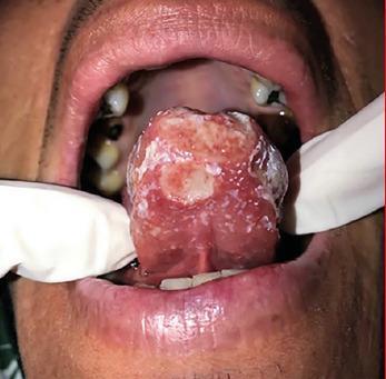
This clinical photograph shows an intraoral view of a patient's tongue with a prominent lesion. The primary pathology is a large, poorly defined ulcerative lesion located at the apex (tip) of the tongue. The lesion displays a variegated appearance, characterized by an erythematous (red) base with irregular areas of white slough and fibrinous pseudomembrane coating. The borders of the ulcer are indistinct and merged with the surrounding inflamed mucosa. The dorsal surface of the tongue shows diffuse erythema and textural changes consistent with chronic inflammation. Background oral structures show secondary clinical findings, including visible dental caries and mild gingival recession. The visual presentation is highly characteristic of granulomatous oral conditions, such as primary or secondary oral tuberculosis, which typically present as non-healing, painful chronic ulcers. The image serves as an educational example of deep fungal or mycobacterial oral manifestations, highlighting the need for biopsy and differential diagnosis from squamous cell carcinoma.
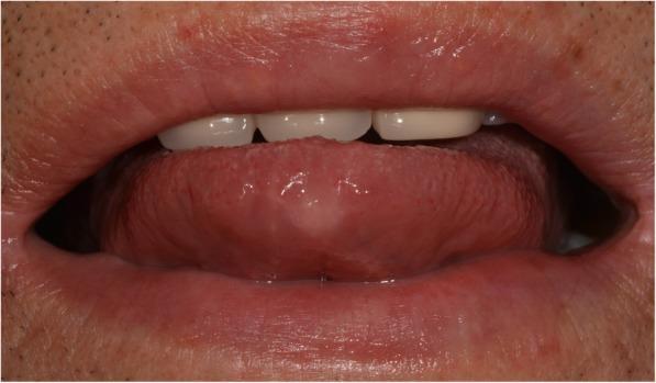
This clinical photograph is an intraoral view of the tongue and anterior oral cavity. The primary finding is a healing lesion located on the anterior dorsal tip of the tongue. Following two months of anti-tuberculosis therapy for lingual tuberculosis, the previously described round, granulomatous ulcer has significantly regressed, now appearing as a subtle, pale, circular area with a flattened surface texture. The surrounding tongue tissue and oral mucosa exhibit a healthy pink color with no signs of active inflammation, erythema, or edema. The dorsal surface of the tongue displays normal filiform and fungiform papillae distribution in the periphery of the resolving site. This image serves as a visual record of clinical improvement and therapeutic response in a case of extrapulmonary tuberculosis presenting as an oral ulcer. The surrounding perioral skin and vermilion border of the lips appear intact and healthy.
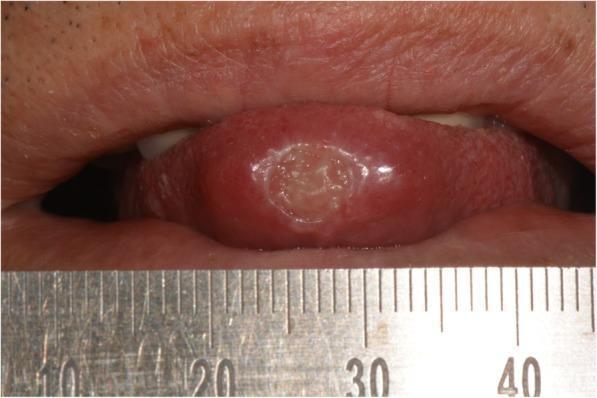
This clinical photograph displays a close-up intraoral view of a focal pathology on the human tongue. A solitary, well-circumscribed, round ulcer is centrally located on the tip of the tongue (apex linguae). The lesion measures approximately 0.7 cm in diameter as indicated by the adjacent surgical ruler. Morphologically, the ulcer features a slightly elevated, whitish, indurated-appearing border and a depressed, granular, pale-yellowish base. The surrounding lingual mucosa appears erythematous and mildly edematous, with the lateral margins of the tongue showing slight impressions from the teeth. This image illustrates a chronic oral ulcer, which in a clinical context represents a differential diagnosis including major aphthous stomatitis, traumatic granuloma, squamous cell carcinoma, or granulomatous infections such as oral tuberculosis. It is a key educational resource for oral pathology, dermatology, and infectious disease training, emphasizing the importance of biopsy for persistent, firm oral lesions.
tuberculosis histology Langhans giant cells caseating necrosis granuloma H&E
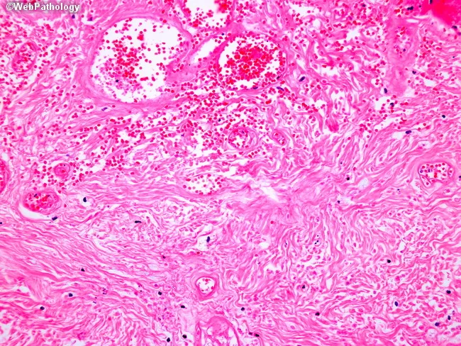
Light microscopy of an H&E-stained formalin-fixed tissue section at low to intermediate magnification reveals granulomatous inflammation characterized by well-formed nodular aggregates of epithelioid histiocytes and occasional Langhans-type multinucleated giant cells embedded in a fibrous stromal matrix. The granulomas appear in close association with accentuated lymphocytic cuffs and scattered plasma cells. Central areas show coagulative or caseating necrosis in some nodules, giving a pale eosinophilic center surrounded by a rim of epithelioid cells. The surrounding dermis/soft tissue shows delicate reticular fibers and vascular congestion with perivascular mononuclear infiltrates. There are no overt bacterial organisms evident with standard H&E, though organisms such as mycobacteria or fungi may require special stains (Ziehl-Neelsen, GMS) to confirm infection. The architecture suggests a granulomatous process rather than acute suppurative inflammation. Diagnostic significance: indicative of granulomatous disease; differential includes infectious etiologies (tuberculosis, fungal infections) and noninfectious conditions (sarcoidosis, foreign-body reaction). Clinical correlation recommended with exposure history, immunologic status, and targeted staining/cultures. Potential use cases: educational demonstration of granuloma morphology, pathology student review, differential diagnosis in infectious and granulomatous diseases, histology atlas reference. This description supports search for terms granuloma, epithelioid histiocytes, Langhans giant cells, caseation, TB, sarcoidosis, histopathology.
This is a light microscopy image of a formalin-fixed, paraffin-embedded tissue section stained with Hematoxylin and Eosin (H&E). The sample reveals a well-formed granulomatous lesion characterized by a surrounding cuff of lymphocytes and numerous epithelioid histiocytes with elongated, crescent-shaped nuclei. Within the granuloma, multinucleated giant cells (Langhans-type) are visible, and a central amorphous pink necrotic area (caseous necrosis) is evident. The tissue architecture demonstrates organized granulomatous inflammation with a necrotic core, consistent with infectious granulomas. The differential includes mycobacterial disease (tuberculosis) and certain fungal infections (histoplasmosis, coccioidomycosis), though non-necrotizing granulomas can occur in sarcoidosis. The staining highlights chromatin-rich nuclei and cytoplasmic eosinophilia; occasional fibroblasts and mild edema extend into the perigranulomatous region. Clinically, granulomatous tissue with caseation is highly suggestive of TB in the appropriate clinical context and warrants further diagnostic workup (acid-fast bacilli staining, fungal stains, culture, PCR). This image serves as teaching material for histopathology education, differential diagnosis of granulomatous inflammation, and correlation with radiologic and microbiologic findings in suspected infectious etiologies. Additional keywords for indexing: epithelioid macrophages, granuloma, necrosis, caseation, tuberculosis, mycobacteria, fungal infection, lymphohistiocytic rim, tissue biopsy, pathology slide, H&E, histology teaching. Clinical correlation with imaging and sputum analysis improves diagnostic yield.
Histopathology image showing a granulomatous lesion composed of epithelioid histiocytes with a surrounding rim of lymphocytes and occasional Langhans-type multinucleated giant cells. Within a giant cell, lamellar inclusions known as Schaumann bodies are evident as concentric rings of protein and mineral material; calcium oxalate crystals may also be present. Asteroid bodies may be seen as stellate inclusions, though they are not required for diagnosis. The granuloma is noncaseating, lacking central necrosis, which favors sarcoidosis over infectious granulomatous processes such as tuberculosis. The tissue architecture shows well-formed granulomas with distinct borders, often adjacent to blood vessels in this section. The image is stained with Hematoxylin and Eosin (H&E), highlighting eosinophilic granulomatous material and basophilic nuclei within giant cells. Clinically, the presence of Schaumann bodies and Langhans giant cells supports a granulomatous inflammatory syndrome but is not pathognomonic for sarcoidosis. Differential considerations include berylliosis, hypersensitivity pneumonitis, fungal or mycobacterial infections, and foreign body reactions. Correlation with chest imaging, serum ACE levels, and clinical features is essential for diagnosis. This slide serves an educational role in recognizing granulomatous histology, giant cell inclusions, and lamellar calcific bodies, informing diagnostic workflows and teaching pathology trainees about sarcoidosis-related granulomatous disease.
Histopathology via light microscopy of an H&E-stained lymph node biopsy demonstrates a granulomatous inflammatory response characterized by an epithelioid cell nodular formation with a surrounding lymphocytic infiltrate. Within the central core, a multinucleated Langhans-type giant cell is present, displaying nuclei arranged in a horseshoe pattern at the periphery. The granuloma is composed predominantly of well-differentiated epithelioid histiocytes with scant cytoplasm and tight cellular packing, producing a palisaded appearance that integrates with a mildly eosinophilic to translucent extracellular matrix. The surrounding paracortex contains a dense lymphoid crowding with scattered plasma cells and occasional eosinophils, consistent with chronic granulomatous inflammation. In this image, the granuloma lacks conspicuous caseous necrosis, a feature that supports noncaseating granulomatous processes; however, logical caveats apply since granulomas in sarcoidosis can mimic infectious etiologies. The presence of Langhans-type giant cells within nonnecrotizing granulomas is classically associated with sarcoidosis but can be seen in tuberculosis, atypical mycobacterial infections, fungal infections, berylliosis, and other granulomatous diseases. Additional diagnostic workup would include acid-fast bacilli staining and fungal stains, microbial cultures, and clinical correlation. Recognizing this histologic pattern guides differential diagnosis toward sarcoid-type granulomatous lymphadenitis and informs subsequent clinical management and exclusion of granulomatous infections. Correlation with imaging and laboratory data essential.
tuberculosis Ziehl Neelsen stain acid fast bacilli red rods sputum
This diagnostic image is a high-power light microscopy field showing a histopathological sample stained with Ziehl-Neelsen or a similar acid-fast stain. The background consists of blue-stained cellular debris, fibrous tissue, and inflammatory infiltrates from a pericardial biopsy. Centrally highlighted within a red circular annotation are several bright red or pink, rod-shaped microorganisms. These characteristics are pathognomonic for acid-fast bacilli (AFB), specifically Mycobacterium tuberculosis. The visual demonstrates the diagnostic findings of tuberculous pericarditis, where the pathogen is directly visualized within the pericardial tissue despite a non-granulomatous histological appearance. This slide serves as an educational example of infectious pathology and the clinical utility of specialized staining in identifying extracellular pathogens in extrapulmonary tuberculosis.
This diagnostic micrograph displays a Modified Ziehl-Neelsen stain (using 1% sulfuric acid) of a clinical specimen, likely pleural fluid. The background is composed of a diffuse light blue counterstain. Predominant in the field are vibrant pink-stained, acid-fast filamentous bacilli. The microorganisms exhibit characteristic slender, elongated morphology with prominent irregular branching patterns and a beaded appearance in some areas. These visual features are highly suggestive of Nocardia species, an opportunistic pathogen. The image demonstrates the microscopic diagnostic approach for identifying aerobic actinomycetes in immunocompromised patients, such as those with HIV. The filamentous nature distinguishes these organisms from other acid-fast bacilli like Mycobacterium tuberculosis, which typically appear as non-branching rods.
A labeled pencil-style histological diagram for BDS exam of oral tuberculosis (H&E). The diagram shows a classic tuberculous granuloma with: (1) central zone of caseating (caseous) necrosis - amorphous pink, granular, structureless material; (2) surrounding ring of epithelioid histiocytes (macrophages with abundant pale cytoplasm and elongated kidney-shaped nuclei); (3) Langhans giant cell - large multinucleated cell with nuclei arranged in horseshoe/peripheral arc pattern at the edge of the cell; (4) outer rim of lymphocytes and plasma cells; (5) peripheral fibrosis/fibrous capsule; (6) small inset showing Ziehl-Neelsen stain with red acid-fast bacilli rods (labeled). Black and white pencil sketch style with clear labels and arrows. Title: "Tuberculous Granuloma - Oral TB (H&E)"
BACTERIAL LESIONS OF THE ORAL CAVITY + ORAL TUBERCULOSIS
BDS 3rd Year - Oral Pathology - University Exam Pattern
ENUMERATION OF BACTERIAL LESIONS OF THE ORAL CAVITY
I. SPECIFIC BACTERIAL INFECTIONS (caused by a single identifiable organism)
- Tuberculosis (Mycobacterium tuberculosis)
- Leprosy (Mycobacterium leprae)
- Primary: chancre
- Secondary: mucous patches, split papules
- Tertiary: gumma
- Noma (Cancrum oris) - fusospirochetal (Fusobacterium + Borrelia vincenti)
- Acute necrotizing ulcerative gingivitis (ANUG / Vincent's angina) - fusospirochetal
II. NON-SPECIFIC / POLYMICROBIAL INFECTIONS
- Periapical abscess / Dentoalveolar abscess
- Periodontal disease / Periodontitis
- Acute necrotizing ulcerative gingivitis (ANUG)
- Ludwig's angina - polymicrobial cellulitis of floor of mouth
- Osteomyelitis of jaws (acute and chronic)
- Dry socket (Alveolar osteitis)
- Cellulitis of face / neck
- Sialadenitis (bacterial parotitis - Staphylococcus aureus)
III. BACTERIAL LESIONS WITH SYSTEMIC DISEASE ASSOCIATIONS
- Scarlet fever - strawberry tongue, Forchheimer spots on palate
- Diphtheria - Corynebacterium diphtheriae (grey pseudomembrane on tonsils)
- Whooping cough - Bordetella pertussis (can cause labial frenulum ulceration)
- Typhoid fever - Salmonella typhi (ulceration of posterior oral cavity)
ORAL TUBERCULOSIS - DETAILED
THE ORGANISM
- Causative agent: Mycobacterium tuberculosis (Koch's bacillus)
- Aerobic, non-motile, non-sporing, non-capsulate
- Acid-fast bacillus (AFB) - has lipid-rich cell wall (mycolic acid) that resists decolorization with acid-alcohol
- Identified by Ziehl-Neelsen (ZN) stain - stains red/pink against blue background
- Reservoir: humans; Transmission: droplet nuclei (airborne)
- India has the highest TB burden globally
PATHOGENESIS OF ORAL TB
Routes of Oral Involvement
- Secondary to pulmonary tuberculosis (the commonest form)
- Mechanism:
- Patient with active pulmonary TB expectorates infected sputum
- Bacilli-laden sputum comes in contact with oral mucosa
- Bacilli enter through breaks/abrasions in mucosa, or through salivary ducts or tooth sockets
- Organism implants and causes local lesion
- No pre-existing pulmonary TB
- Organism directly inoculated into oral mucosa
- Associated with primary complex = oral lesion + regional lymphadenopathy (cervical nodes)
- Disseminated / miliary TB
- Multiple small lesions
Immune Response (Pathogenesis at tissue level)
M. tuberculosis enters tissue
↓
Phagocytosed by macrophages (but resists killing due to mycolic acid wall)
↓
Macrophages become EPITHELIOID CELLS (elongated, pale, low phagocytic activity)
↓
CD4+ T-helper cells recruited → release IFN-γ, TNF-α (Type IV / Cell-mediated immunity)
↓
Epithelioid cells fuse → LANGHANS GIANT CELLS (multinucleated)
↓
Accumulation of epithelioid cells + giant cells + lymphocytes = GRANULOMA
↓
Central necrosis (tissue killing + hypersensitivity reaction) = CASEATION
↓
TUBERCULOUS (CASEATING) GRANULOMA = TUBERCLE
ORAL MANIFESTATIONS OF TUBERCULOSIS
SITES OF ORAL INVOLVEMENT (in decreasing order of frequency)
- Tongue - most common (especially tip, dorsum, lateral borders)
- Palate (hard and soft)
- Gingiva
- Lips
- Buccal mucosa
- Jaws (mandible > maxilla) - tuberculous osteomyelitis
- Salivary glands (rare) - parotid most common
- Tonsils and lymph nodes (cervical lymphadenopathy)
A. ORAL MUCOSAL TUBERCULOSIS (Most common oral manifestation)
Clinical Features
-
Appearance:
- Typically a single, chronic, non-healing, painful ulcer
- Irregular, ragged, undermined (overhanging) edges
- Floor of ulcer: granular, covered with yellowish-grey slough or fibrinous exudate
- Base may be indurated or soft
- Surrounding mucosa: erythematous, edematous
- May appear serpiginous (snake-like) outline
-
Pain:
- Often disproportionately painful (characteristic of TB ulcer)
- Pain worse on eating and speaking
- Radiating pain possible
-
Duration: Weeks to months - non-healing (key feature)
-
Associated systemic features (in secondary TB):
- Chronic cough, hemoptysis
- Fever (evening rise), night sweats
- Weight loss, anorexia, fatigue
- History of contact with TB patient
-
Regional lymphadenopathy: cervical nodes enlarged, may be matted
-
In primary oral TB:
- Ulcer + ipsilateral cervical lymphadenopathy = primary complex
Clinical Photographs - Oral TB Ulcer:
B. TUBERCULOUS OSTEOMYELITIS OF JAWS
- Mandible more commonly affected than maxilla
- More common in posterior mandible (body, ramus)
- Rare; usually secondary to pulmonary TB or hematogenous spread
Clinical Features
- Swelling of jaw (hard, bony, non-tender initially)
- Sinus formation - chronic draining sinus with thin, watery discharge
- Loosening of teeth in the affected area
- Trismus (if ramus involved)
- Pathological fracture in severe cases
- Regional lymphadenopathy
Radiological Features of Jaw TB
- Ill-defined radiolucency - patchy bone destruction
- Multiple small foci coalescing
- No periosteal reaction (unlike pyogenic osteomyelitis)
- "Moth-eaten" appearance
- Sequestrum formation (devitalized bone)
C. TUBERCULOUS CERVICAL LYMPHADENITIS (Scrofula)
- Most common extrapulmonary TB manifestation globally
- Most common site for TB in head and neck region
- Cervical lymph nodes (posterior triangle) most commonly involved
- Nodes: initially firm, rubbery; then soften due to caseation → fluctuant
- "Cold abscess" = fluctuant node WITHOUT signs of acute inflammation (no heat, no redness)
- Collar-stud abscess: abscess breaks through deep fascia → subcutaneous cold abscess
- May form sinus (discharging sinus on skin) - "TB collar stud abscess"
- Mantoux test positive
HISTOLOGICAL FEATURES OF ORAL TUBERCULOSIS
THE TUBERCULOUS GRANULOMA (Tubercle) - Key to Diagnosis
1. Central Caseating Necrosis
- Central zone of amorphous, eosinophilic, granular, cheesy (caseous) necrotic material
- Resembles cottage cheese / cream cheese macroscopically
- Structureless - no cell outlines visible (unlike liquefactive necrosis)
- Represents tissue death + hypersensitivity reaction
- Caseation is the hallmark of TB (distinguishes from sarcoidosis which has non-caseating granulomas)
2. Epithelioid Histiocytes (Epithelioid Cells)
- Ring of elongated macrophages with:
- Abundant pale pink cytoplasm
- Elongated, pale, kidney-shaped (reniform) nuclei
- Indistinct cell borders (cells appear to merge / syncytium)
- These are activated macrophages that have been "transformed" into epithelioid cells
3. Langhans Giant Cells - PATHOGNOMONIC
- Large multinucleated giant cells (up to 50+ nuclei)
- Nuclei arranged in horseshoe / peripheral arc / peripheral ring pattern at the edge of the cell
- Cytoplasm is abundant and pale pink
- Formed by fusion of multiple epithelioid cells
- Distinguishes TB from foreign body giant cells (where nuclei are scattered randomly throughout cytoplasm)
4. Lymphocytic Cuffing
- Rim of lymphocytes and plasma cells surrounding the epithelioid zone
- Represents the adaptive immune response (T-cells, B-cells)
5. Peripheral Fibrosis
- Outer zone of dense fibrous connective tissue (fibroblasts + collagen)
- Represents healing/walling off response
Summary of Granuloma Structure (Center → Periphery):
CASEATION (central) → EPITHELIOID CELLS → LANGHANS GIANT CELL
→ LYMPHOCYTES / PLASMA CELLS → FIBROSIS (peripheral)
H&E Histology Photomicrograph - Tuberculous Granuloma:
DIAGRAM FOR EXAM (H&E Pencil Diagram)
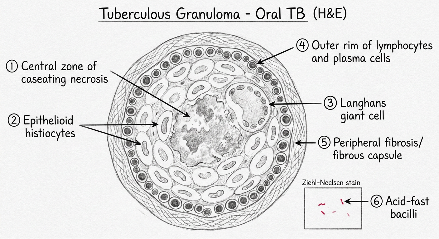
HOW TO DRAW TUBERCULOUS GRANULOMA (H&E):
Step 1: Center - draw amorphous pink structureless material = CASEATING NECROSIS
(no cell outlines, just granular pink material)
Step 2: Around it - draw elongated cells with kidney-shaped nuclei = EPITHELIOID CELLS
(pale cytoplasm, no sharp cell borders)
Step 3: Among epithelioid cells - draw one large cell with many nuclei
arranged at the PERIPHERY in a horseshoe = LANGHANS GIANT CELL
(nuclei arranged like arc/horseshoe at edge of cytoplasm)
Step 4: Outside the epithelioid zone - many small dark round cells = LYMPHOCYTES
+ some larger cells with eccentric nuclei = PLASMA CELLS
Step 5: Outermost zone - wavy lines = PERIPHERAL FIBROSIS
INSET (ZN stain):
→ Blue background
→ Thin red rods = ACID FAST BACILLI (Mycobacterium tuberculosis)
→ Label: "ZN stain - AFB positive (red rods)"
LABELS (with arrows):
→ Central caseating necrosis (caseous necrosis)
→ Epithelioid histiocytes (elongated, reniform nuclei)
→ Langhans giant cell (peripheral / horseshoe nuclei arrangement)
→ Lymphocytic cuffing
→ Plasma cells
→ Peripheral fibrous capsule
→ [Inset]: Acid-fast bacilli (Ziehl-Neelsen stain)
SPECIAL STAINS USED IN TB DIAGNOSIS
| Stain | Purpose | Appearance |
|---|---|---|
| Ziehl-Neelsen (ZN) stain | Identify acid-fast bacilli | Bacilli stain red against blue background |
| Auramine-rhodamine | Fluorescence stain for AFB | Yellow-green fluorescent bacilli |
| PAS stain | Not specific for TB | Used in fungal differential |
| H&E | Identify granuloma structure | Standard |
DIAGNOSIS OF ORAL TUBERCULOSIS
| Investigation | Finding |
|---|---|
| Biopsy + H&E | Caseating granuloma with Langhans giant cells |
| ZN stain / AFB stain | Red acid-fast bacilli (may be scanty or absent) |
| Mantoux (tuberculin) test | Positive (>10 mm induration in immunocompetent) |
| Chest X-ray | Pulmonary TB lesions (in secondary oral TB) |
| Culture (Gold standard) | M. tuberculosis growth on Lowenstein-Jensen medium (4-8 weeks) |
| Gene Xpert (PCR) | Rapid detection of MTB + rifampicin resistance |
| FNAC (lymph nodes) | Caseating granulomas in cervical nodes |
| CBC + ESR | Raised ESR, lymphocytosis |
DIFFERENTIAL DIAGNOSIS OF ORAL TB ULCER
| Feature | Oral TB | OSCC | Aphthous Ulcer | Syphilitic Ulcer |
|---|---|---|---|---|
| Pain | Severe | Painless (late) | Very painful | Painless (chancre) |
| Edges | Ragged, undermined | Everted, raised | Smooth, punched out | Indurated, rolled |
| Base | Granular, slough | Indurated | Yellowish floor | Clean, "wash-leather" |
| Duration | Weeks-months (chronic) | Months | 10-14 days | 3-6 weeks |
| Lymph nodes | Matted, non-tender | Hard, fixed | Tender | Rubbery, non-tender |
| Histology | Caseating granuloma + Langhans cells | Malignant squamous nests + keratin pearls | Nonspecific | Plasma cell infiltrate |
| AFB stain | Positive | Negative | Negative | Negative |
TREATMENT OF ORAL / PULMONARY TUBERCULOSIS
| Drug | Mnemonic | Phase |
|---|---|---|
| H - Isoniazid (INH) | H | Intensive + Continuation |
| R - Rifampicin | R | Intensive + Continuation |
| Z - Pyrazinamide | Z | Intensive phase only |
| E - Ethambutol | E | Intensive phase only |
- Intensive phase (2 months): HRZE (all 4 drugs)
- Continuation phase (4 months): HR (2 drugs)
- Total duration: 6 months for pulmonary TB
- Extrapulmonary TB (including oral): 6-9 months
- Response: Oral lesion dramatically resolves with anti-TB treatment (diagnostic confirmation)
QUICK REVISION TABLE
| Feature | Oral Tuberculosis |
|---|---|
| Causative organism | Mycobacterium tuberculosis (Koch's bacillus) |
| Most common type | Secondary (to pulmonary TB) |
| Most common oral site | Tongue (tip / dorsum / lateral border) |
| Characteristic ulcer | Chronic, non-healing, painful, irregular, undermined edges, granular base |
| Histology | Caseating granuloma: caseating necrosis + epithelioid cells + Langhans giant cells + lymphocytes + fibrosis |
| Pathognomonic cell | Langhans giant cell (peripheral / horseshoe nuclei) |
| Special stain | Ziehl-Neelsen (AFB stain) - bacilli stain red |
| Gold standard diagnosis | Culture on Lowenstein-Jensen medium |
| Treatment | 2HRZE + 4HR (6-month DOTS regimen) |
Exam Tips:
- Langhans giant cell - nuclei in horseshoe / peripheral arc - THE most important cell to describe and draw
- Caseation necrosis differentiates TB granuloma from sarcoidosis (non-caseating) - always state this
- Distinguishing Langhans vs Foreign-body giant cell: Langhans = peripheral nuclei; Foreign body = central/scattered nuclei
- ZN stain - for oral TB: bacilli may be very scanty (paucibacillary) - culture is gold standard
- Undermined edges is the classical descriptor of the TB ulcer (vs everted edges of OSCC)
- In India context: any chronic non-healing oral ulcer in a patient with cough + weight loss = suspect oral TB until proven otherwise
C/f , h/f , and r/f of Paget’s disease
Paget's disease bone histology mosaic pattern cement lines H&E
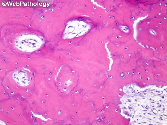
Light microscopy of an H&E stained bone biopsy reveals features characteristic of late-stage Paget disease of bone. The architecture shows thick, irregular plate-like trabeculae arranged in a mosaic or jigsaw pattern and interposed by abundant fibrous vascular tissue, reflecting extensive remodeling. Cement lines are basophilic and conspicuous, delimiting woven and lamellar bone segments and producing a dark, zigzag border between remodeling units. The marrow space is variably replaced by fibrous connective tissue and new vasculature, with relative paucity of normal hematopoietic elements. Osteoid and mineralized bone are variably deposited; osteoblastic and osteoclastic activity may be reduced in the quiescent late phase, yet turnover remains high overall relative to normal bone. The lesion exhibits a mix of eosinophilic osteoid and pink mature bone, with widening of trabecular plates and cortical thickening in surrounding regions. These features explain radiographic findings such as bone expansion, thickened cortex, and coarse trabeculae seen in Paget disease. Clinically, the image is most consistent with a long-standing, deforming process in an elderly patient and supports diagnostic conclusions in combination with serum markers of bone turnover and imaging studies. Differential considerations include fibrous dysplasia and osteosarcoma with pagetoid remodeling, but the presence of classic cement lines favors Paget disease.
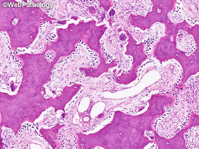
This is a histopathology image of Paget disease of bone (osteitis deformans) in the mixed osteolytic-osteoblastic phase, assessed on a bone biopsy by bright-field light microscopy after Hematoxylin and Eosin staining. The tissue shows disorganized osseous remodeling with mosaic bone architecture. Irregular, thickened trabeculae of newly formed woven bone are interspersed with older lamellar bone and are separated by prominent blue cement lines, creating a jigsaw-like mosaic pattern typical of PDB. Osteoblasts line resorption bays and actively form bone on the surfaces, evidencing elevated osteoblastic activity, while osteoclast-mediated resorption bays from the earlier lytic phase may be present in adjacent areas. The overall appearance reflects concurrent osteoblastic apposition and osteoclastic resorption, with expanded, immature bone formation and cortical thickening in affected regions. The image may lack large malignant features, though rapid remodeling can mimic neoplasia; there is no cytologic atypia. Diagnostic significance: the mosaic pattern with cement lines is highly characteristic of PDB and helps differentiate from fibrous dysplasia, osteomyelitis, or osteosarcoma in ambiguous cases. Clinically, histology complements radiographs and bone turnover markers and informs management decisions between observation, bisphosphonate therapy, and fracture risk assessment. This histology correlates with high bone turnover markers and radiographic sclerosis characteristic of Paget disease.
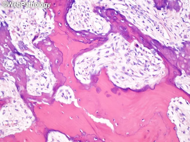
Imaging modality: light microscopy on hematoxylin and eosin stained bone biopsy section. The specimen shows features typical of Paget disease of bone (osteitis deformans) with mixed osteolytic and osteoblastic remodeling. Histology reveals osteoclast-mediated resorption cavities in the lytic phase, later filled by patches of newly formed woven bone. A mosaic pattern is evident, where woven trabeculae are separated from established lamellar bone by numerous irregular blue cement lines. The woven bone is relatively disorganized, more cellular, and accompanied by pronounced osteoblastic activity and increased osteoid deposition. The surrounding lamellar bone is thickened and sclerotic, with irregular trabecular geometry and augmented vascularity in the marrow spaces. Overall turnover is accelerated, with coexisting resorptive and formative processes. Clinically, these findings correlate with bone enlargement, deformity, warmth, and potential fragility fractures. Diagnostic significance is high for PDB when a mosaic cement line pattern is observed in the appropriate clinical setting. Differential considerations include fibrous dysplasia, osteosarcoma, osteomyelitis, and metastatic bone disease displaying mixed lytic-sclerotic changes. This histologic pattern supports diagnosis and informs management, including bisphosphonate therapy and skeletal distribution assessment in advanced disease. Correlation with radiographic findings and serum alkaline phosphatase enhances clinical decision making and surveillance during bisphosphonate courses for skeletal health.
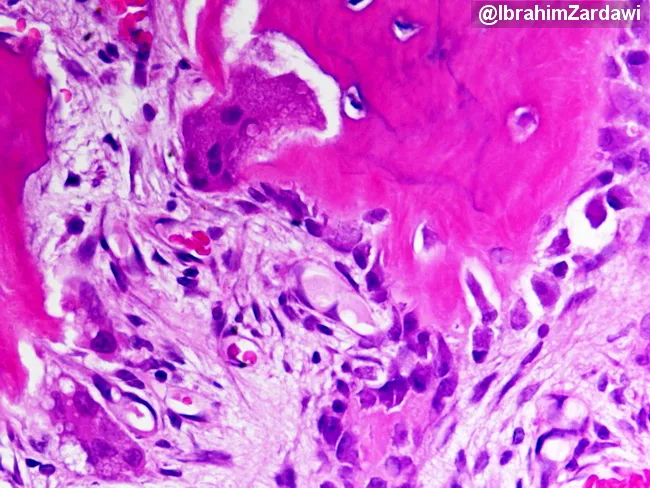
Imaging modality: Light microscopy of hematoxylin and eosin (H&E)–stained bone tissue section. Primary subject: Paget disease of bone (osteitis deformans) in a mixed osteolytic and osteoblastic phase, demonstrated by plate-like trabeculae with exuberant remodeling. Specimen: bone biopsy/surgical bone specimen. Perspective: histologic section examined under finite magnification; features documented on a transverse/longitudinal plane of trabecular bone. Visual description: broad, thickened, disorganized lamellar bone forming plate-like trabeculae; mosaic pattern of lamellar bone; cement lines appearing as irregular blue/gray intertrabecular bands; active osteoblasts line bone formation surfaces; multinucleated osteoclasts erode adjacent bone; increased vascularity and fibrous tissue may be present at the marrow interface. Pathology: accelerated bone turnover with concurrent osteolysis and osteogenesis; signs of high-output remodeling; regions of sclerosis interspersed with radiolucent zones. Diagnostic significance: the mosaic lamellar bone with cement lines is characteristic of Paget disease and helps distinguish it from osteomyelitis, fibrous dysplasia, or metastasis; correlation with clinical markers like alkaline phosphatase and imaging findings strengthens diagnosis. Potential clinical applications: confirm suspected Paget disease, guide bisphosphonate therapy, monitor treatment response, educate trainees on osteoclast–osteoblast coupling, and differentiate high-turnover bone disorders in musculoskeletal pathology. This histology correlates with radiographs showing mixed lytic and sclerotic lesions and is essential for confirming disease activity and guiding therapy response monitoring.
Paget's disease jaw radiograph cotton wool appearance skull
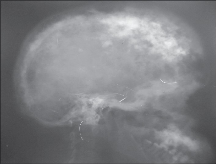
**Imaging Modality:** Lateral plain film radiograph (X-ray). **Anatomical Region:** Skull (cranium). **Observed Pathology:** Advanced Paget's disease of bone (osteitis deformans). **Characteristic Visual Features:** The image demonstrates a classic "cotton wool appearance" characterized by multifocal, ill-defined sclerotic patches interspersed with radiolucent areas throughout the calvarium. There is marked thickening of the diploic space and expansion of the outer table of the skull. The normal cortical margins of the inner and outer tables are blurred due to disorganized bone remodeling. **Key Diagnostic Features:** - **Cotton wool spots:** Focal areas of opacification representing the sclerotic phase of Paget's disease. - **Diploic thickening:** Significant widening of the cranial vault. - **Bone expansion:** Generalized enlargement of the skull landmarks compared to normal anatomical proportions. **Clinical Significance:** These radiographic findings are pathognomonic for the mixed lytic and sclerotic stages of Paget’s disease involving the axial skeleton, specifically the calvarium.
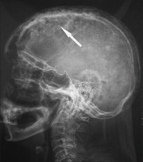
**Imaging Modality:** Lateral plain film radiograph (X-ray). **Anatomical Region:** Lateral view of the skull (calvaria) and upper cervical spine. **Observed Pathology:** The image demonstrates classic radiologic features of Paget's disease of bone (osteitis deformans) affecting the skull. **Characteristic Visual Features:** * **Cotton-Wool Appearance:** The calvaria exhibits generalized, patchy, mixed radiolucent and radiopaque areas, creating a characteristic "cotton-wool" appearance. * **Cortical Thickening:** Significant thickening of the inner and outer tables of the skull is evident, particularly in the frontal and parietal regions. * **Diploic Space Expansion:** There is a marked widening of the diploic space with loss of the normal distinct margins between the tables. * **Distribution:** Findings are diffuse throughout the vault of the skull. **Key Diagnostic Features:** The combination of calvarial thickening and the multifocal sclerotic patches (cotton-wool spots) is highly suggestive of the sclerotic (late) phase of Paget’s disease, distinguishing it from metastatic disease or primary bone malignancies. White arrow indicates a prominent area of mixed bone density within the parietal bone.
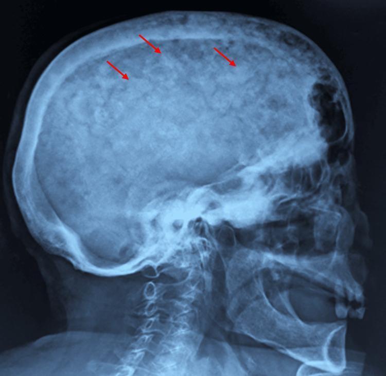
This lateral skull X-ray demonstrates a classic 'cotton wool' appearance of the cranium, a hallmark radiographic finding of Paget's disease of bone (osteitis deformans). The image shows diffuse thickening of the calvarium with a characteristic mottled texture. Multiple, ill-defined sclerotic patches are visible throughout the skull (highlighted by red arrows), representing areas of disordered, high-density bone formation interspersed with radiolucent areas of osteolysis. There is a noticeable widening of the diploic space and a loss of distinction between the inner and outer tables of the skull. The irregular mineralization replaces the normally smooth, homogeneous trabecular pattern of the cranial vault. These visual features are clinically significant as they reflect the late sclerotic phase of Paget's disease, where excessive and disorganized bone remodeling leads to structural deformities and potential neurological complications due to foraminal narrowing or cranial vault thickening.
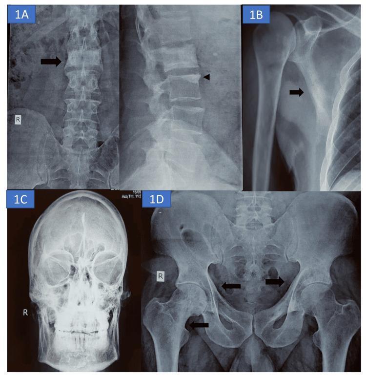
This composite of four conventional radiographs illustrates multi-focal skeletal manifestations of Paget’s disease of bone (PDB). Panel A displays anteroposterior and lateral views of the lumbar spine, highlighting a diffuse sclerosis of the L2 vertebral body, characteristic of an 'ivory vertebra,' with associated coarsened trabeculae and degenerative changes at the L3 endplate. Panel B shows a right shoulder radiograph with prominent cortical thickening along the lateral margin of the scapula. Panel C presents a frontal skull radiograph demonstrating diffuse widening, thickening, and sclerosis of the diploic space, often termed a 'cotton wool' appearance. Panel D is an AP radiograph of the pelvis and proximal femurs, revealing bilateral thickening of the iliopectineal lines (brim sign) and coarsened trabecular patterns within the inferomedial right femoral neck. These findings collectively represent the mixed lytic and sclerotic phases of Paget's disease, focusing on bony expansion and architectural remodeling across the axial and appendicular skeleton.
A labeled pencil-style histological diagram for BDS exam of Paget's disease of bone (H&E). The diagram shows: (1) thick, irregular bone trabeculae arranged in a jigsaw/mosaic pattern; (2) prominent irregular "cement lines" (reversal lines / blue basophilic wavy lines separating bone units) - the hallmark; (3) active osteoclasts - large multinucleated cells in Howship's lacunae on bone surface (more nuclei than normal, up to 100 nuclei); (4) active osteoblasts lining bone surfaces; (5) fibrovascular marrow stroma replacing fatty marrow; (6) woven bone mixed with lamellar bone. Black and white pencil sketch with clear labels and arrows. Title: "Paget's Disease of Bone - Histology (H&E)" - BDS exam diagram style.
PAGET'S DISEASE OF BONE (OSTEITIS DEFORMANS)
BDS 3rd Year - Oral Pathology - University Exam Pattern
DEFINITION
First described by Sir James Paget (1877). Alternate name: Osteitis deformans (because of bone inflammation and deformity)
ETIOLOGY
1. Genetic Factors
- Positive family history in 15-25% of patients (autosomal dominant with variable penetrance)
- SQSTM1 gene (p62 protein / sequestosome-1) mutation - most common; disrupts NF-κB signaling in osteoclasts
- TNFRSF11A gene (encodes RANK) - mutations cause familial expansile osteolysis
- TNFRSF11B gene (encodes Osteoprotegerin / OPG) - juvenile Paget's disease
- Seven-fold increased risk among first-degree relatives
2. Viral Hypothesis
- Paramyxovirus (measles virus, canine distemper virus, Respiratory Syncytial Virus) suspected
- Viral nucleocapsid-like inclusions found in giant pagetic osteoclasts (nuclear inclusions)
- Virus may act as trigger in genetically predisposed individuals
3. Epidemiology
- Age: >40 years (rare before 40); prevalence ~3% over age 40
- Sex: Males > Females (slight)
- Common in Western Europe (UK, France, Germany), Australia, New Zealand
- Rare in Asia, Africa, India (Caucasian populations predominantly affected)
- Prevalence is decreasing globally (reason unknown)
PATHOGENESIS
PHASE 1: OSTEOLYTIC (Hot/Active phase)
↓
Giant, abnormal osteoclasts with many more nuclei than normal (up to 100 nuclei)
Increased RANKL / decreased OPG → excessive osteoclast recruitment
Rapid, aggressive bone resorption
Radiograph: radiolucent "advancing lytic wedge" (flame/blade-of-grass sign in long bones)
PHASE 2: MIXED OSTEOLYTIC-OSTEOBLASTIC
↓
Compensatory osteoblastic new bone formation - but chaotic and disorganized
Woven bone deposited → remodeled repeatedly → MOSAIC pattern
Marrow replaced by fibrovascular tissue (hypervascularity)
Bone enlarged, thickened but structurally WEAK
Radiograph: mixed lytic + sclerotic appearance
PHASE 3: OSTEOSCLEROTIC / BURNED-OUT (Late/Quiescent phase)
↓
Osteoclastic activity decreases
Sclerotic, dense, thick, deformed bone remains
Radiograph: dense "cotton wool" appearance
CLINICAL FEATURES
General Features
Bones Commonly Affected (in decreasing order)
- Pelvis (most common - 70%)
- Lumbar vertebrae
- Skull / Cranium (jaw involvement important for dental students)
- Femur
- Tibia
- Thoracic vertebrae
- Humerus
- Jaws (maxilla > mandible)
Symptoms by System:
A. Bone Pain
- Most common presenting symptom
- Deep, aching, constant bone pain
- Worse at night (increased bony vascularity)
- Sites: pelvis, back, leg, skull
B. Skeletal Deformities
- Bowing of long bones - tibia (saber-shin deformity), femur (shepherd's crook deformity)
- Enlargement of skull - frontal bossing, increased hat size (patients report hat no longer fits)
- "Simian posture" - enlarged skull + bowing of limbs + flexion of spine → patient resembles a gorilla
- Kyphosis (dorsal spine involvement)
- Short stature in severe, long-standing cases
C. Neurological Complications (due to bone expansion compressing nerves)
- Deafness / Hearing loss - most common neurological complication (compression of 8th cranial nerve / cochlea by temporal bone involvement)
- Cranial nerve palsies - other cranial nerves compressed
- Headaches (skull involvement)
- Spinal cord compression - back pain, paraparesis
- Platybasia - softening of base of skull → brainstem compression (rare)
- Blindness (optic nerve compression - rare)
D. Cardiovascular Complications
- Pagetic bone is highly vascular → arteriovenous shunting
- Increased cardiac output (heart works harder to perfuse vascular bone)
- High-output cardiac failure (rare, in extensive disease with >15-35% skeleton involved)
- Overlying skin may feel warm to touch (due to hypervascularity)
E. Secondary Osteoarthritis
- Bowing + deformity → abnormal joint loading
- Secondary OA of hip, knee
F. Pathological Fractures
- Weakened pagetic bone fractures easily
- "Chalk-stick fractures" in long bones (horizontal fractures through softened bone)
- Femoral shaft, subtrochanteric region most common
G. Malignant Transformation (rare but important)
- < 0.5-1% of patients
- Osteosarcoma (most common) - suspect when new severe pain develops in known pagetic bone
- Chondrosarcoma, fibrosarcoma
- Prognosis of pagetic osteosarcoma is very poor
H. Biochemical Findings
- Serum Alkaline Phosphatase (ALP) - markedly elevated (reflects osteoblastic activity) - best marker of disease activity and treatment response
- Serum calcium and phosphorus: normal (unlike hyperparathyroidism)
- Serum PTH: normal
- Urinary hydroxyproline: elevated (reflects bone collagen breakdown)
- Urinary N-telopeptide / C-telopeptide: elevated (bone resorption markers)
ORAL / JAW MANIFESTATIONS (Important for BDS)
- Progressive jaw enlargement - painless, slow
- Spacing / drifting of teeth - malocclusion
- Difficulty wearing dentures - dentures become loose and ill-fitting as jaw enlarges
- "Leontiasis ossea" - lion-face appearance in severe, diffuse craniofacial involvement
- Hypercementosis - excessive cementum deposition on roots of teeth in affected jaw
- Tooth loss - due to root resorption, hypercementosis, or secondary infection
- Obliteration of lamina dura on dental radiographs
- Osteomyelitis susceptibility after extractions (hypervascular bone → poor healing)
- Maxillary sinus obliteration by expanding pagetic bone
- Nerve compression symptoms - paresthesia, pain
RADIOLOGICAL FEATURES
General Radiological Principles - THREE STAGES:
Stage 1 - Osteolytic Phase
- "Osteoporosis circumscripta" (in skull) - well-defined osteolytic area usually starting at frontal/occipital bone and spreading posteriorly
- "Blade of grass" or "Flame sign" (in long bones) - V-shaped advancing lytic front at one end of bone
- Bone appears lucent/radiolucent
Stage 2 - Mixed Phase
- Both radiolucent and radiopaque areas
- Cortical thickening, bone expansion
- Coarsened trabecular pattern
Stage 3 - Sclerotic Phase (Most characteristic images)
- Dense, expanded bones
- Classic named signs (see below)
Named Radiological Signs of Paget's Disease:
| Sign | Bone | Appearance |
|---|---|---|
| "Cotton wool" appearance | Skull | Most classic - multiple, ill-defined radiopaque patches interspersed with radiolucent areas giving cotton wool texture |
| "Picture frame" vertebra | Spine | Cortical thickening of all four margins of vertebral body with central osteoporosis |
| "Ivory vertebra" | Spine | Entire vertebra uniformly radiopaque / sclerotic |
| "Brim sign" | Pelvis | Thickening of iliopectineal (arcuate) line |
| "Blade of grass" / "Flame sign" | Long bones (tibia, femur) | V-shaped advancing lytic front |
| "Chalk-stick fracture" | Long bones | Transverse (horizontal) fracture |
| "Shepherd's crook deformity" | Femur | Bowing of proximal femur |
| "Saber shin" | Tibia | Anterior bowing of tibia |
Skull X-ray showing "Cotton Wool" appearance:
Multiple skeletal manifestations:
Jaw Radiological Features:
- Enlarged maxilla/mandible with loss of normal trabecular pattern
- "Ground glass" or "cotton wool" appearance in jaws
- Hypercementosis - bulbous, irregular roots visible on OPG
- Obliteration of lamina dura
- Obliteration of inferior dental canal (IAN canal) in mandible
- Coarsened trabeculae
HISTOLOGICAL FEATURES
The HALLMARK: Mosaic Pattern of Bone
Histological Findings:
1. MOSAIC PATTERN (Most Important Feature)
- Bone trabeculae show a jigsaw puzzle / mosaic pattern
- Multiple irregular fragments of bone cemented together by conspicuous cement lines (reversal lines)
- These basophilic (blue) irregular cement lines mark where previous osteoclastic resorption stopped and osteoblastic formation resumed
- The overall appearance resembles a crazy-paving pattern or jigsaw puzzle
2. CEMENT LINES (Reversal Lines) - Hallmark
- Irregular, basophilic (blue/purple), wavy lines separating different generations of bone
- Also called "reversal lines" or "arrest lines"
- Represent the multiple alternating cycles of resorption and formation
- These lines are the single most diagnostic histological feature
3. ENLARGED OSTEOCLASTS
- In the lytic phase: giant osteoclasts with many more nuclei than normal (normal = 3-5; Pagetic = up to 100 nuclei)
- Osteoclasts sit in Howship's lacunae (resorption pits on bone surface)
- Viral nuclear inclusions may be visible in osteoclasts (on electron microscopy)
4. OSTEOBLASTIC ACTIVITY
- Plump osteoblasts lining bone surfaces (active bone formation)
- Coexist with osteoclasts in the mixed phase
5. FIBROVASCULAR STROMA
- Normal fatty marrow replaced by loose fibrovascular connective tissue
- Increased vascularity (explains warmth of overlying skin)
- This feature explains why pagetic bone bleeds profusely on cutting
6. WOVEN BONE + LAMELLAR BONE MIXTURE
- Haphazardly arranged collagen in woven bone (coarse, irregular)
- Interspersed with lamellar bone segments
- Together they create the mosaic appearance
7. LATE PHASE (Quiescent)
- Osteoclasts and osteoblasts reduced / absent
- Thick, sclerotic, disorganized bone with prominent mosaic pattern remains
- Marrow: fibrous tissue
H&E Photomicrographs:
EXAM DIAGRAM (H&E Pencil Diagram)
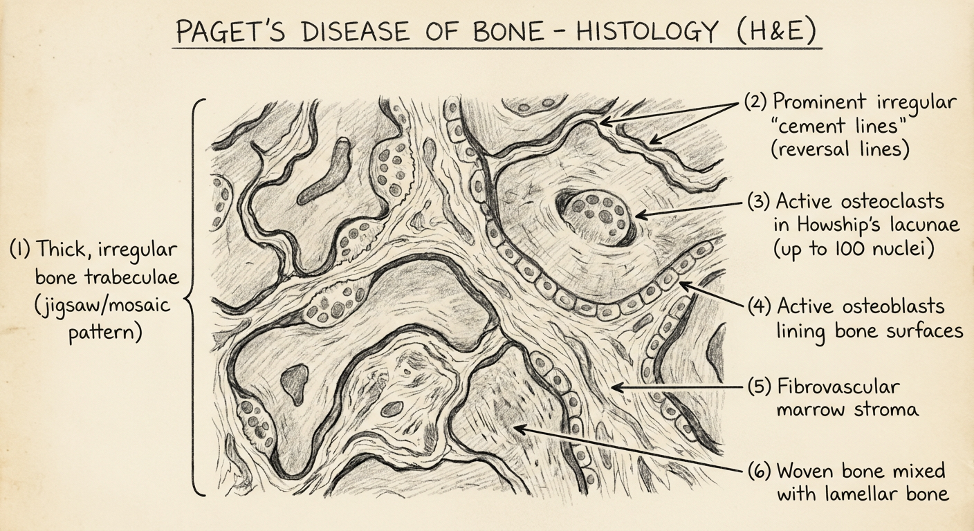
HOW TO DRAW PAGET'S DISEASE HISTOLOGY (H&E):
Step 1: Draw irregularly shaped bone fragments (trabeculae) - thickened, irregular
Step 2: Between the bone fragments - draw dark wavy/zigzag BLUE lines
= CEMENT LINES / REVERSAL LINES (the HALLMARK - draw prominently)
These create the MOSAIC / JIGSAW PUZZLE pattern
Step 3: On bone surfaces - draw large cells with MANY NUCLEI (5-10 in diagram)
sitting in resorption pits = ENLARGED OSTEOCLASTS in Howship's lacunae
Step 4: Also on bone surfaces - draw a row of plump cells = OSTEOBLASTS
Step 5: In the space between trabeculae - draw loose, vascular tissue
with blood vessels = FIBROVASCULAR MARROW (replacing fat)
Step 6: Show a mixture of lamellar and woven bone within trabeculae
LABELS:
→ Irregular bone trabecula (thickened, disorganized)
→ Cement lines / reversal lines (irregular, basophilic, wavy)
→ MOSAIC / JIGSAW PATTERN (label the overall pattern)
→ Enlarged giant osteoclast (numerous nuclei) in Howship's lacuna
→ Active osteoblasts lining bone surface
→ Fibrovascular stroma (replacing fatty marrow)
→ Woven bone (irregular collagen arrangement)
DIFFERENTIAL DIAGNOSIS
| Feature | Paget's Disease | Fibrous Dysplasia | Hyperparathyroidism (Brown tumor) |
|---|---|---|---|
| Age | >40 years | Children/young adults | Any age |
| Histology | Mosaic pattern + cement lines | Chinese letter woven bone + fibrous stroma | Giant cells + hemorrhage + hemosiderin |
| ALP | Very elevated | Elevated | Normal or slightly elevated |
| Calcium | Normal | Normal | Elevated (hypercalcemia) |
| Radiograph | Cotton wool (skull), picture frame (vertebra) | Ground glass, ill-defined | Brown cystic lesions, subperiosteal resorption |
| Jaw | Cotton wool, hypercementosis | Ground glass, ill-defined borders | Giant cell (brown) tumor jaw |
TREATMENT
Medical Treatment:
- Bisphosphonates (first-line) - inhibit osteoclastic resorption
- Zoledronic acid (IV) - single infusion, most potent, long-lasting
- Alendronate, risedronate (oral)
- Etidronate (older)
- Calcitonin (nasal spray or SC injection) - inhibits osteoclasts; second-line
- Analgesics - NSAIDs for bone pain
- Calcium + Vitamin D supplementation (to prevent hypocalcemia after treatment)
Surgical Treatment:
- Joint replacement (total hip/knee) for severe OA
- Fracture fixation (prophylactic intramedullary nail for long bones at risk)
Monitoring:
- Serum ALP - used to monitor treatment response (falls with effective treatment)
- Bone scintigraphy (isotope bone scan) - most sensitive for detecting all active sites
QUICK REVISION TABLE
| Feature | Paget's Disease of Bone |
|---|---|
| Other name | Osteitis deformans |
| Etiology | Unknown; SQSTM1 gene + paramyxovirus |
| Age | >40 years (males > females) |
| Common bones | Pelvis > Lumbar spine > Skull > Femur > Tibia |
| Jaw | Maxilla > Mandible; hypercementosis |
| Key histology | Mosaic pattern with cement lines (reversal lines) |
| Key radiology | Cotton wool (skull), picture frame vertebra, brim sign |
| Key lab | Elevated serum ALP (bone formation); normal Ca, PO4 |
| Complication | Osteosarcoma (<1%), deafness, high-output cardiac failure |
| Treatment | Bisphosphonates (zoledronic acid); calcitonin |
Exam Tips:
- "Mosaic pattern with cement lines / reversal lines" = the single most important histological phrase for Paget's disease - state it prominently, draw it clearly
- "Cotton wool appearance" = skull X-ray - most asked radiological feature
- Pagetic osteoclasts have far more nuclei than normal osteoclasts (up to 100 vs 3-5) - mention this
- ALP elevated but serum calcium is NORMAL - key differentiator from hyperparathyroidism
- Serum ALP = best marker to monitor treatment response
- Dental significance: hypercementosis, teeth become fused to jaw (ankylosed), extractions are difficult and risky (profuse bleeding due to hypervascularity; osteomyelitis risk)
- Malignant transformation → osteosarcoma - always mention as a complication
Developmental anomalies of tongue
fissured tongue scrotal tongue clinical appearance
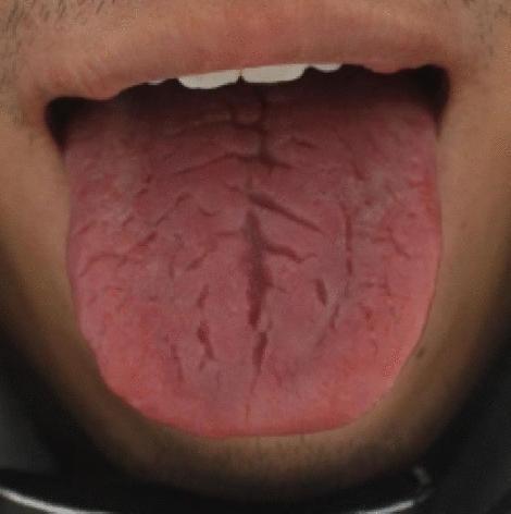
This clinical photograph shows a close-up, dorsal view of a human tongue, illustrating the condition known as fissured tongue (lingua fissurata). The tongue is pinkish-red and exhibits a distinct morphological pattern characterized by numerous grooves and deep fissures across its surface. A prominent central longitudinal groove runs along the midline from the posterior region toward the tip. Branching out laterally from this main fissure is a complex, asymmetric network of secondary cracks varying in depth and width. These fissures give the tongue a characteristic cracked or 'scrotal' appearance. The surrounding oral mucosa appears healthy, and there are no obvious signs of inflammation, ulceration, or abnormal coating in the visible area. In medical education and Traditional Chinese Medicine (TCM) diagnostics, such features are often indexed for tongue image analysis and machine learning applications to quantify crack visibility (FCI) and depth (FDI) indices for disease correlation.
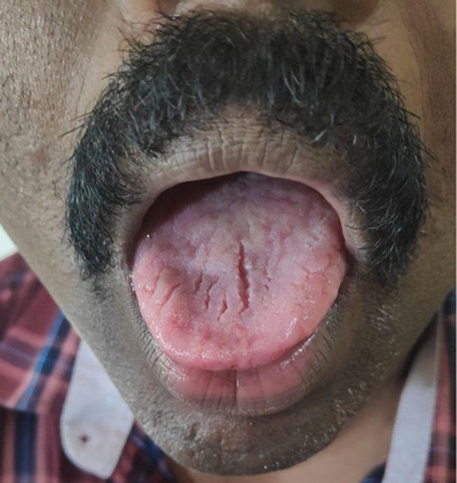
This clinical photograph shows an intraoral view of the tongue in a patient with a known diagnosis of myasthenia gravis (MG). The primary finding is a distinctly furrowed or fissured appearance of the dorsal surface of the tongue, characterized by multiple deep, longitudinal crevices and grooves. The tongue exhibits noticeable atrophy, evidenced by a reduction in muscular bulk and a flattened, wrinkled texture. The coloration is predominantly pale pink with localized areas of mild erythema. Such fissuring (scrotal tongue appearance) and lingual atrophy are rare clinical manifestations of MG, likely associated with bulbar muscle involvement and chronic weakness. The image serves as an educational example of atypical oral presentations in neuromuscular disorders, highlighting the importance of thorough physical examination in diagnosing and monitoring systemic disease progression.
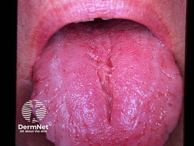
Clinical photograph of the dorsum of the tongue showing fissures. The tongue displays multiple longitudinal grooves with a grooved, rugose surface and interspersed smooth areas; mucosa is pink and moist, with no ulceration or mass effect evident. This presentation corresponds to fissured tongue (lingua fissura), also termed scrotal tongue, a benign morphological variant of the oral mucosa. The fissuring pattern can be diffuse or regional, often more prominent on the central dorsum and midline, and may be accentuated by tongue movement. In many individuals it is asymptomatic, but some report mild irritation or burning, particularly with spicy or acidic foods. Associated phenomena include geographic tongue (benign migratory glossitis) or, less commonly, syndromic contexts such as Melkersson-Rozensky; however, most cases occur in isolation and lack systemic disease. Differential diagnoses include inflammatory glossitis, candidal fissures in immunocompromised patients, nutritional deficiencies (iron, ferritin, vitamin B12, folate), or dermatologic tongue variants. Clinically significant findings include absence of focal swelling, ulceration, or neoplasia. The diagnostic significance lies in recognizing a benign mucosal variant and distinguishing it from inflammatory or infectious processes. Management focuses on reassurance, meticulous oral hygiene, avoidance of irritants, and symptomatic relief if irritation is present; consider nutritional assessment if symptoms persist.
geographic tongue benign migratory glossitis erythematous patches white border clinical
This clinical photograph displays an intraoral view of the tongue's dorsal and lateral surfaces, showcasing two distinct pathologies. On the dorsal surface, there are multiple well-demarcated erythematous patches surrounded by a thin, raised, serpiginous white border, classic for benign migratory glossitis (geographic tongue). On the posterolateral margin of the tongue, there is a distinct, solitary, rounded yellowish papule approximately 5 mm in diameter. This lesion, which was histologically diagnosed as an oral lymphoepithelial cyst, appears smooth and submucosal. The image serves as an educational tool for differential diagnosis of oral mucosal lesions, illustrating the coexistence of a common inflammatory condition (geographic tongue) and a rarer benign cystic lesion (lymphoepithelial cyst) in the same patient. This is relevant for clinical specialties such as oral medicine, dermatology, and otolaryngology.
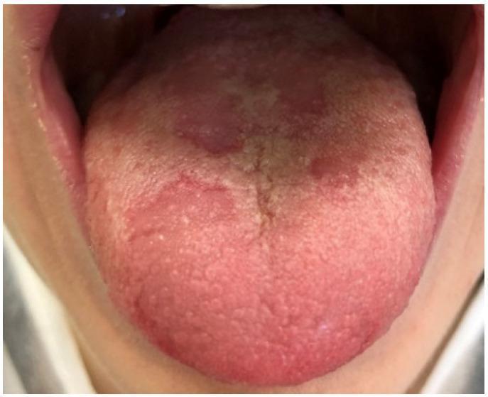
This clinical intraoral photograph displays the dorsal surface of the tongue in an 11-year-old patient, demonstrating classic features of geographic tongue, also known as benign migratory glossitis. The image shows multiple, well-demarcated, erythematous patches resulting from the atrophy of filiform papillae. These smooth, red zones contrast sharply with the surrounding healthy mucosa, which appears more textured and pinkish-white due to intact papillae. Some of the erythematous areas are bordered by slightly elevated, yellowish-white circinate lines. The lesions are distributed irregularly across the anterior and middle portions of the tongue, exhibiting the characteristic map-like or 'geographic' appearance. There is no evidence of ulceration or acute inflammation beyond the papillar atrophy. This condition is a common pediatric oral mucosal lesion and is often diagnosed clinically based on its migratory nature and unique morphology. It is used in dental and medical education to illustrate benign inflammatory disorders of the tongue and the visual presentation of papillary loss.
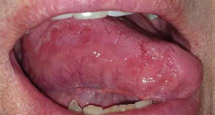
This clinical photograph shows an intraoral view of the tongue, demonstrating classic features of benign migratory glossitis, also known as geographic tongue. The dorsal and lateral surfaces of the tongue exhibit multiple irregular, well-circumscribed erythematous patches. These areas of depapillation appear smooth and glossy due to the loss of filiform papillae. The red lesions are bordered by slightly raised, serpiginous, whitish-yellow margins, creating a characteristic map-like appearance. The surrounding oral mucosa, including the lips and gingiva, appear healthy and pink. The teeth show some slight crowding and yellow-white enamel. The image serves as a high-quality educational example of a common inflammatory condition of the tongue, emphasizing the distinct visual contrast between the smooth atrophic regions and the keratinized borders. Key educational concepts include oral pathology identification, the migratory nature of these lesions, and clinical differentiation from other forms of glossitis or oral candidiasis.
median rhomboid glossitis tongue midline rhomboid erythematous
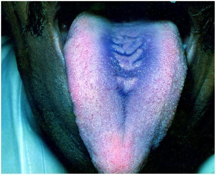
This clinical photograph displays the dorsal surface of a human tongue, demonstrating a well-defined lesion characteristic of Median Rhomboid Glossitis. The primary finding is an elongated, rhomboid-shaped area of erythema and depapillation located in the midline of the posterior third of the tongue. The central affected region appears more intensely pigmented with a blue-purple hue and exhibits a corrugated or fissured texture with several horizontal ridges. This contrasts with the surrounding light pink, granular appearance of the normal filiform papillae. The lesion represents a localized area of atrophy, often associated with chronic erythematous candidiasis. A prominent longitudinal median lingual sulcus is also visible extending from the apex toward the posterior region. This image is used for dental and dermatological education to distinguish benign midline tongue lesions from other forms of glossitis or oral malignancies.
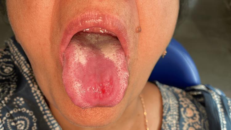
An intraoral clinical photograph showing the dorsal surface of the tongue of a patient with median rhomboid glossitis. The image demonstrates a well-circumscribed, erythematous, and depapillated area centrally located on the midline of the tongue, anterior to the circumvallate papillae. The lesion exhibits a smooth, atrophic texture compared to the surrounding normal filiform papillae. Within this central erythematous zone, there is a small, focal area of ulceration with minor pinpoint bleeding. Faint, irregular white patches are visible along the lateral and posterior margins of the depapillated area, suggestive of secondary candidal infection (pseudomembranous candidiasis). The surrounding oral mucosa and lips appear clinically normal. This visual presentation is characteristic of a 'kissing lesion' associated with oral thrush, showing evidence of early healing or partial response to antifungal therapy as indicated by the reduction in the size of the central ulcer and thinning of the white coating.
ankyloglossia tongue tie short lingual frenulum
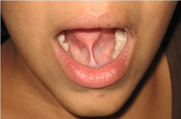
Clinical photograph of the oral cavity demonstrating ankyloglossia (tongue-tie). The image shows a short, thick, and fibrous lingual frenulum that restricts the free movement of the tongue. The frenulum attaches high on the ventral surface of the tongue, near the apex, and extends to the floor of the mouth. This tension creates a notched or 'heart-shaped' appearance of the tongue tip upon attempted protrusion or elevation. The surrounding mucosa is healthy and pink, with visible sublingual vasculature and the caruncles of the submandibular ducts located at the base of the frenulum. This presentation is consistent with a Kotlow Class III ankyloglossia, a congenital anatomical variation that can impact speech, deglutition, and oral hygiene. The photograph serves as a pre-operative assessment for a potential frenectomy.
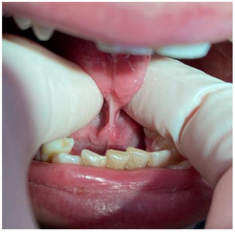
This clinical intraoral photograph demonstrates a case of severe ankyloglossia, commonly referred to as a 'tongue-tie.' The image captures the ventral surface of the tongue being elevated by gloved fingers to reveal a short, thick, and fibrous lingual frenulum. This connective tissue band attaches the underside of the tongue close to its apex to the floor of the mouth, significantly restricting the tongue's range of motion. The frenulum appears taut and under tension, creating a visible pull on the sublingual tissues. The surrounding anatomy includes the lower mandibular incisors, which show signs of crowding and mild extrinsic staining. The floor of the mouth exhibits healthy pink mucosa with visible vasculature, although the focal point is the pathologically short frenulum, estimated at less than 7 mm based on clinical severity grading. This visual is characteristic of Class III or IV ankyloglossia, which can impact speech, deglutition, and oral hygiene in pediatric and adult patients.
black hairy tongue elongated filiform papillae
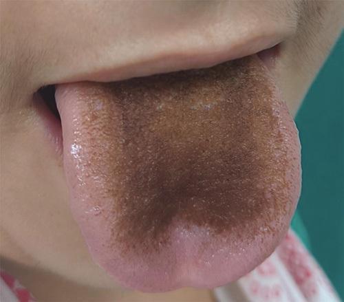
Clinical photograph of the human oral cavity showing the dorsal surface of the tongue. The image demonstrates Lingua Villosa Nigra, also known as black hairy tongue. There is a prominent, well-demarcated area of dark brown to blackish discoloration concentrated on the posterior and central dorsal regions, sparing the lateral borders and the tip. Morphologically, the filiform papillae exhibit significant hypertrophy and elongation, creating a distinctively textured, carpet-like or 'hairy' appearance. This condition is a benign clinical finding often associated with antibiotic use (such as linezolid), poor oral hygiene, or smoking, resulting from the accumulation of keratin and pigment-producing bacteria on the elongated papillae. The surrounding oral mucosa and lips appear healthy, providing a clear contrast to the central pathology. This image is used in medical education to illustrate drug-induced side effects and dermatological manifestations of the oral mucosa.
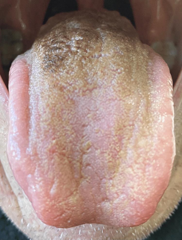
This clinical photograph displays a superior (dorsal) view of a human tongue exhibiting signs of Lingua Villosa Nigra, commonly known as black hairy tongue. The visual findings demonstrate significant hypertrophy and elongation of the filiform papillae across the posterior and central dorsum. These elongated papillae show a varying degree of discoloration, ranging from a dark brown-black hue in the most posterior region to a tan-brown or yellowish-brown coating toward the mid-tongue. The anterior tip and lateral borders of the tongue appear relatively spared, retaining a more natural pink color with visible but normal-sized papillae. The texture of the affected area appears thickened and 'hairy' or 'furry' due to the accumulation of keratin and chromogenic debris. This diagnostic image illustrates a typical adverse drug reaction or oral hygiene-related manifestation, specifically highlighting the characteristic distribution of papillary changes and extrinsic staining on the dorsal surface of the tongue.
A labeled pencil-style diagram for BDS exam showing histology of geographic tongue (benign migratory glossitis) H&E. The diagram shows: (1) areas of filiform papillae loss (atrophy/depapillation) - smooth flat epithelium; (2) adjacent areas with normal filiform papillae; (3) epithelium shows spongiosis (edema between cells) and neutrophil exocytosis forming Munro microabscesses in the superficial epithelium; (4) subepithelial lamina propria with mixed inflammatory infiltrate - neutrophils, lymphocytes, plasma cells; (5) normal fungiform papilla nearby. Black and white pencil sketch style, labeled with arrows. Title: "Geographic Tongue Histology (H&E)" - BDS exam diagram style.
DEVELOPMENTAL ANOMALIES OF THE TONGUE
BDS 3rd Year - Oral Pathology - University Exam Pattern
CLASSIFICATION / ENUMERATION
I. ANOMALIES OF SIZE
- Macroglossia - abnormally large tongue
- Microglossia - abnormally small tongue
- Aglossia - absence of tongue (very rare)
II. ANOMALIES OF FORM / SHAPE
- Bifid (cleft) tongue - partial or complete division at tip
- Ankyloglossia (Tongue-tie) - restricted tongue movement due to short frenulum
- Fissured tongue (Scrotal / Plicated tongue)
- Lobulated tongue - abnormal lobulation
III. ANOMALIES OF SURFACE / PAPILLAE
- Geographic tongue (Benign migratory glossitis / Erythema migrans)
- Median rhomboid glossitis (central papillary atrophy)
- Black hairy tongue (Lingua villosa nigra) - though acquired, included in surface anomalies
- Smooth tongue (Bald tongue / Glossodynia) - atrophic glossitis
IV. ANOMALIES OF POSITION
- Lingual thyroid - ectopic thyroid tissue at base of tongue (foramen cecum region)
- Thyroglossal cyst (associated with incomplete descent of thyroid)
V. CYSTS OF THE TONGUE
- Dermoid cyst of tongue
- Lymphoepithelial cyst of tongue
DETAILED DESCRIPTION OF MAJOR CONDITIONS
1. MACROGLOSSIA
Definition
Classification
- Primary / Congenital: Lymphangioma, hemangioma, muscular hypertrophy
- Secondary / Acquired: Acromegaly, hypothyroidism (cretinism), amyloidosis, angioedema, Down syndrome, Beckwith-Wiedemann syndrome, neurofibromatosis, rhabdomyosarcoma
Clinical Features
- Large tongue that protrudes from mouth
- Difficulty in speech (dysarthria), swallowing (dysphagia), chewing
- Scalloped margins (crenated / scalloped tongue) - indentations from lower teeth
- Open-bite tendency
- Prognathic mandible in long-standing cases
- Drooling of saliva
- Mouth breathing
Common Causes to Remember in BDS Context:
- Down syndrome (Trisomy 21) - relative macroglossia due to hypotonia + small oral cavity
- Lymphangioma - most common cause of true macroglossia in children
- Amyloidosis - classic cause in systemic disease
- Acromegaly (excess GH) - adult-onset enlargement
- Hypothyroidism - myxedematous infiltration
2. ANKYLOGLOSSIA (Tongue-Tie)
Definition
Etiology
- Failure of normal apoptosis/degeneration of the lingual frenulum during development
- Familial tendency in some cases
Clinical Features
- Short, tight, fibrous lingual frenulum attaching to the ventral surface of tongue tip to floor of mouth
- Restricted tongue elevation and protrusion
- "Heart-shaped" tongue tip on protrusion (notching at tip due to frenulum pull)
- Difficulty with breastfeeding in neonates
- Speech problems: difficulty producing dental consonants (T, D, N, L) and sibilants (S, Z)
- Poor oral hygiene (tongue cannot sweep oral cavity)
- Diastema between mandibular central incisors (in severe cases)
- Often asymptomatic; discovered incidentally
Kotlow's Classification:
| Class | Free tongue length | Severity |
|---|---|---|
| I | 12-16 mm | Mild |
| II | 8-11 mm | Moderate |
| III | 3-7 mm | Severe |
| IV | <3 mm | Complete ankyloglossia |
Clinical Photograph - Ankyloglossia:
Treatment: Frenectomy or Frenuloplasty (laser or surgical)
3. FISSURED TONGUE (Scrotal / Plicated Tongue / Lingua Plicata)
Definition
Synonyms: Scrotal tongue, Plicated tongue, Furrowed tongue, Lingua fissurata
Etiology
- Developmental / hereditary (most common explanation)
- Associated with Down syndrome, Melkersson-Rosenthal syndrome (fissured tongue + facial palsy + granulomatous cheilitis - triad)
- Can be seen with geographic tongue (common association)
- Increases in prevalence with age
Clinical Features
- Multiple grooves/fissures on the dorsal surface - central deep longitudinal fissure + branching lateral secondary fissures
- Fissures may be shallow or deep (up to 6 mm deep)
- Bilateral and roughly symmetric pattern
- Tongue appears wrinkled or "brain-like" ("scrotal appearance" - resembles scrotal skin)
- Usually asymptomatic
- Debris and bacteria can accumulate in deep fissures → soreness, burning sensation with spicy foods
- No treatment required; good oral hygiene advice
Clinical Photograph - Fissured Tongue:
Histological Features
- Deep clefts/fissures lined by stratified squamous epithelium (surface epithelium dips into grooves)
- Normal epithelium along the fissure walls
- Mild chronic inflammatory infiltrate in lamina propria (if secondary irritation)
- Filiform papillae may be absent along the floor of deep fissures
4. GEOGRAPHIC TONGUE (Benign Migratory Glossitis / Erythema Migrans)
Definition
Synonyms: Benign migratory glossitis, Erythema migrans, Wandering rash of tongue
Etiology
- Unknown; considered a developmental/inflammatory condition
- Associated with: psoriasis (histologically identical), atopy/allergy, stress, nutritional deficiency, hormonal changes
- Positive family history in some cases
- Often associated with fissured tongue
Clinical Features
- Multiple well-demarcated erythematous patches on dorsal and lateral tongue
- Filiform papillae atrophy within patches → smooth, red, depapillated zones
- White-yellow elevated serpiginous (wavy/snake-like) border surrounding each red zone - the white border represents the advancing edge of desquamation
- "Map-like" appearance - like a geographic map; patches vary in size and shape
- Migratory - lesions change shape, location, and size over days/weeks - healing in one area and appearing in another ("wandering")
- Usually asymptomatic; some patients report sensitivity to hot, spicy, or acidic foods
- Affects any age, slightly more common in children and females
- Fungiform papillae are preserved (appear as red dots within depapillated zones)
Clinical Photographs - Geographic Tongue:
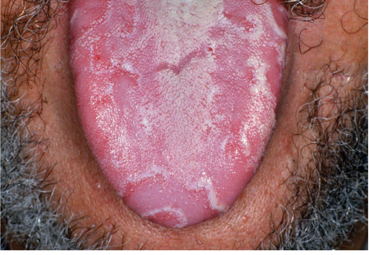
Histological Features of Geographic Tongue
-
Zone of active depapillation (red zone):
- Loss/atrophy of filiform papillae - epithelium flattened, thin
- Thinned epithelium with loss of normal keratinization
- Spongiosis (intercellular edema) in spinous layer
- Exocytosis of neutrophils into epithelium
- Munro microabscesses - small collections of neutrophils within superficial epithelium (identical to psoriasis)
-
Zone of advancing white border:
- Hyperparakeratosis (thickened, parakeratinized surface)
- Epithelium thickened at the white border
- Neutrophil infiltration migrating upward
-
Lamina propria (connective tissue):
- Mixed chronic inflammatory infiltrate: lymphocytes, plasma cells
- Neutrophils near advancing border
- Dilated blood vessels (explains erythema)
-
Fungiform papillae: preserved (appear on histology as normal)
H&E Diagram - Geographic Tongue:
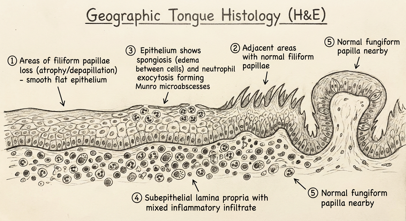
HOW TO DRAW GEOGRAPHIC TONGUE HISTOLOGY (H&E):
Step 1: Draw epithelium with 2 zones:
LEFT side: NORMAL thick epithelium with tall filiform papillae on surface
(hyperparakeratosis at white border edge)
RIGHT side: DEPAPILLATED thin epithelium - no papillae, smooth flat surface
Step 2: In the transition zone (white border): thickened parakeratotic epithelium
Step 3: In the thinned epithelium zone: small clusters of neutrophils
= MUNRO MICROABSCESSES (small dark cell collections in superficial epithelium)
Step 4: Epithelial intercellular spaces widened = SPONGIOSIS
Step 5: Below epithelium in lamina propria: scattered lymphocytes + plasma cells
+ dilated blood vessels
LABELS:
→ Atrophic epithelium (loss of filiform papillae)
→ Hyperparakeratotic advancing border (white border)
→ Munro microabscesses (neutrophils in epithelium)
→ Spongiosis (intercellular edema)
→ Exocytosis of neutrophils
→ Chronic inflammatory infiltrate in lamina propria
→ Dilated capillaries
5. MEDIAN RHOMBOID GLOSSITIS (Central Papillary Atrophy)
Definition
Clinical Features
- Oval / rhomboid-shaped smooth patch in the midline of posterior 1/3 of tongue
- Depapillated - no filiform papillae; smooth, pink-red or sometimes nodular/lobulated surface
- Well-defined borders
- Location: anterior to the sulcus terminalis / circumvallate papillae in the midline
- Usually asymptomatic
- Associated with Candida albicans (erythematous candidiasis) in many cases
- May show a corresponding "kissing lesion" on the hard palate (contact area when tongue presses on palate)
- More common in smokers, denture wearers, HIV patients
Clinical Photograph - Median Rhomboid Glossitis:
Histological Features
- Loss of filiform papillae (smooth surface)
- Hyperplastic or atrophic epithelium (varies)
- Candida hyphae may be visible in superficial epithelium (PAS stain)
- Chronic inflammatory infiltrate in lamina propria
- Dilated capillaries
Treatment: Antifungal therapy (clotrimazole, nystatin, fluconazole) if candidal association
6. BLACK HAIRY TONGUE (Lingua Villosa Nigra)
Definition
Etiology / Predisposing Factors
- Antibiotics (most common cause - especially broad-spectrum: tetracycline, linezolid)
- Poor oral hygiene
- Smoking / tobacco use
- Excessive tea/coffee
- Chronic oxidizing mouth rinses (hydrogen peroxide)
- Radiation therapy to head/neck
- Xerostomia
Pathogenesis
- Disruption of oral flora → overgrowth of chromogenic bacteria + fungi
- Elongation of filiform papillae (failure of normal desquamation)
- Pigment accumulation from bacteria (Nocardia, chromogenic bacteria) → black/brown color
Clinical Features
- Elongated, hair-like filiform papillae on posterior dorsal tongue
- Discoloration: black (most dramatic), brown, yellow, green depending on etiology
- Sensation of gagging / tickling at back of throat
- Mild halitosis
- Usually asymptomatic; resolves when causative factor removed
Clinical Photograph - Black Hairy Tongue:
Histological Features
- Marked elongation of filiform papillae (can reach 1-3 cm)
- Hyperparakeratosis of papillary surface
- Accumulation of keratinous debris between papillae
- Bacteria and fungal hyphae visible within keratin
- Chronic inflammatory infiltrate in lamina propria
Treatment
- Remove causative factor (stop antibiotics, improve hygiene)
- Gentle tongue brushing
- Good hydration
- Antifungal if candidal component
7. LINGUAL THYROID
Definition
Embryology
- Thyroid develops from endoderm at foramen cecum (junction of anterior 2/3 and posterior 1/3 of tongue)
- Normally descends via thyroglossal duct to its final position in the neck
- Arrest of descent → lingual thyroid
Clinical Features
- Smooth, round, pinkish-red, midline mass at base of tongue (posterior tongue)
- May be the only functioning thyroid tissue in the body (in 70% of cases)
- Symptoms: dysphagia, dysphonia, dyspnea (if large)
- Hypothyroidism may develop (if lingual thyroid is the only thyroid)
- Enlarges during puberty, pregnancy, menopause (due to TSH stimulation)
- Females > Males
- Rarely: bleeding, pain
Investigations
- Technetium-99m or I-131 scan - confirms presence of thyroid tissue
- TSH levels - may be elevated
- Do NOT perform biopsy without first confirming whether it is the only thyroid tissue (CRITICAL)
Histological Features
- Normal thyroid follicles with colloid
- Lined by cuboidal to columnar follicular cells
- No encapsulation (unlike ectopic thyroid in neck)
Treatment
- Conservative if small and asymptomatic + euthyroid
- Thyroid hormone replacement if hypothyroid → may reduce size
- Surgical excision if large + obstructive (only if other thyroid tissue confirmed present)
SUMMARY TABLE - QUICK REVISION
| Condition | Key Feature | Association | Treatment |
|---|---|---|---|
| Macroglossia | Enlarged tongue, scalloped margins | Down syndrome, lymphangioma, amyloidosis | Treat cause; surgery if severe |
| Ankyloglossia | Short frenulum, heart-shaped tip, restricted movement | Familial | Frenectomy |
| Fissured tongue | Deep grooves on dorsum; scrotal appearance | Melkersson-Rosenthal, Down syndrome, geographic tongue | Reassurance, hygiene |
| Geographic tongue | Map-like erythematous patches, white border, migratory | Psoriasis, fissured tongue | Reassurance; avoid triggers |
| Median rhomboid glossitis | Rhomboid depapillated zone, midline posterior tongue | Candida infection | Antifungals |
| Black hairy tongue | Elongated dark filiform papillae | Antibiotics, smoking | Remove cause, hygiene |
| Lingual thyroid | Midline base of tongue mass | Only thyroid in 70% | Do NOT biopsy first; scan first |
IMPORTANT EXAM POINTS
| Condition | Key Histological Feature |
|---|---|
| Geographic tongue | Munro microabscesses (identical to psoriasis) + loss of filiform papillae + spongiosis |
| Fissured tongue | Deep fissures lined by stratified squamous epithelium |
| Median rhomboid glossitis | Depapillation + candidal hyphae (PAS stain) |
| Black hairy tongue | Elongated filiform papillae + bacteria/fungi in keratin |
| Lingual thyroid | Thyroid follicles with colloid at base of tongue |
Exam Tips:
- Geographic tongue = most asked for histology; key terms: Munro microabscesses + spongiosis + neutrophil exocytosis + depapillation - same as psoriasis
- Fissured tongue + geographic tongue often co-exist - a common short question
- Melkersson-Rosenthal syndrome triad = fissured tongue + facial palsy + granulomatous cheilitis
- Lingual thyroid - NEVER biopsy without scanning first (may be the only thyroid tissue)
- Ankyloglossia = "tongue-tie" - frenectomy is the treatment; causes speech problems (T, D, N, L, S, Z sounds)
- Median rhomboid glossitis - classically described as "developmental" (tuberculum impar remnant) but now associated with Candida - mention both in exams