
give post surgical management of sports player
post surgical rehabilitation sports athlete return to play
Post-Surgical Management of Sports Players
1. Immediate Post-Operative Care (Day 0 - Week 1)
Pain Management
- Local anesthetic (with liposomal bupivacaine considered for extended relief) liberally applied at the surgical site
- Multimodal analgesia: Acetaminophen + NSAIDs + ice to minimise opioid use
- Adductor canal block (purely sensory) preferred over femoral nerve motor block after knee surgery - provides equivalent pain relief with faster quadriceps strength recovery
Wound & Limb Care
- Cryotherapy (ice bags) used liberally for swelling and comfort; note that evidence for reducing hospital stay or improving ROM is limited
- Elevation of the operated limb
- Compression dressings as needed
- Early mobilisation (partial weight bearing with crutches is allowed immediately after isolated ACL reconstruction)
Bracing
- Knee immobilisation in a fully extended brace started immediately after ACL surgery - prevents flexion contracture and protects the weakened quadriceps, while still allowing flexion
- Continuous passive motion (CPM) machines are NOT used routinely - literature does not support added benefit
2. Early Rehabilitation Phase (Weeks 1-4)
Range of Motion
- Active and passive knee extension with heel elevated and knee supported posteriorly
- Progressive restoration of full flexion as tolerated
- Early intensive rehabilitation prevents arthrofibrosis and restores strength sooner
Weight Bearing & Ambulation
- Crutches typically discontinued by 3 to 4 weeks post-operatively
- Weight-bearing status may be modified if concurrent meniscal repair or articular cartilage procedures were performed
Strengthening - Phase 1
- Quadriceps sets and straight-leg raises - do not stress the graft
- Early hamstring emphasis - hamstrings act in concert with the ACL to prevent anterior tibial translation
- Blood flow restriction (BFR) training: An air tourniquet is placed proximally on the limb during low-intensity exercises - promotes muscular hypertrophy/attenuates atrophy. Works via recruitment of fast-twitch fibres in hypoxia + catecholamine/growth hormone stimulation (similar to heavy conventional exercise)
Neuromuscular / Proprioceptive Training
- Instituted within the first 2 weeks
- Neuromuscular training improves knee function and pain reduction
- Strength training improves hamstring recovery
- Post-op program should combine both modalities
Electrical Muscle Stimulation (EMS)
- Does NOT significantly decrease muscle atrophy after ACL surgery
- No long-term strengthening benefit
- May be used sparingly for muscle re-education in patients with poor quadriceps activation
3. Progressive Strengthening Phase (Weeks 4-8)
Exercise Progression
| Exercise Type | Rationale | Caution |
|---|---|---|
| Closed kinetic chain (CKC) - squats, leg press (weight-bearing) | Loads the joint axially, joint contours stabilise the knee, less patellofemoral pain, lower arthrometer side-to-side differences, higher patient satisfaction | Preferred over OKC early |
| Open kinetic chain (OKC) - resisted knee extension | Puts strain on ACL, especially in the last few degrees of extension without weight bearing | Worrisome early; can be introduced carefully later |
| Progressive resistance exercises, split squats, stationary bike | Restore functional strength | Intensity progressed as tolerated |
Core Muscle Injury / Sports Hernia Protocol (Fischer's, Table 225.4)
| Phase | Timeline | Activities |
|---|---|---|
| Phase 1 | Days 0-7 | Walking 3-6 mph up to 20 min, light stretching of thigh and groins |
| Phase 2 | Weeks 2-4 | Hip ROM, incline treadmill walking, stationary bike (progressive), wall sits, abdominal drawing-in manoeuvre |
| Phase 3 | Weeks 3-4 | Light jogging, scar mobilisation with deep tissue massage, low-level sport-specific exercises, pool exercises |
| Phase 4 | Weeks 4-5 | Progressive resistance, split squats, slide board, increased running intensity, lower abdominal strengthening |
| Phase 5 | Weeks 5-6 | Weight room strength/conditioning, low-level practice, core stabilisation |
| Phase 6 | Weeks 6-8 | Full-speed drills, scrimmage with contact, end-stage exercises, medical clearance for RTS |
4. Return to Sport (RTS) Criteria
Objective Functional Tests
- Hop testing is the key functional measure:
- Single-leg hop for distance
- Single-leg triple hop for distance
- Single-leg timed hop
- Single-leg crossover hop for distance
- Vertical jump test
- A Limb Symmetry Index (LSI) score of ≥85% compared to the normal limb is required for release to RTS
Strength Criteria
- Minimum 80% return of thigh (quadriceps and hamstring) strength
- Ability to perform sport-specific agility tasks
Timeline
- Return to sport should be delayed at least 6 months after ACL reconstruction to allow graft maturation
- Animal studies suggest the graft should be spared significant loads for 6 to 12 months
- Early return to high-level sport is a significant risk factor for ACL graft failure
- Graft maturation may continue up to 18 months post-op
- For core muscle injuries: RTS generally within 5-8 weeks, longer for bilateral repairs or adductor procedures
Key Statistics (Campbell's, 2026)
- Only 44% of patients returned to competitive sport despite 90% having normal/near-normal outcome scores - highlighting the gap between functional recovery and actual RTS
5. Adjunct Considerations
Home vs Clinic Rehabilitation
- Home-based programs shown to be equally effective and score higher on quality-of-life measures
- However, evidence comes largely from recreational athletes, not high-level competitive athletes - supervised programs are still preferred at elite level
Functional Bracing
- Controversial after ACL surgery
- Some data suggest benefit for in vivo knee kinematics and graft protection
- Limited evidence supports routine brace use to reduce reinjury rates
- May be considered for collision sports (football, rugby, etc.)
Psychological Readiness
- Fear of reinjury is a major barrier to RTS; psychological readiness assessment (e.g., ACL-RSI score) should accompany functional testing
Minimally Invasive Surgery Advantage
- Laparoscopic repairs (TEP/TAPP) for core muscle injuries show >90% return to sport in <1 month in multiple series - faster than open repair for early recovery, though long-term outcomes are comparable (Fischer's Mastery of Surgery)
6. Complications to Watch For
| Complication | Signs | Action |
|---|---|---|
| Arthrofibrosis | Stiff joint, loss of extension | Intensive early ROM therapy |
| Muscle atrophy | Thigh girth loss | Early strengthening, BFR training |
| Graft failure/re-tear | Pain, instability, positive pivot shift | Imaging, possible revision |
| Wound infection | Erythema, warmth, discharge | Antibiotics, wound care |
| DVT | Calf swelling, pain | Prophylaxis, Doppler USS |
- Campbell's Operative Orthopaedics, 15th Ed (2026), Chapter 50 - [ACL Rehabilitation Protocol] - Knee Surgery, p. 2722-2723
- Fischer's Mastery of Surgery, 8th Ed - Chapter 225: Athletic Pubalgia/Core Muscle Injury - Postoperative Rehabilitation, p. 6138-6139
- Recent systematic review: Prehabilitation before ACL reconstruction (PMID: 40276858, Knee Surg Sports Traumatol Arthrosc, 2025) confirms short- and long-term benefits of structured pre/post-op rehab
- Open kinetic chain exercises in early ACL rehab (PMID: 39985872, Phys Ther Sport, 2025) - meta-analysis showing safety and benefit when introduced appropriately
Search for latest RTS testing protocols and prognostic factors in return-to-play studies0 marks question
return to sport testing criteria athlete
return to play prognostic factors sports injury
PMID: 39892026
PMID: 39163752
PMID: 38216071
PMID: 40665533
PMID: 41724878
PMID: 41851593
PMID: 39977365
PMID: 41138195
Latest RTS Testing Protocols & Prognostic Factors in Return-to-Play Studies
Overview: The RTS Decision Framework
- Return to participation - cleared to train non-competitively
- Return to sport - training at full intensity
- Return to performance - competing at pre-injury level
1. Lower Extremity RTS Testing Protocols
A. Knee / ACL (Most Evidence-Dense Area)
- 85% of RTS literature focuses on knee pathology; 82% specifically on ACL reconstruction
- No uniform/standardised RTS criteria exist for any lower extremity joint
Strength Testing
| Test | Details | Key Finding |
|---|---|---|
| Isokinetic dynamometry | Most studied test (73% of studies); measures quad and hamstring torque at set angular velocities | Gold standard for strength symmetry; expensive, not always accessible |
| Isometric knee extension | Simpler, portable | 50% of healthy controls fail; questions the 85% LSI threshold |
| Isometric knee flexion | Hamstring assessment | 37% of healthy controls fail |
| Limb Symmetry Index (LSI) ≥85% | Standard threshold | PROBLEM: many healthy people have inherent asymmetry >10%, so threshold may be inappropriate |
Hop Testing Battery (Functional Performance)
| Test | Description |
|---|---|
| Single-leg hop for distance | Most common; measures explosive power |
| Triple hop for distance | 3 consecutive hops on one leg |
| Crossover triple hop | Hop across a line 3 times |
| 6-metre timed hop | Speed + single-leg coordination |
| Drop vertical jump | Assesses dynamic valgus biomechanics on landing |
B. Soccer / Football Muscle Injuries
- Strength symmetry: between-limb knee flexor/extensor strength symmetry (most cited)
- No pain during soccer-specific actions
- Range of motion tests: Active Knee Extension (AKE), passive/active straight leg raise, Askling-H test (highest certainty)
- Subjective readiness assessment
- Completing at least one full team training session
- Pain assessment during adductor-loaded movements
- Completing at least one full team training session
- Restoring strength levels
- Hip adductor squeeze test normalisation
2. Upper Extremity RTS Testing
| Test | Reliability | Validity | Notes |
|---|---|---|---|
| Single Arm Shot Put (SASP) | ICC >0.90 consistently | Correlates with UE isokinetic torque | Best reliability; overhead athletes |
| Closed Kinetic Chain Upper Extremity Stability Test (CKCUEST) | ICC 0.73-0.98 (good-excellent) | Predicts future UE injury risk; discriminates injured vs uninjured | Most versatile; shoulder + elbow |
| Upper Quarter Y-Balance (UQY) | Mixed (ICC 0.47-0.97) | No significant correlation with strength in several studies | Least reliable; use cautiously |
| Athletic Shoulder (ASH) Test (emerging) | ICC 0.94-0.98 (excellent) | High concurrent validity | Isometric strength in 3 positions prone |
3. Prognostic Factors for RTS After ACL Reconstruction
Positive Prognostic Factors (favour RTS)
| Factor | Domain | Notes |
|---|---|---|
| Fewer concomitant meniscal injuries | Structural/surgical | Isolated ACL recon has better RTS rates |
| Shorter time between injury and surgery | Surgical timing | Early surgery preserves neuromuscular function |
| Higher hop test scores at RTS testing | Physical | Functional capacity measure |
| Better physical functioning pre/post-op | Physical | Baseline fitness matters |
| Higher muscle strength | Physical | Quad and hamstring symmetry |
| Greater psychological readiness (ACL-RSI) | Psychological | Strongest modifiable prognostic factor |
4. Psychological Readiness - The Emerging Priority
- ACL-RSI scale: effect size 1.55 (95% CI: 1.24-1.87) - the most effective predictor of RTS success
- Hop tests and LSI were only moderately effective
- Flexion-LSI was least effective
- Recommendation: adopt a multi-dimensional approach integrating both psychological and physical assessments
5. Practitioner Decision-Making in RTP (Real-World Gap)
- Surgeons (47% of studies): rely predominantly on injury and post-operative timelines
- Physiotherapists: prioritise functional assessment and strength (≥50%)
- MDTs: more likely to include psychological readiness (50%) and sport-specific testing (38.5%)
- Only 20% of studies referenced formal RTP frameworks or guidelines
- Athlete-centred shared decision-making is the recommended approach
- Biggest influencing factors: type of sport and athlete's ambition (cited in 26.4% of all studies)
6. Recommended Multi-Dimensional RTS Test Battery (Current Evidence Synthesis)
| Domain | Recommended Tests | Threshold |
|---|---|---|
| Muscle strength | Isokinetic quad/hamstring at 60°/s and 180°/s | LSI ≥90% quad; ≥90% hamstring (stricter than older 85%) |
| Functional performance | Single-leg hop for distance + triple hop + crossover hop | LSI ≥90% (interpret with caution - healthy people also fail) |
| Neuromuscular control | Drop vertical jump (knee valgus angle) | <5° valgus asymmetry |
| Range of motion | Active knee extension, SLR (hamstring); hip ROM (adductors) | Full symmetric ROM |
| Psychological readiness | ACL-RSI scale | Score ≥65 (recommended threshold) |
| Subjective function | IKDC questionnaire | ≥85% score |
| Sport-specific testing | Cutting, agility, sprint tests relevant to the athlete's sport | No pain, no hesitation |
| Clinical clearance | Full team training participation | At least 1 full training session without symptoms |
7. Key Evidence Gaps & Research Needs
- No standardised criteria exist for ankle, hip, or upper extremity RTS - almost all evidence is ACL/knee-centric
- LSI 85% threshold is questionable: healthy individuals regularly fail, suggesting the threshold needs recalibration by sport, sex, and age
- Functional tests (hop, strength) have poor predictive validity in isolation - only multimodal batteries reduce reinjury risk
- Prognostic models have moderate accuracy (~70%) - insufficient to replace clinical judgement
- Pediatric and female athlete populations are under-studied in RTS testing literature
- Long-term outcomes beyond 12 months post-RTS are rarely tracked
Summary of Key Papers
| PMID | Study | Journal | Year | Key Takeaway |
|---|---|---|---|---|
| 39892026 | van Haren et al. | Ann Phys Rehabil Med | 2025 | 6 prognostic factors for RTS; very low certainty |
| 39163752 | Smiley et al. | The Knee | 2024 | No standardised lower extremity RTS criteria exist |
| 38216071 | Gill et al. | Arthroscopy | 2024 | Hop/strength tests inconsistent at predicting reinjury |
| 40665533 | Lo et al. | J Sports Sci | 2025 | ACL-RSI is most predictive RTS criterion (ES 1.55) |
| 39977365 | Wood et al. | Am J Sports Med | 2025 | 5-42% of healthy athletes fail standard RTS tests |
| 41851593 | Pecci et al. | Sports Med | 2026 | Muscle injury RTP criteria for soccer - strength symmetry + no pain |
| 41724878 | Pontillo et al. | Sports Med Open | 2026 | First UE RTS review: CKCUEST + SASP best validated tests |
| 41138195 | Chetty et al. | Eur J Sport Sci | 2025 | Real-world RTP: <20% use formal frameworks; shared decision-making best |
post operative rehab only of exercise of shoulder and knee phase wise
ACL knee rehabilitation exercises phases progression
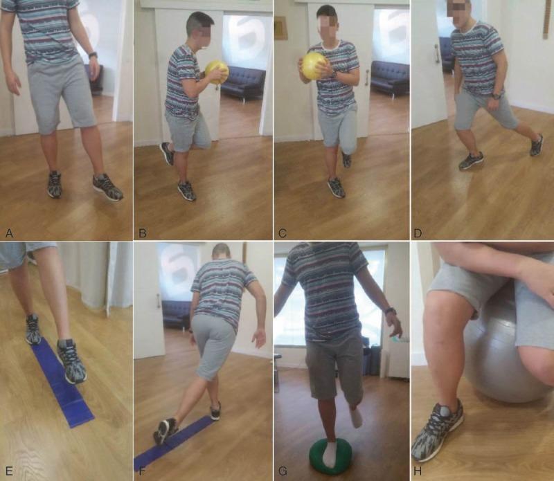
This composite of clinical photographs illustrates a progressive proprioceptive exercise protocol designed for anterior cruciate ligament (ACL) rehabilitation. The sequence (A-H) demonstrates a progression from stable environments to increasing neuromuscular challenge through instability. (A-D) show initial stages in a monopodial (single-leg) stance on a solid floor, incorporating varying visual inputs, trunk rotations with a weighted ball, and eccentric squatting/lunging maneuvers to challenge dynamic balance. (E-F) progress to using a blue linear instability strip to guide movement directions and combinations. (G) utilizes a small green foam instability platform to reduce the base of support and increase proprioceptive demand on the ankle and knee. (H) shows the final stage of the protocol, involving monopodial exercises in a seated (sedestation) position on a large stability ball, which emphasizes core stability and limb control by removing stable pelvic support. The protocol is intended for physical therapy and orthopedic rehabilitation to improve joint position sense and motor control post-ACL injury.
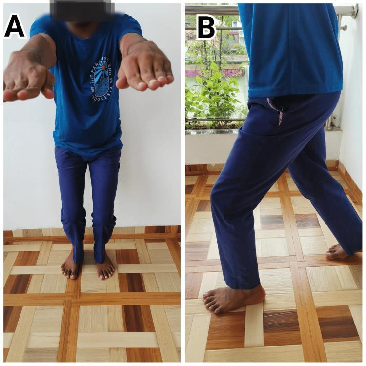
Two-panel clinical photograph demonstrating therapeutic rehabilitation exercises for a patient following Anterior Cruciate Ligament Reconstruction (ACLR). The images illustrate components of the Schlechter Protocol of Youth Sports and Orthopedics. Image A (left) shows the patient from a frontal view performing a mini squat. The patient is standing with feet shoulder-width apart, knees slightly flexed, and arms extended forward at shoulder level for balance. Image B (right) shows the patient from a lateral profile performing a forward lunge. The lead leg is flexed at the knee with the foot flat, while the trailing leg is extended posteriorly, demonstrating dynamic lower extremity stability and weight-bearing progression. These exercises are critical in post-surgical orthopedic rehabilitation to restore quadriceps strength, improve neuromuscular control, and regain joint range of motion. The educational focus is on the correct execution of closed-chain kinetic exercises used in the intermediate phases of post-operative ACL recovery.

A sequence of nine clinical photographs illustrating the biomechanical phases of a non-contact anterior cruciate ligament (ACL) injury during athletic activity. The progression captures a subject transitioning from high-velocity running and jumping into a rapid deceleration and landing phase. Key visual features include an initial flight phase with hip and knee flexion, followed by a single-leg landing and immediate transition into a deep crouched (squat) position. The images highlight critical high-risk kinematics: sudden deceleration, a shift in the center of gravity as the trunk leans forward, and stabilization maneuvers where the arms extend to maintain balance. This series demonstrates the common injury mechanism involving landing and rapid change of direction, emphasizing the postural instability and explosive forces (anterior tibial translation and rotational loads) that contribute to ACL rupture. The content is suitable for orthopedic education, sports medicine analysis, and physiotherapy training regarding injury prevention and mechanisms.
shoulder rotator cuff rehabilitation exercises phases
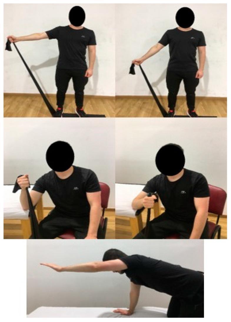
A clinical series of five photographs depicting a physical therapy Eccentric Exercise (EE) protocol for rotator cuff and shoulder rehabilitation. The images demonstrate three distinct exercises targeting specific musculature. The top row shows two frames of a standing patient performing resistance band shoulder abduction, targeting the supraspinatus; the band is anchored under the feet to provide resistance during the concentric abduction and eccentric adduction phases. The middle row consists of two frames showing a seated patient performing shoulder external rotation with a resistance band to target the infraspinatus, emphasizing the eccentric return to internal rotation. The bottom image shows the patient in a quadruped position on a treatment table, performing shoulder flexion to enhance global shoulder stability and serratus anterior activation. This visual guide illustrates clinical protocols for managing tendinopathies such as supraspinatus tendinosis through controlled mechanical loading.
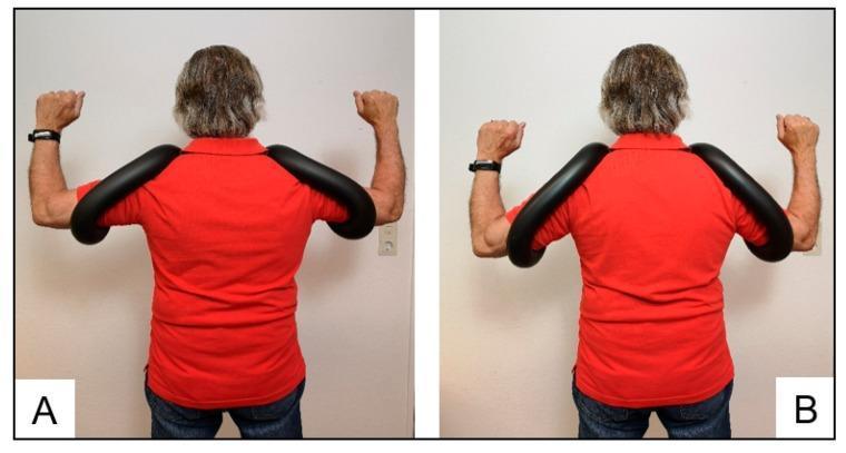
Clinical photograph (posterior view) of a patient demonstrating the use of two variations of a manual shoulder-training device, Schulterhilfe®, designed for rotator cuff rehabilitation. The images compare arm positions during therapeutic exercise. Panel A (Standard) shows the arms supported in 90-degree glenohumeral abduction with the horseshoe-shaped, rubber-covered frame positioned behind the neck. Panel B (Akut) displays the arms in a lower 45-degree abduction position, with the device frame positioned anterior to the neck, suitable for patients with limited joint flexibility or acute subacromial pain. The device is a thick, synthetic, padded tube that stabilizes the upper arms to reduce deltoid muscle activation and increase subacromial space during external rotation exercises. This setup is intended to isolate the infraspinatus and teres minor muscles while minimizing intraarticular pressure. The patient is shown in a standing position, wearing a red shirt, with the forearms flexed and fists clenched as if performing external rotation movements.
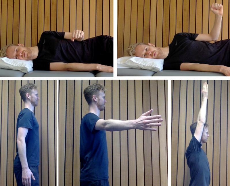
A composite clinical photograph illustrating therapeutic rehabilitation exercises for the shoulder joint. The top two panels demonstrate side-lying external rotation: the starting position shows the patient on their side with the upper elbow flexed to 90 degrees and the forearm resting across the abdomen; the ending position shows the forearm rotated upward away from the body, maintaining elbow flexion. The bottom three panels demonstrate shoulder elevation from a standing position: the sequence progresses from a neutral anatomical position (arm at side), through approximately 90 degrees of forward flexion, to full overhead elevation (approximately 150-180 degrees). These exercises are typically used in physical therapy protocols for rotator cuff strengthening, range of motion recovery, and scapular stabilization. The visual emphasizes proper form, range of motion, and body mechanics for orthopedic rehabilitation and telerehabilitation monitoring.
Post-Operative Rehabilitation: Phase-Wise Exercise Protocols
SHOULDER & KNEE - Campbell's Operative Orthopaedics, 15th Ed (2026)
PART 1 - KNEE: ACL Reconstruction Rehabilitation
STAGE I: 0 - 2 Weeks (Protection & Early Motion)
| # | Exercise |
|---|---|
| 1 | Patellar mobilisations - emphasise superior/inferior glides |
| 2 | Multicomponent brace (MCB) 0-90° |
| 3 | Quadriceps sets / Straight Leg Raises (SLR) all planes - emphasise SLR without extension lag |
| 4 | Prone / standing hamstring curls |
| 5 | Passive extension - emphasise full extension; prone hangs, pillow under heel |
| 6 | Passive, active & AAROM knee flexion - wall slides, sitting slides, prone towel pulls |
| 7 | Edema control - compression pump |
| 8 | Electrical stimulation for muscle re-education if poor quad set |
| 9 | PWB 50-75% with crutches, or WBTT without crutches if MCB locked in full extension |
| 10 | Sleep in brace locked in extension |
STAGE II: 2 - 4 Weeks (Progressive Loading)
| # | Exercise |
|---|---|
| 1 | MCB full ROM |
| 2 | Progress ROM to 120° by week 4 |
| 3 | Progress SLR and prone/standing hamstring curls with weights |
| 4 | Stationary bike for ROM; begin low-resistance program when ROM adequate |
| 5 | Stool scoots |
| 6 | FWB with crutches; discontinue when ambulating without limp |
| 7 | Double-leg BAPS (Biomechanical Ankle Platform System), progress to single leg |
| 8 | Double-leg press with light weight / high repetitions |
| 9 | Wall sits at 45° with tibia vertical, progress time |
| 10 | Lateral step-ups (4 inches) when able to do single-leg quarter squat |
| 11 | Hip machine and hamstring machine when able to SLR with 10 lb |
| 12 | Treadmill (forward and backward) with emphasis on normal gait |
| 13 | Knee extension 90-60° (submaximal) with manual resistance by therapist |
STAGE II: 4 - 6 Weeks (Closed-Chain Progression)
| # | Exercise |
|---|---|
| 1 | Progress to full ROM by 6 weeks |
| 2 | Isokinetic hamstring progression (Kin-Com dynamometer - isotonic/isokinetic) |
| 3 | Kin-Com dynamometer quad work 90-40° isotonics with anti-shear pad |
| 4 | Stairmaster (forward and backward) |
| 5 | Progress closed-chain exercises |
| 6 | At 6 weeks: Kin-Com quad work 90-40° isokinetics (start higher speed, work on endurance) |
| 7 | Aquatic exercises |
STAGE II: 8 - 10 Weeks (Dynamic Training Begins)
| # | Exercise |
|---|---|
| 1 | Progress all above exercises |
| 2 | Slow-form running with sport cord (forward and backward) |
| 3 | Isokinetic quad work at multiple speeds (60, 90, 120°/sec) |
| 4 | Lunges |
| 5 | At 10 weeks: Fitter board, slide board |
STAGE III: 12 - 16 Weeks (Strength & Full ROM)
| # | Exercise |
|---|---|
| 1 | Full-range isotonics on Kin-Com dynamometer (begin moving anti-shear pad distally) |
| 2 | Knee extension machine - low weight / high repetitions |
| 3 | Lateral sport cord drills (slow, controlled) |
| 4 | Kin-Com dynamometer test hamstrings; discontinue isokinetic hamstrings if ≥90% symmetry |
| 5 | Progress isokinetic quadriceps to full extension by 16 weeks |
STAGE IV: 16 - 20 Weeks (Plyometrics & Jogging)
| # | Exercise |
|---|---|
| 1 | Kin-Com test for quadriceps; retest hamstrings if necessary |
| 2 | Plyometric program: shuttle, mini-trampoline, jump rope |
| 3 | Jogging program begins |
STAGE V: 20 - 36 Weeks (Sport-Specific Training)
| # | Exercise |
|---|---|
| 1 | Agility training |
| 2 | Sport-specific drills: carioca, 45° cutting, figure-of-eight |
| 3 | Retest quad if necessary |
STAGE VI: Return to Sport (36 Weeks)
- Motion >130°
- Hamstrings >90% symmetry
- Quadriceps >85% symmetry
- Sport-specific agility testing passed
- Maintenance exercises 2-3×/week ongoing
PART 2 - SHOULDER REHABILITATION
2A. ROTATOR CUFF REPAIR - Minor Tear (<2 cm)
PHASE I - Protection & PROM (Weeks 1-6)
- Sling at all times for 3 weeks, then as needed for comfort
- Sutures removed at 7-10 days
- Pendulum exercises
- AROM wrist and hand
- HEP 2-3×/day
| Exercise | ROM Limit |
|---|---|
| PROM - Forward elevation (FL) | <90° |
| PROM - External rotation (ER) | Neutral to 20° |
| PROM - Internal rotation (IR) | <30° |
| Shoulder pulley - FL | As tolerated |
| Cane exercises supine - FL, ER | Gentle |
| Scapular mobility: protraction/retraction, elevation/depression | Full |
| E-stim / TENS for pain as needed | - |
| Exercise | ROM Goal |
|---|---|
| PROM FL | Progress to full |
| PROM ER | To 30° (wk3) → 40° (wk4) |
| Wand & overhead pulley exercises | FL progression |
| Cane exercises supine - ER | Gradual |
| Grades II-III joint mobilisations | Capsular mobility |
| Wall walks - FL and scaption | Progressive |
| Submaximal (25%) isometrics - ER, IR, FL, ABD at side | No pain |
- PROM ≥150° FL | PROM ≥65° ER | PROM ≥160° scaption
- Able to comb hair (dominant arm) | Open/close car door
- Lifting ≤1 lb
PHASE II - Progressive AROM & Strengthening (Weeks 7-10)
| Exercise | Details |
|---|---|
| Progress AAROM/AROM | Full-range progression |
| Isotonics (light wt / high rep): | |
| - Biceps curls, triceps extension | Standing |
| - Shoulder shrugs | Standing |
| - Supine scapular protraction | Supine |
| - Reverse Codman | Supine |
| UBE (Upper Body Ergometer) | Minimal resistance |
| Supine manual resistance PNF patterns | D1/D2 diagonals |
| Functional activities: clothespin, cupboard placing | ADL-based |
| Exercise | Details |
|---|---|
| Isotonics (light wt, high rep): | |
| Standing - FL & ABD to 90°, ER & IR with tubing | |
| Prone - rows, horizontal ABD 90° & 120°, extension | |
| Side-lying - IR & ER with towel roll under axilla | |
| Increased resistance with UBE | Progressive |
| Wall push-ups with a plus → progress to table → chair → bench → floor | Closed chain |
| Weighted PNF patterns D1 and D2 |
PHASE III - Advanced Strengthening & Return to Sport (Weeks 11-16)
| Exercise | Details |
|---|---|
| Progress all isotonic exercises | |
| Bench press (light weight, short range) | Wk 12 |
| Lateral pull-downs to chest | Wk 12 |
| Incline chest press | Wk 12 |
| Short arc, high TheraBand ER & IR at 90° ABD | Wk 12 |
| Plyometrics (from 3 months): chest pass, Plyoball chop toss, overhead throw | Wk 11+ |
| Return to throwing/racquet sports at 3 months | If normal strength, normal GH mechanics, no pain |
| Isokinetic evaluation if necessary | Wk 16 |
2B. ROTATOR CUFF REPAIR - Moderate Tear (2-5 cm)
Key Differences vs Minor Tear:
| Phase | Minor Tear (<2 cm) | Moderate Tear (2-5 cm) |
|---|---|---|
| Phase I duration | 1-6 weeks | 1-6 weeks (same) |
| Sling duration | 3 weeks | 3-6 weeks (longer) |
| AROM begins | Wk 5-6 | Wk 7 (delayed) |
| PROM IR aggressive | Avoid throughout Phase I | Same |
| Lifting restriction | ≤1 lb (Phase I) | ≤1 lb (Wks 1-6) |
| Plyometrics | 3 months | 4 months (delayed) |
| Throwing/racquet sports | 3 months | 4 months |
- Wean from sling
- Initiate true ABD stretch and IR without overpressure
- AROM <90° (at 6 weeks)
- Supine TheraBand ER stretch (as needed)
- Cane exercises supine - FL, scaption, ER neutral, ER in 90° scaption
2C. BANKART REPAIR (Labral/Instability Repair)
PREOPERATIVE GOALS:
- Independent with post-op exercise program
- Independent with preoperative strengthening (isometrics + isotonics in pain-free, stable range)
PHASE I - Weeks 1-2
| Exercise | Notes |
|---|---|
| Pendulum exercises | Gravity-assisted ROM |
| Elbow, forearm, wrist AROM | Distal mobility |
| Wrist isotonics + grip exercises | Maintain hand strength |
| Sling at all times | Immobilisation |
PHASE I - Weeks 3-4 (PT 4×/week begins ~day 15)
| Exercise | ROM Limit |
|---|---|
| PROM - FL | <160° |
| PROM - Scaption | <150° |
| PROM - ER neutral | 30° (wk3) → 40° (wk4) |
| PROM - IR in 45° scaption | <60° |
| Gentle AAROM with cane - FL, ER | As above |
| Table slides in FL | Progress ROM |
| Scapular mobility: protraction/retraction, elevation/depression | Full |
PHASE I - Weeks 5-6 (PT 4×-3×/week)
| Exercise | ROM |
|---|---|
| PROM - FL | <170° |
| PROM - Scaption | <160° |
| PROM - ER in 45° scaption | <60° |
| PROM - IR in 45° scaption | To 60° |
| AAROM - cane, pulley, wall walks | FL |
| Submaximal (25%) isometrics at side - IR, ER, ABD | No loading |
| Submaximal manual resistance scapular protraction/retraction + elevation/depression | |
| AROM - prone extension + rows, supine protraction, reverse Codman | Light |
PHASE II - Weeks 7-8 (Goals by 8 weeks)
| Exercise |
|---|
| Progress AAROM/AROM |
| Isotonics (light wt/high rep): biceps, triceps, shrugs, scapular protraction, reverse Codman |
| UBE with minimal resistance |
| Supine manual resistance PNF D1/D2 |
| Functional ADL-based tasks |
PHASE II - Weeks 9-12 (PT 1×/2-3 weeks)
| Exercise |
|---|
| Isotonics: standing FL+ABD to 90°, ER+IR with tubing |
| Prone: rows, horizontal ABD 90° & 120°, extension |
| Side-lying: IR & ER with towel roll under axilla |
| Increased resistance UBE |
| Wall push-ups with a plus → progressively lower surfaces |
| Weighted PNF D1/D2 |
| Functional exercises - throwing lunges, standing PNF |
PHASE III - Weeks 11-16 (Sport Return)
| Exercise | Timeline |
|---|---|
| Progress isotonics | Ongoing |
| Bench press (light, short arc) | Wk 12 |
| Lateral pull-downs to chest | Wk 12 |
| Incline chest press | Wk 12 |
| TheraBand ER+IR at 90° ABD | Wk 12 |
| Plyometrics - chest pass, Plyoball chop toss, overhead throw | 4 months |
| Return to throwing/racquet sports | 4 months (normal strength + GH mechanics + no pain) |
2D. Shoulder Arthroplasty (TSA/RSA) Protocol
| Phase | Timeline | Exercises |
|---|---|---|
| Phase I | POD 1 - 6 weeks | AAROM/PROM only: forward elevation in scapular plane to 90°, IR to chest, ER 0-20°, pendulum exercises 5×/day, AAROM elbow/wrist/hand |
| Phase II | 6-12 weeks | Continue AAROM/PROM: full FL, ER to 30°; wand and overhead pulley; isometric strengthening for flexion, extension, ER, ABD in neutral |
| Phase III | 12 weeks+ | AROM + dynamic strengthening: TheraBand progressions; progress to home program |
Summary Comparison: Phase Progression at a Glance
| Phase | Knee (ACL) | Shoulder (RC Minor) |
|---|---|---|
| Phase I (0-2 wks) | Quad sets, SLR, PROM, edema control | Pendulum, wrist AROM, sling |
| Phase II (2-6 wks) | Bike, wall sits, leg press, BAPS, treadmill | PROM progression, scapular exercises, submaximal isometrics |
| Phase III (6-12 wks) | Isokinetics, closed-chain, aquatic, running | Isotonics (light), UBE, PNF patterns, wall push-ups |
| Phase IV (12-20 wks) | Full isotonics, plyometrics, jogging | Advanced isotonics, bench press, lateral pull-downs |
| Phase V (20-36 wks) | Sport-specific agility drills, cutting, carioca | Plyometrics, throwing, racquet sports |
| RTS Clearance | Quad >85%, Ham >90%, ROM >130°, agility pass | MMT 5/5 all planes, normal GH mechanics, no pain |
give what to do to reduce pain instantly in tfcc
triangular fibrocartilage complex TFCC pain management treatment
TFCC conservative treatment injection corticosteroid wrist
PMID: 39418544
PMID: 41416321
TFCC injury immediate pain relief treatment immobilization injection taping 2024 2025
https://pmc.ncbi.nlm.nih.gov/articles/PMC10921179
Immediate & Conservative Pain Relief in TFCC Injury
What is TFCC Pain?
- Forearm rotation (pronation/supination)
- Gripping and weight bearing through the wrist
- Ulnar deviation
- Twisting motions (racquet sports, gymnastics, golf, gymnastics, combat sports)
STEP 1: Immediate (First 48-72 Hours) - PRICE Protocol
1. Protection - Immobilisation (Most Important)
- Apply a short-arm sugar-tong splint or Muenster cast (forearm in neutral rotation, wrist in neutral)
- The forearm must be locked in neutral rotation - this is key because rotation is the primary pain generator
- Duration: 2-4 weeks for acute injuries
- Why neutral rotation? It unloads the TFCC and allows the vascular peripheral zone to begin healing
- Avoid: ulnar deviation, pronation, supination while immobilised
2. Rest & Activity Modification
- Complete stop of sport activity for 3-6 weeks depending on severity
- Stop all activities involving axial wrist loading: pushups, gymnastics, weightlifting, throwing, racquet sports, gym, yoga
- For daily life: avoid gripping, twisting jar lids, turning keys, lifting with pronated forearm
3. Ice (Cryotherapy)
- Apply ice pack wrapped in cloth to the ulnar wrist for 15-20 minutes
- Repeat every 2 hours in the first 48-72 hours
- Reduces acute inflammatory oedema and provides immediate analgesic effect
- Do NOT apply directly to skin (risk of ice burn)
4. Compression
- Elastic bandage or compression wrist wrap over the ulnar side
- Reduces swelling and provides proprioceptive joint support
- Kinesio taping (see below) also serves this purpose
5. Elevation
- Keep the wrist elevated above heart level in the first 24-48 hours to reduce oedema
STEP 2: Pharmacological Pain Relief
NSAIDs - First-Line Analgesic
| Route | Options | Notes |
|---|---|---|
| Oral | Ibuprofen 400-600 mg TDS with food; Naproxen 500 mg BD | Take for 5-7 days; avoid if GI issues |
| Topical | Diclofenac gel / Ibuprofen gel applied over ulnar wrist | Fewer systemic side effects; apply 3-4×/day |
- NSAIDs reduce prostaglandin-mediated inflammation and provide pain relief within 1-2 hours of first dose
- Topical NSAIDs are especially useful for localised TFCC pain with fewer GI side effects
Analgesics
- Paracetamol/Acetaminophen 500-1000 mg every 6-8 hours as background analgesia
- Can be combined with NSAIDs for additive effect
- Avoid opioids - not indicated for this soft tissue injury
STEP 3: Kinesio Taping (Immediate Functional Support + Pain Relief)
Technique for TFCC Taping:
- Position: Wrist in neutral rotation, slight radial deviation
- Anchor tape on dorsal mid-forearm
- Apply "I" strip along the ulnar border of the wrist with ~50% tension over the TFCC region
- Add a "fan" or "Y" strip around the ulnar styloid for decompression
- Finish with a stabilising strip over the DRUJ for rotational support
- Lifts skin microscopically - reduces pressure on sub-dermal nociceptors
- Provides proprioceptive feedback to reduce neuromuscular guarding
- Supports DRUJ stability without full immobilisation - useful for athletes who need to continue playing
- Can be combined with a rigid splint for maximum protection
STEP 4: Corticosteroid Injection (If Acute Inflammatory Pain is Severe)
| Detail | Recommendation |
|---|---|
| Steroid | Betamethasone or Methylprednisolone (preferred - Bell et al., Cureus, 2025 - PMID: 41416321) |
| Guidance | Ultrasound-guided preferred for accuracy |
| Target site | DRUJ joint space or distal ulnocarpal compartment |
| Frequency | Maximum 4 injections/year, minimum 3 months between injections |
| Effect | Reduces acute inflammatory pain within 24-48 hours |
| Caution | Risk of cartilage damage, skin depigmentation, infection if repeated |
STEP 5: Physical Therapy / Occupational Therapy (Week 2 Onwards)
Pain Modalities (Passive PT):
| Modality | Effect |
|---|---|
| Ice/Heat alternation | Analgesic + vasodilatory |
| Therapeutic ultrasound | Deep tissue heating, promotes healing in peripheral (vascular) TFCC zone |
| TENS / Electrical stimulation | Gate control pain relief |
| Soft tissue massage | Reduces muscle guarding around DRUJ |
| Joint mobilisation (Grade I-II) | Reduces pain inhibition |
Active Exercises (Pain-Free Range Only):
- Gentle finger and elbow AROM to prevent disuse stiffness
- Wrist flexion/extension in neutral forearm rotation (not into ulnar deviation)
- Avoid: forearm rotation exercises until pain-free
Summary: Instant Pain Relief Priority Order
IMMEDIATE (0–48 hours)
   ↓
1. IMMOBILISE in neutral rotation (sugar-tong splint)
2. ICE 15-20 min every 2 hours
3. NSAID oral (ibuprofen) + topical (diclofenac gel)
4. KINESIO TAPE for support + proprioception
5. ELEVATE & COMPRESS the wrist
   ↓
DAYS 3–14
   ↓
6. Continue splint 2–4 weeks
7. Paracetamol + NSAIDs scheduled
8. Consider CORTICOSTEROID INJECTION if pain severe (US-guided)
   ↓
WEEKS 2–6
   ↓
9. Physical therapy: modalities + gentle AROM
10. Activity modification + OT guidance
What to Absolutely AVOID (These Worsen TFCC Pain Immediately)
| Action | Why It Hurts |
|---|---|
| Forearm rotation (pronation/supination) | Directly loads and shears TFCC |
| Push-ups / weight bearing through wrist | Axial compression on TFCC |
| Gripping with forearm pronated | Maximum TFCC stress position |
| Wrist ulnar deviation under load | Compresses ulnocarpal joint on TFCC |
| Continuing sport without bracing | Repetitive mechanical damage, prevents healing |
| Forced stretching into rotation | Can extend a partial tear into a complete tear |
Quick Clinical Note: Palmer Classification & Treatment Relevance
| Type | Description | Conservative vs Surgery |
|---|---|---|
| 1A (most common) | Central avascular disc tear | Conservative first (no blood supply = poor healing) |
| 1B | Peripheral ulnar avulsion (vascular zone) | Conservative first; surgery if fails (good healing potential) |
| 1C | Ulnocarpal ligament avulsion | Cast/surgery depending on instability |
| 1D | Radial attachment tear | Usually surgical |
| 2A-2E | Degenerative | Conservative first; ulnar shortening if severe |
- Type 1B peripheral tears respond best to conservative management (and have the best surgical healing outcomes too) - reviewed in PMC10921179
- Central 1A tears are avascular - conservative management reduces pain but the tear does not biologically heal; debridement may eventually be needed
modalities for this
TFCC wrist physiotherapy modalities ultrasound electrotherapy laser
PMID: 37490953
PMID: 33927808
physiotherapy modalities TFCC wrist pain ultrasound TENS laser IFT manual therapy evidence 2024
https://www.journalijar.com/uploads/2025/08/68ad7a4c52912_IJ…
shockwave therapy wrist TFCC fibrocartilage pain
PRP platelet rich plasma TFCC wrist cartilage injection
wrist physiotherapy ultrasound TENS electrotherapy treatment
PMID: 39391675
Physiotherapy Modalities for TFCC Injury
Classification of Modalities
| Category | Modalities |
|---|---|
| Thermal | Cryotherapy, Moist heat, Contrast bath, Paraffin wax |
| Electrotherapy | TENS, IFT (Interferential therapy), NMES, Galvanic stimulation |
| Mechanical | Therapeutic ultrasound, Extracorporeal Shockwave Therapy (ESWT) |
| Electromagnetic / Photobiomodulation | Low-Level Laser Therapy (LLLT), PEMF |
| Manual | Joint mobilisation, Soft tissue techniques, Taping |
| Regenerative Injections | Corticosteroid, Hyaluronic acid, PRP |
1. CRYOTHERAPY (Ice / Cold Therapy)
- Vasoconstriction → reduces haematoma and oedema
- Decreases nerve conduction velocity (slows pain signal transmission) → gate control analgesia
- Reduces local metabolic rate → limits secondary cell death (cryoprotection)
- Decreases muscle spasm around the DRUJ
- Ice pack wrapped in damp cloth directly over the ulnar styloid/TFCC region
- 15-20 minutes per session
- Every 2 hours in first 48-72 hours
- Then 2-3 times/day as needed
2. THERMOTHERAPY (Heat)
| Type | Method | Use |
|---|---|---|
| Moist hot pack | Hydrocollator pack 10-15 min | Chronic stiffness, before exercise |
| Paraffin wax bath | 52-54°C, dip method, 3-4 layers | Chronic TFCC + stiff wrist joints |
| Contrast bath | Alternate hot (40°C) 3 min / cold (15°C) 1 min × 4 cycles | Subacute, reduces oedema + improves circulation |
- Vasodilation → increases blood flow and tissue oxygen delivery
- Increases collagen extensibility → improves ROM before stretching exercises
- Reduces pain via thermal pain-gate modulation
3. THERAPEUTIC ULTRASOUND (TUS)
- Thermal effects: Deep tissue heating (up to 5 cm depth), increases collagen extensibility, accelerates enzyme activity, promotes vasodilation
- Non-thermal effects: Cavitation and acoustic streaming - increases cell membrane permeability, promotes protein synthesis, stimulates fibroblast activity and collagen production
- Promotes remyelination, suppresses pro-inflammatory gene expression
- Critical for peripheral (vascular) TFCC tears where healing is biologically possible
| Parameter | Acute/Sub-acute | Chronic |
|---|---|---|
| Frequency | 3 MHz (superficial) | 1 MHz (deeper penetration) |
| Mode | Pulsed (1:4 ratio - 20% duty cycle) | Continuous |
| Intensity | 0.5 - 1.0 W/cm² | 1.0 - 2.0 W/cm² |
| Duration | 5 min | 7-10 min |
| Area | Ulnar fovea / DRUJ region | Same |
| Coupling | Ultrasound gel or underwater (water immersion for small wrist area) | |
| Frequency of sessions | Daily for 7-10 days | 3×/week for 3-4 weeks |
4. TENS (Transcutaneous Electrical Nerve Stimulation)
- Conventional/High-frequency TENS (80-100 Hz): Activates Aβ sensory fibres → gates pain at dorsal horn (spinal gate control) → fast-onset analgesia
- Acupuncture/Low-frequency TENS (2-4 Hz): Stimulates Aδ and C-fibres → triggers endorphin and enkephalin release → longer-lasting analgesia
- Reduces central sensitisation
| Parameter | Value |
|---|---|
| Mode | Conventional TENS (acute pain) / Burst-mode TENS (chronic) |
| Frequency | 80-100 Hz (conventional) or 2-4 Hz (burst/acupuncture) |
| Pulse width | 50-100 μs |
| Intensity | Strong but comfortable tingling; sub-motor threshold |
| Electrode placement | One pad over ulnar styloid (pain site), one on dorsal forearm 5 cm proximal; or bilateral pad placement flanking the DRUJ |
| Duration | 20-30 min per session |
| Frequency | Twice daily for acute pain; once daily for chronic |
5. INTERFERENTIAL THERAPY (IFT)
- Two medium-frequency currents (4000 Hz and 4100 Hz) cross at the target tissue producing a beat frequency of 100 Hz in the deep tissues
- Penetrates deeper than TENS with less skin resistance
- Reduces oedema through muscle pumping effect (at low beat frequencies)
- Analgesia at 80-150 Hz beat frequency; muscle stimulation and oedema reduction at 20-50 Hz
| Parameter | Value |
|---|---|
| Beat frequency | 80-120 Hz (analgesia) |
| Electrode placement | 4-pole coplanar or cross-fiber over ulnar wrist |
| Duration | 15-20 min |
| Sessions | 3-5×/week for 2-3 weeks |
6. LOW-LEVEL LASER THERAPY (LLLT) / Photobiomodulation
- Photons absorbed by mitochondrial cytochrome c oxidase → increases ATP production
- Stimulates: macrophage activation, mast cell degranulation, fibroblast activation, angiogenesis, increased phagocytic activity, photodissociation of oxyhemoglobin
- Anti-inflammatory: reduces prostaglandin E2, IL-1β, TNF-α
- Promotes collagen and extracellular matrix synthesis - important for fibrocartilage repair
- Analgesic: modulates nerve conduction and reduces nociceptor sensitivity
| Parameter | Value |
|---|---|
| Wavelength | 830 nm (infrared - deeper penetration) or 650 nm (red) |
| Power density | 50-100 mW/cm² |
| Energy density | 1-4 J/cm² per point |
| Application | Point-by-point over TFCC / ulnar fovea; contact method |
| Number of points | 3-5 points around ulnar wrist |
| Duration | 10-15 min total |
| Sessions | 3×/week for 3-4 weeks |
7. EXTRACORPOREAL SHOCKWAVE THERAPY (ESWT)
- Acoustic pressure waves stimulate neovascularisation (VEGF upregulation) - critical because central TFCC is avascular and normally cannot heal
- Promotes extracellular matrix remodelling
- Anti-inflammatory: enhances nitric oxide signalling, modulates nociceptive pathways
- Activates stem cells and growth factors in fibrocartilage tissue
| Parameter | Value |
|---|---|
| Type | Focused ESWT |
| Energy flux density | 0.25 mJ/mm² |
| Impulses per session | 1,500 shockwaves |
| Frequency | 4 Hz |
| Target | Ulnar fovea (TFCC area), patient seated forearm in neutral rotation |
| Coupling medium | Ultrasound gel |
| Sessions | 4 sessions, 1× per week for 4 weeks |
| Adjunct | Night-time ulnar gutter splint + ice 20 min twice daily |
8. PULSED ELECTROMAGNETIC FIELD THERAPY (PEMF)
- Electromagnetic fields stimulate chondrocyte and fibroblast activity
- Promotes calcium ion transport and collagen synthesis
- Anti-inflammatory and analgesic effects
- Used in cartilage / fibrocartilage regeneration protocols
- 10-20 Gauss, pulsed at 5-20 Hz
- 20-30 min/session, daily for 3-4 weeks
- Applied directly over the DRUJ
9. MANUAL THERAPY MODALITIES
a. DRUJ Mobilisation (Maitland/Mulligan)
- Grade I-II: Oscillatory movements within resistance-free range → reduces pain inhibition
- Grade III-IV: Stretches into resistance → improves ROM
- Mulligan DRUJ mobilisation with movement (MWM): Passive DRUJ glide (dorsal-volar) with active forearm rotation → immediate pain reduction and ROM improvement
- Specific technique: stabilise radius, glide ulnar head dorsally/volarly while patient rotates forearm
b. Soft Tissue Massage / Myofascial Release
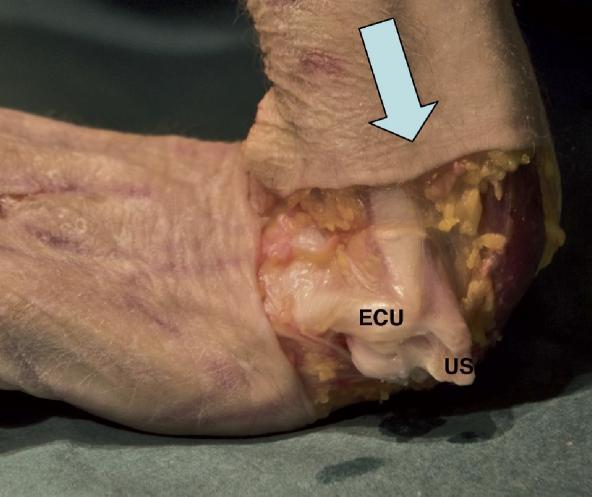
- Deep tissue massage to extensor carpi ulnaris (ECU), flexor carpi ulnaris (FCU), and pronator quadratus
- These muscles guard the DRUJ and generate compressive pain when in spasm
- Scar mobilisation post-operatively (surgical scar on ulnar wrist adheres to deeper tissues early)
c. Neural Mobilisation
- Ulnar nerve neurodynamic mobilisation (nerve slider/tensioner technique)
- The dorsal sensory branch of the ulnar nerve is commonly irritated in TFCC injury (runs adjacent to TFCC)
- Reduces neural mechanosensitivity and associated burning/tingling pain
10. PRP (Platelet-Rich Plasma) Injection
- Concentrated autologous platelets release growth factors: PDGF, TGF-β, VEGF, EGF, IGF-1
- Promotes fibrocartilage cell proliferation and extracellular matrix synthesis
- Stimulates neovascularisation in the avascular central disc zone
- Anti-inflammatory cytokine modulation
- 3-5 mL autologous PRP prepared from 15-30 mL venous blood (centrifuged)
- Injected under ultrasound guidance into the DRUJ / TFCC region
- 1-3 injections at 4-6 week intervals
Phase-Wise Modality Prescription Summary
| Phase | Timing | Primary Modalities | Goal |
|---|---|---|---|
| Acute | Day 0-3 | Cryotherapy, TENS, Immobilisation/splinting | Pain relief, oedema control |
| Sub-acute | Day 4 - Week 3 | Pulsed TUS (3 MHz), IFT, TENS, Contrast bath, Manual therapy (Grade I-II) | Reduce inflammation, begin tissue healing |
| Chronic/Repair | Week 3-8 | Continuous TUS (1 MHz), LLLT, PEMF, ESWT (if recalcitrant), Paraffin wax | Tissue repair, collagen remodelling, ROM |
| Functional | Week 6 onward | Manual therapy (Grade III-IV), Proprioceptive training, Strengthening, LLLT | Restore strength, DRUJ stability, return to sport |
Contraindications to Note
| Modality | Contraindication |
|---|---|
| Heat / TUS | Acute phase (<48 hrs), active infection |
| ESWT | Acute TFCC, active inflammation, coagulopathy, pregnancy, over metal implants |
| LLLT | Over malignancy, over eyes, pregnancy |
| TENS/IFT | Pacemaker, over metal implants, over open wounds |
| PRP | Anticoagulant therapy, thrombocytopenia, active infection |