
Pancreatitis
Reading File
Pancreatitis is inflammation of the pancreas, classified as acute or chronic, with causes ranging from gallstones and alcohol to metabolic and autoimmune conditions. Key features include epigastric pain radiating to the back, elevated amylase/lipase, and systemic complications.
Searching sources...
Finding Sources
Searching PubMed
"pancreatitis"[MeSH Terms] AND "management"[MeSH Terms]
Reading File
Finding Sources
Reading File
Finding Sources
Finding Sources
Finding Sources
Reading File
Finding Sources
Reading File
Reading File
Reading File
Reading File
Reading File
Reading File
Reading File
Finding Sources
Reading File
Reading File
I now have comprehensive data from multiple authoritative sources. Let me compile the full answer.
Loading Image
Here is a comprehensive overview of pancreatitis drawn from multiple authoritative medical textbooks.
Pancreatitis
Anatomy Overview
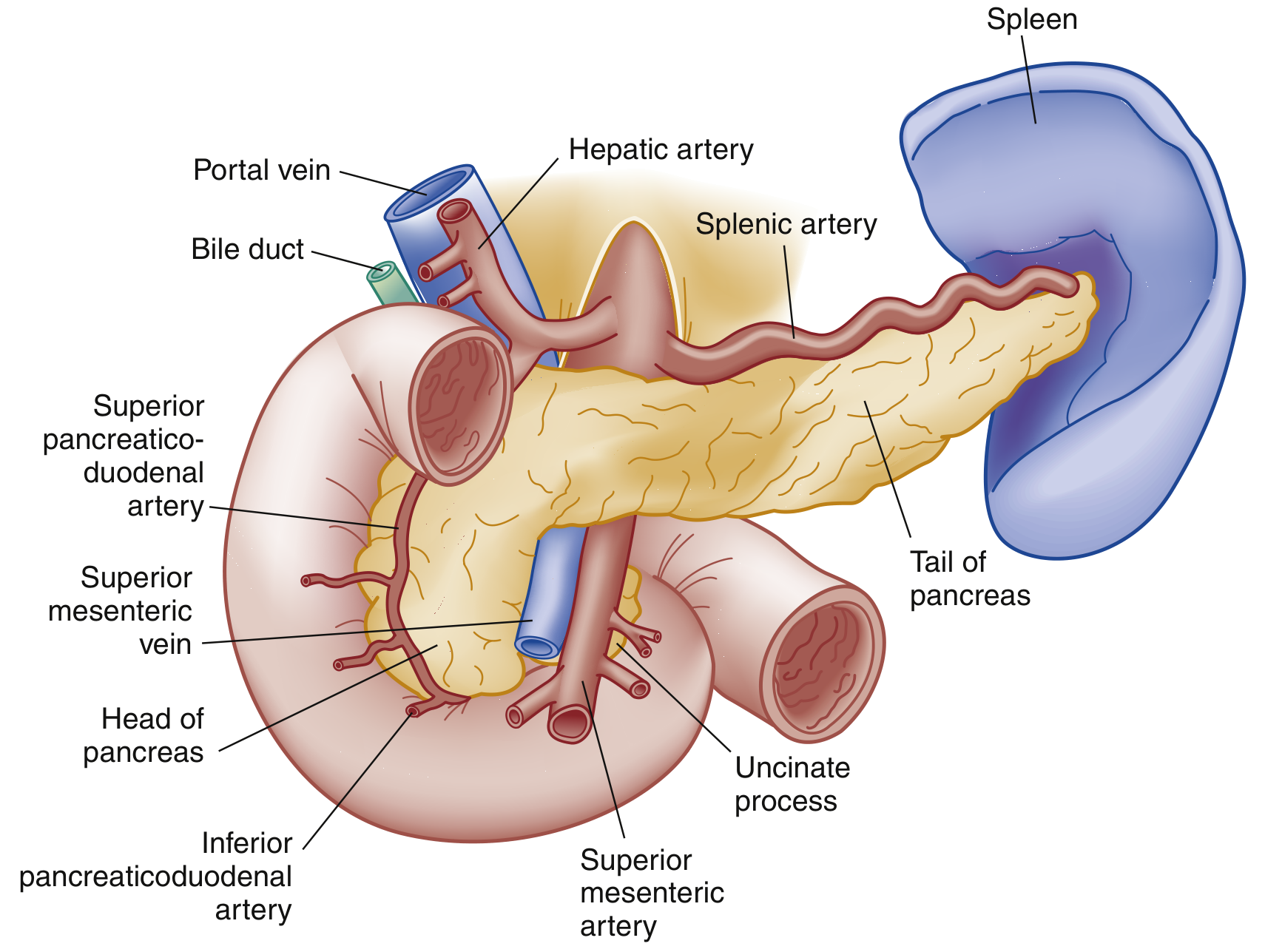
The pancreas is a retroperitoneal organ with three segments: head (nestled in the C-loop of the duodenum), body (posterior to the stomach), and tail (abutting the splenic hilum). The main pancreatic duct (duct of Wirsung) joins the common bile duct at the ampulla of Vater, draining through the sphincter of Oddi into the duodenum. Exocrine function includes secretion of digestive enzymes (trypsinogen, lipase); endocrine function includes insulin, glucagon, and somatostatin.
Pathophysiology
Injury begins with an inciting event - duct obstruction by a gallstone, alcohol, or another toxin. Cellular injury disrupts normal membrane trafficking, triggering inappropriate activation of trypsinogen and increased trypsin production, which in turn activates other digestive enzymes. Autodigestion and activation of the inflammatory cascade (macrophages, neutrophils) lead to further tissue destruction. Cytokine release causes vascular permeability changes - resulting in edema, hemorrhage, and necrosis. When the autoimmune response becomes heightened, SIRS, sepsis, and shock can develop. Bacterial translocation from intestinal flora can cause bacteremia. Extrapancreatic complications include ARDS and acute kidney injury. - ROSEN's Emergency Medicine, p. 1265
Etiology
Gallstones (40-70%) and chronic alcohol use (25-35%) account for the majority of cases. Other important causes include:
Metabolic/Toxic
- Hypertriglyceridemia (TG > 1000 mg/dL)
- Hypercalcemia
- Uremia
- Scorpion venom
Mechanical/Obstructive
- Post-ERCP (iatrogenic)
- Pancreas divisum, annular pancreas
- Ampullary or pancreatic tumors
- Duodenal diverticulum
- Trauma
Infectious
- Viral: mumps, coxsackievirus, HIV, CMV, EBV, varicella
- Bacterial: TB, Salmonella, Mycoplasma, Legionella
- Parasitic: Ascaris
Vascular
- Vasculitis, embolism, hypoperfusion/ischemia, hypercoagulability
Other
-
Medications (valproate, L-asparaginase, 6-mercaptopurine, prednisone in children)
-
Hereditary/genetic (SPINK1, CFTR mutations)
-
Autoimmune
-
Idiopathic
-
Current Surgical Therapy 14e, p. 604 | ROSEN's Emergency Medicine, p. 1267
Acute Pancreatitis
Diagnosis
Acute pancreatitis is diagnosed by 2 of 3 criteria:
- Abdominal pain characteristic of acute pancreatitis
- Serum lipase or amylase >3× the upper limit of normal
- Characteristic findings on abdominal imaging
Lipase is preferred over amylase - it is more sensitive and specific. Lipase peaks quickly and stays elevated for ~1-2 weeks; amylase stays elevated only 3-5 days. Importantly, amylase may be falsely normal in alcohol- and hypertriglyceridemia-induced pancreatitis. Neither enzyme elevation correlates with severity.
ALT is particularly specific for biliary pancreatitis (positive predictive value ~95%). Triglyceride and calcium levels help identify metabolic causes. A CBC and BMP assess for SIRS and organ failure. - ROSEN's Emergency Medicine, p. 1267
Imaging:
- Abdominal ultrasound is the first-line study to evaluate for biliary etiology (gallstones, common bile duct dilatation).
- CT with IV contrast is NOT routinely required for diagnosis. It is indicated when: (1) diagnosis is uncertain, (2) alternative serious pathology needs exclusion, or (3) complications are suspected after 48-72 hours of non-response to therapy. Contrast CT identifies necrosis as non-enhancing parenchyma, fluid collections, and vascular complications.
- MRI/MRCP is useful for detecting stones, pancreatic duct disruption, or necrosis without radiation - preferred in renal impairment.
2012 Revised Atlanta Severity Classification
| Grade | Organ Failure | Local/Systemic Complications | Mortality |
|---|---|---|---|
| Mild | Absent | Absent | Very rare (<5%) |
| Moderately Severe | Transient (<48 hrs) | Present, without persistent OF | Low |
| Severe | Persistent (>48 hrs) | Present | High (36-50%); extremely high with infected necrosis |
Other scoring systems (Ranson criteria, APACHE II, BISAP, CTSI) are validated for severity prediction and risk stratification; each has different strengths but similar overall predictive accuracy. - Current Surgical Therapy 14e, p. 604
Local Complications
| Complication | Definition | Timing |
|---|---|---|
| Interstitial edematous pancreatitis | Diffuse pancreatic edema without necrosis | <4 weeks |
| Acute peripancreatic fluid collection (APFC) | Non-encapsulated homogeneous peripancreatic fluid, no necrosis | <4 weeks |
| Pancreatic pseudocyst | Encapsulated fluid, well-defined wall, minimal/no necrosis | >4 weeks |
| Necrotizing pancreatitis | Necrosis of parenchyma and/or peripancreatic tissue | <4 weeks |
| Acute necrotic collection (ANC) | Fluid + necrosis, non-encapsulated | <4 weeks |
| Walled-off necrosis (WON) | Mature, encapsulated necrotic collection | >4 weeks |
Most APFCs and pseudocysts resolve spontaneously. Infected necrosis is recognized on CT by rim-enhancement and gas within the collection. Other local complications include hemorrhage, gastric outlet obstruction, splenic/portal vein thrombosis, pseudoaneurysm, and colonic infarction.
Systemic complications include acute kidney injury, ARDS, septic shock, and abdominal compartment syndrome. - Current Surgical Therapy 14e, p. 605
Management of Acute Pancreatitis
Fluid Resuscitation
Early and intensive fluid resuscitation is the cornerstone of treatment. Lactated Ringer's (LR) is preferred over normal saline - it is more physiologic and has anti-inflammatory effects (reduces risk of SIRS). Initial goal-directed therapy: 5-10 mL/kg/h for the first 2 hours, then 1.5-3 mL/kg/h for 12-24 hours, targeting 2-4 L over 24 hours. Goals include heart rate normalization, MAP ≥ 65 mmHg, and urine output. Severe cases require ICU monitoring with CVP targets of 8-12 mmHg. Over-resuscitation should be avoided (can cause ARDS, abdominal compartment syndrome). - Current Surgical Therapy 14e, p. 606
Pain Management
No single analgesic is superior. Acetaminophen 1 g every 6 hours is recommended for initial control. Opioids can be used but risk ileus. Low-dose ketamine (0.1-0.3 mg/kg IV) is an opioid-sparing alternative. NSAIDs should be avoided in critically ill patients (risk of AKI). Antiemetics for symptom relief.
Nutrition
- Early oral feeding (<24 hours) when tolerated is now recommended over keeping patients NPO
- If oral is not tolerated: nasogastric or nasojejunal tube feeds are safe
- Gastric and jejunal feeding have similar outcomes in severe AP
- Early enteral nutrition (EN) significantly reduces infectious complications, organ failure, surgical interventions, and mortality vs. parenteral nutrition (PN)
- Two meta-analyses showed EN results in a 2-fold reduction in pancreatic infectious complications and a 2.5-fold reduction in mortality vs. PN
- For ICU patients or those with severe SIRS on mechanical ventilation: initiate NG/NJ feeds within 24-36 hours
- Sleisenger & Fordtran's GI and Liver Disease, p. 2935-2939
Antibiotics
- Prophylactic antibiotics are NOT indicated in acute pancreatitis, even severe cases - they have not been shown to reduce infected necrosis or improve mortality
- Antibiotics should be started only when: infected pancreatic necrosis is documented (CT findings + positive culture), or clear evidence of sepsis/infection (cholangitis, pneumonia, bacteremia)
- Carbapenems are preferred for infected necrosis (better pancreatic penetration vs. cephalosporins or fluoroquinolones)
- ERCP is indicated only for cholangitis or biliary obstruction, not routinely for gallstone pancreatitis
Interventions (Necrotizing Pancreatitis - "Step-Up Approach")
Historical open necrosectomy carried a 50% mortality rate. The modern step-up approach is now standard:
- Step 1 - Conservative management ± antibiotics to stabilize
- Step 2 - Percutaneous or endoscopic drainage of necrotic collection
- Step 3 - Minimally invasive surgical or endoscopic necrosectomy only if needed
Key timing principles:
-
Wait at least 4 weeks before any intervention to allow WON maturation, demarcation, and liquefaction - reduces hemorrhage and fistula risk
-
Surgical intervention in the first 2 weeks carries 75% mortality; at 2-4 weeks 45%; beyond 30 days ~8%
-
Urgent percutaneous drainage may be required earlier if clinical deterioration with infected necrosis
-
Emergent surgery is reserved for intestinal ischemia, perforated viscus, uncontrolled bleeding, or abdominal compartment syndrome
-
Current Surgical Therapy 14e, p. 606-607
Chronic Pancreatitis
Chronic pancreatitis is a progressive fibroinflammatory syndrome leading to permanent structural changes and impaired exocrine and endocrine function. Key features include:
- Exocrine insufficiency: malabsorption, steatorrhea, weight loss
- Endocrine insufficiency: pancreatogenic (type 3c) diabetes
- Pain: central feature, often debilitating; jejunal feeding can reduce pain and narcotic use
- Oxidative stress is implicated in pathophysiology; antioxidant supplementation (selenium, ascorbic acid, β-carotene, α-tocopherol, methionine) has shown mixed results in RCTs
Recurrent acute pancreatitis can progress to chronic pancreatitis through repeated cycles of inflammation and fibrosis. - Sleisenger & Fordtran's GI and Liver Disease | ROSEN's Emergency Medicine
Key Teaching Points Summary
| Feature | Details |
|---|---|
| Most common causes | Gallstones (40-70%), alcohol (25-35%) |
| Diagnostic criteria | 2 of 3: pain, lipase/amylase >3× ULN, imaging |
| Preferred enzyme | Lipase (more sensitive and specific than amylase) |
| First imaging | Abdominal ultrasound (biliary etiology) |
| CT timing | NOT routine; use at 48-72h if no improvement |
| Preferred IV fluid | Lactated Ringer's (not normal saline) |
| Antibiotics | Not prophylactic; only for proven infection |
| Nutrition | Early oral/enteral; EN better than PN |
| Necrosectomy timing | ≥4 weeks after symptom onset |
| Chronic CP features | Fibrosis, exocrine/endocrine insufficiency, pain |
This is a shared conversation. Sign in to Orris to start your own chat.