
Give a long on Herpes simplex virus causing vesicobullous lesion for writing in final year exam
Reading File
Now let me run the three-layer search in parallel for comprehensive coverage.
Finding Sources
Finding Sources
Finding Sources
Excellent results. Now let me read the most relevant dermatology textbook sections in parallel.
Finding Sources
Reading File
Reading File
Now let me get the main Fitzpatrick chapter on HSV and Dermatology 5e content:
Reading File
Reading File
Finding Sources
Now let me read the Andrews' section and Dermatology 5e section, and the key Fitzpatrick pathology/histology section:
Reading File
Reading File
Reading File
Excellent - I now have very rich content. Let me get the key histopathology and treatment sections from Fitzpatrick and also check for the relevant images:
Reading File
Reading File
I now have comprehensive content from multiple authoritative dermatology textbooks. Let me also fetch the key clinical images to include in the answer:
Loading Image
Loading Image
Reading File
I now have comprehensive, detailed information from multiple authoritative textbooks. Let me now compile the complete final year exam answer.
Herpes Simplex Virus (HSV) as a Cause of Vesicobullous Lesions
1. INTRODUCTION
Herpes Simplex Virus (HSV) is one of the most prevalent human DNA viral pathogens and the most common infectious cause of vesicobullous lesions on the skin and mucous membranes. It belongs to the family Herpesviridae, subfamily Alphaherpesvirinae. Two distinct serotypes exist:
- HSV-1: Primarily responsible for orofacial and labial lesions
- HSV-2: Primarily responsible for genital lesions, though either type can infect either site
HSV-1 seroprevalence is 30-60% in children, rising to >60% in adults aged 30-39 years. HSV-2 seroprevalence is ~19% in adults aged 30-39 years in the United States. Crucially, many more individuals are infected than show clinical disease - approximately 60% of HSV-2-infected persons have unrecognized clinical lesions.
(Fitzpatrick's Dermatology, 9e, Chapter 164)
2. VIROLOGY & STRUCTURE
HSV is a double-stranded DNA virus with four structural components:
| Component | Description |
|---|---|
| Core | Linear dsDNA genome (~152 kb) |
| Capsid | Icosahedral, 162 capsomeres |
| Tegument | Amorphous proteinaceous layer |
| Envelope | Lipid bilayer with glycoprotein spikes (gB, gC, gD, gE, gG, gH) |
Key glycoproteins relevant to pathogenesis:
- gB and gD: Mediate viral attachment and fusion with host cell
- gC: Binds complement component C3b (immune evasion)
- gE: Acts as IgG Fc receptor (immune evasion)
- gG: Determines type-specific serology (gG-1 for HSV-1, gG-2 for HSV-2)
3. PATHOGENESIS - HOW THE VESICLE FORMS
Understanding the pathogenesis is central to understanding why HSV produces a vesicobullous lesion.
3a. Primary Infection
- Inoculation: Virus enters through abraded skin or mucous membrane via direct contact
- Local replication: The virus replicates in keratinocytes of the epidermis and dermis
- Cytopathic effects: The infected keratinocytes undergo:
- Ballooning degeneration - cells swell due to intracellular edema
- Acantholysis - loss of intercellular adhesion
- Reticular degeneration - the cell cytoplasm collapses into a network
- Vesicle formation: Separation of necrotic keratinocytes from each other and from the basal layer creates an intraepidermal vesicle filled with serous fluid, free-floating virally-infected keratinocytes, and inflammatory cells
- Neurotropism: The virus then enters local sensory nerve terminals and ascends by retrograde axonal transport to the dorsal root ganglia or trigeminal ganglia, where it establishes latency
(Dermatology 2-Volume Set 5e; Cummings Otolaryngology)
3b. Latency
- Viral DNA persists as a circular episome within neuronal nuclei
- No viral proteins are expressed except Latency Associated Transcripts (LATs)
- The virus is "hidden" from immune surveillance
- Latency is maintained by HSV-specific CD8+ T lymphocytes that cluster around latently infected neurons
3c. Reactivation
Triggered by:
- Emotional stress, UV light (sunlight), fever, menstruation, trauma, fatigue, immunosuppression
- The virus reactivates, undergoes anterograde transport down the axon, and re-infects the skin at or near the original site, producing recurrent vesicobullous lesions
4. HISTOPATHOLOGY - THE BASIS OF VESICLE FORMATION
The histopathology of HSV vesicles is characteristic and forms the basis of Tzanck smear diagnosis:
- Intraepidermal vesicle: The vesicle is located within the epidermis (as opposed to subepidermal in bullous pemphigoid)
- Ballooning degeneration: Infected keratinocytes are greatly enlarged with pale, "ballooned" cytoplasm and marginated chromatin
- Reticular degeneration: Fluid accumulates within the cytoplasm, rupturing cell walls into a network
- Multinucleated giant cells (Tzanck cells): Fusion of adjacent infected keratinocytes under viral glycoprotein mediation produces large cells with 2-8+ nuclei arranged in a "grape-like" cluster with "nuclear molding" and ground-glass chromatin
- Cowdry Type A intranuclear inclusion bodies: Eosinophilic intranuclear inclusions surrounded by a clear halo (pathognomonic)
- Acantholysis: Loss of intercellular bridges secondary to reticular degeneration
- Dermal inflammatory infiltrate: Lymphocytes and histiocytes in the dermis
The vesicle is intraepithelial and filled with serum and free-floating, virally infected keratinocytes and inflammatory cells. (Cummings Otolaryngology, Chapter 87)
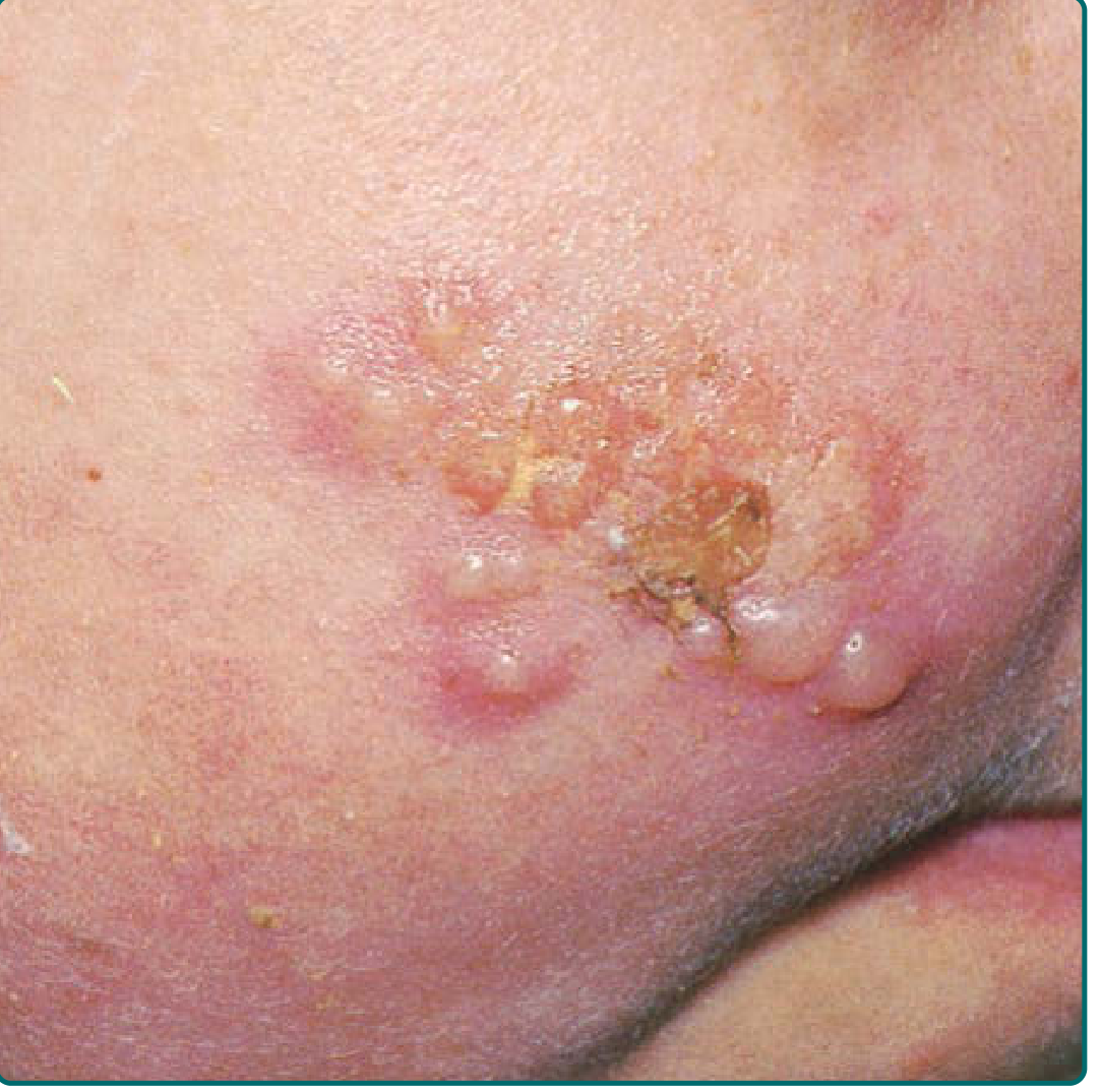
5. CLINICAL PRESENTATIONS OF HSV VESICOBULLOUS LESIONS
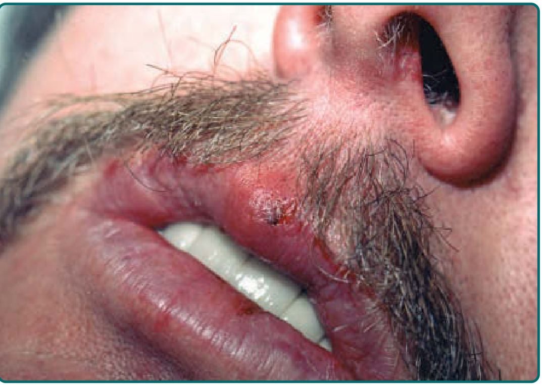
5a. Herpes Labialis (Cold Sore / Fever Blister) - Most Common
Site: Vermilion border of the lip (outer one-third of lower lip most commonly); less commonly nose, chin, cheek
Progression of lesion (the classic 5 stages):
- Prodrome: Tingling, burning, pruritus, or pain at the site (precedes lesions by 45-60% of episodes, lasting up to 48 hours)
- Erythema and papule: Area becomes erythematous; grouped papules appear
- Vesicle: Classic thin-walled, grouped vesicles on an erythematous base - this is the "vesicular stage"
- Ulcer/Erosion: Vesicles rupture to form shallow, painful ulcers/erosions
- Crust: A honey-colored crust forms; heals without scarring in 5-15 days
5b. Primary Herpetic Gingivostomatitis
- Classic primary infection presentation in children (HSV-1)
- Prodrome: Fever, malaise, myalgia, cervical lymphadenopathy
- Lesions: Clusters of small thin-walled vesicles on both keratinized (hard palate, gingiva) and non-keratinized (buccal mucosa, tongue) surfaces
- Key feature: Gingival involvement with erythematous, boggy, tender gingiva with free gingival margin involvement - a key diagnostic criterion
- Vesicles are short-lived on mucosa, rapidly rupturing to form shallow, painful ulcers
- Duration: 7-14 days; viral shedding continues for weeks after clinical resolution
5c. Genital Herpes
- HSV-2 (predominantly) or HSV-1
- Primary episode: Multiple grouped vesicles on erythematous base on the penis, vulva, vagina, cervix, or perineum; accompanied by fever, dysuria, inguinal lymphadenopathy. Vesicles rapidly ulcerate. More severe and prolonged than recurrences.
- Recurrent episodes: A prodrome of tingling/burning precedes 1-3 grouped vesicles that ulcerate and heal within 5-10 days. Recurrences are less severe than primary episodes.
5d. Herpetic Whitlow
- HSV infection of the distal finger (usually from autoinoculation via oral herpetic infections in children; occupational in healthcare workers from patients' oral secretions)
- Presents as grouped vesicles with deep pain and erythema of the fingertip
- Can mimic bacterial paronychia but is NOT fluctuant and should not be incised
(Tintinalli's Emergency Medicine)
5e. Eczema Herpeticum (Kaposi Varicelliform Eruption - KVE)
A distinct and serious complication:
- Widespread dissemination of HSV over skin with a pre-existing skin disorder (most commonly atopic dermatitis, also pemphigus, burns, Darier disease, cutaneous T-cell lymphoma)
- Mechanism: Impaired epidermal barrier + Th2 immune shift + decreased antimicrobial peptides
- Lesions: Hundreds of umbilicated vesicles arising acutely over eczematous areas, with fever and regional lymphadenopathy
- In severely immunocompromised patients (pemphigus + systemic immunosuppression), can be fatal from S. aureus septicemia or visceral dissemination
- Risk factors include: severe atopic dermatitis, high IgE, eosinophilia, age of AD onset <5 years, use of topical calcineurin inhibitors
(Andrews' Diseases of the Skin, 13e)
5f. Herpes Gladiatorum
- Grouped vesicles and erosions on the neck, face, and arms of wrestlers and rugby players (direct skin-to-skin contact)
- Caused by HSV-1
5g. Neonatal Herpes
Neonatal herpes is an emergency with three clinical forms:
- Skin, eye, mouth (SEM) disease: Vesicles on skin (often on the presenting part), eye lesions, oral vesicles - best prognosis
- Encephalitis: Seizures, lethargy, bulging fontanel - mortality 64% with treatment; only ~30% develop normally
- Disseminated disease: Multi-organ failure with hepatitis, pneumonitis, DIC; mortality 30% with treatment
- Important: >20% of neonates with neurologic and disseminated disease do NOT develop cutaneous vesicles
- Transmitted primarily from maternal genital infection at delivery; primary maternal HSV carries 25-50% transmission risk; recurrent maternal HSV <3% risk
(Fitzpatrick's Dermatology, 9e; Andrews' Diseases of the Skin)
5h. HSV in Immunocompromised Patients
As immunity wanes (HIV, transplant, malignancy):
- Frequency of recurrences increases
- Vesicles evolve into chronic, extensive, deep, painful ulcers (perianal region, genitalia, tongue are favored)
- Atypical presentations: folliculitis, verrucous plaques, hypertrophic anogenital masses simulating neoplasia
- Can disseminate to viscera (esophagitis, pneumonitis, hepatitis, encephalitis)
- Acyclovir resistance (usually due to thymidine kinase mutation) more common (~5% vs ~1% in immunocompetent)
(Dermatology 2-Volume Set 5e)
6. IMMUNOLOGY
Innate Immunity
- Keratinocytes express Toll-like receptors (TLR-2, TLR-9) that detect HSV
- TLR signaling induces Type I interferons (IFN-α/β), which inhibit viral mRNA translation and promote NK cell activation
- NK cells kill infected cells via perforin/granzyme and release IFN-γ
- Langerhans cells in the epidermis are important antigen-presenting cells
Adaptive Immunity
- CD8+ T cells are the primary defense - they kill infected cells through perforin and Fas pathways
- CD4+ T cells release IFN-γ, recruit CD8+ cells, and help B cells produce antibodies
- Humoral immunity: Antibodies reduce virus titers at inoculation sites; maternal IgG protects neonates
HSV Immune Evasion
- ICP47 protein: Binds TAP (Transporter associated with Antigen Processing), blocking MHC class I peptide loading - infected cells "hide" from CD8+ T cells
- gC: Binds complement C3b, blocking complement activation
- gE: Functions as IgG Fc receptor, blocking antibody-dependent cellular cytotoxicity
- VHS (Virion Host Shutoff) protein: Degrades host mRNA, prevents DC activation
- HSV decreases langerin expression and increases apoptosis of Langerhans cells
(Dermatology 2-Volume Set 5e, Block 18)
7. DIAGNOSIS
Clinical Diagnosis
- Characteristic grouped vesicles on an erythematous base, often with prodromal symptoms
- History of recurrence at same site is strongly suggestive
Laboratory Methods
| Test | Sensitivity/Specificity | Comment |
|---|---|---|
| Tzanck Smear | 60-90% accuracy | Rapid, bedside; scrape base of fresh vesicle, Giemsa or Wright stain; shows multinucleated giant cells; does NOT distinguish HSV from VZV; NOT sensitive for crusted/ulcerated lesions |
| Viral Culture | ~60-70% from fresh genital vesicles | Specific; best for vesicular-stage lesions; results in 48-96 hours; enables antiviral sensitivity testing |
| PCR | 4x more sensitive than culture | Gold standard and preferred method; works on dried or fixed tissue; types HSV-1 vs HSV-2; preferred for CNS disease and late-stage lesions |
| Direct Fluorescent Antibody (DFA) | More accurate than Tzanck | Identifies virus type; results in hours |
| Serology (type-specific) | - | Based on gG antigen differences; useful for counseling, pregnancy screening, primary vs recurrent differentiation; NOT useful for active lesion diagnosis |
| Skin biopsy + IP staining | High accuracy | Shows intraepidermal vesicle + ballooning degeneration + Cowdry A inclusions; immunoperoxidase confirms HSV type |
Key point: Only fresh, acute vesicular lesions should be used for Tzanck smear. PCR is the investigation of choice for CNS, neonatal, and late-stage lesions.
(Fitzpatrick's Dermatology 9e; Andrews' Diseases of the Skin)
8. DIFFERENTIAL DIAGNOSIS OF VESICOBULLOUS LESIONS
| Condition | Key Distinguishing Features |
|---|---|
| Herpes Zoster (VZV) | Dermatomal distribution; unilateral; more painful; Tzanck positive (cannot distinguish from HSV on Tzanck alone) |
| Varicella | Disseminated "dew drop on rose petal"; lesions in different stages simultaneously |
| Impetigo | Honey-colored crusts; no grouped vesicle stage; Gram-positive cocci on culture |
| Hand-Foot-Mouth Disease | Coxsackievirus A16; oval greyish vesicles on palms, soles, oral cavity |
| Herpangina | Coxsackievirus; vesicles only on soft palate/posterior oropharynx |
| Aphthous Stomatitis | No preceding vesicular stage; no skin lesions; not infectious |
| Pemphigus Vulgaris | Flaccid bullae; Nikolsky sign positive; suprabasal acantholysis; IgG against desmoglein 3 |
| Erythema Multiforme | Target lesions; often HSV-triggered (but EM is NOT due to direct viral infection) |
9. HSV AND ERYTHEMA MULTIFORME
A clinically important association:
- Recurrent HSV infection is the most common precipitating event in recurrent erythema multiforme (EM)
- The pathogenesis is immunological (not direct viral lesions at the EM site) - HSV DNA fragments are found in EM lesions, triggering a CD4+ Th1 immune response
- Lesions: symmetric, acral distribution; target lesions on elbows, knees, face, nailfolds
- Mild mucosal involvement (mouth only)
- Self-limited, lasts ~3 weeks; heals without scarring
- Treatment: Suppressive acyclovir reduces recurrent HSV-triggered EM episodes
10. TREATMENT
Antiviral Agents (Mechanism: Inhibit viral DNA polymerase)
Acyclovir (prototype):
- Acyclovir is a guanosine analogue prodrug
- Monophosphorylated by viral thymidine kinase (TK) - highly selective for infected cells
- Then further phosphorylated by cellular kinases to acyclovir triphosphate
- Inhibits viral DNA polymerase and causes chain termination
- Oral bioavailability is poor (~20%)
Valacyclovir: L-valyl ester prodrug of acyclovir; oral bioavailability ~55%; preferred oral agent
Famciclovir: Prodrug of penciclovir; good bioavailability; similar mechanism
| Indication | Regimen |
|---|---|
| Primary genital herpes | Acyclovir 400 mg TID ×10 days, or Valacyclovir 1g BID ×10 days |
| Recurrent genital herpes (episodic) | Valacyclovir 500 mg BID ×3 days |
| Suppressive therapy | Valacyclovir 500 mg OD (reduces frequency of recurrences by 70-80%) |
| Herpes labialis | Topical acyclovir or penciclovir cream; oral valacyclovir 2g BID ×1 day |
| Neonatal herpes | IV acyclovir 20 mg/kg q8h ×14-21 days; suppressive oral acyclovir for 6 months post-treatment |
| Immunocompromised/Severe | IV acyclovir 5-10 mg/kg q8h |
| Acyclovir-resistant HSV | Foscarnet (inhibits viral DNA polymerase without TK activation), Cidofovir, Trifluridine (topical) |
| Eczema herpeticum | Systemic acyclovir/valacyclovir; IV acyclovir in severe cases |
Suppressive vs Episodic Therapy
- Episodic therapy: Initiated within 24 hours of prodrome - shortens duration/severity of each episode
- Suppressive/prophylactic therapy: Daily antiviral for patients with frequent recurrences (>6/year) or to reduce transmission to seronegative partners
Topical Acyclovir
- Modest benefit only for herpes labialis; not recommended for genital herpes
(Fitzpatrick's Dermatology 9e; Andrews' Diseases of the Skin; Washington Manual of Therapeutics)
11. COMPLICATIONS
| Complication | Details |
|---|---|
| Eczema herpeticum | Dissemination in atopic dermatitis; may be fatal |
| Herpetic encephalitis | Most common cause of sporadic fatal encephalitis in adults; temporal lobe involvement; treat with IV acyclovir |
| Herpetic keratoconjunctivitis | Leading cause of corneal blindness; dendritic corneal ulcers; stromal keratitis |
| Neonatal herpes | 65% mortality without treatment |
| Erythema multiforme | Post-herpetic immune reaction |
| Aseptic meningitis | More common with HSV-2 genital infection |
| Sacral radiculopathy | Urinary retention, constipation with genital HSV-2 |
| Bell's palsy | HSV-1 reactivation in facial nerve ganglia |
| Disseminated HSV | In immunosuppressed - hepatitis, pneumonitis, multi-organ failure |
| Acyclovir resistance | Especially in immunocompromised; TK mutation |
| HIV augmentation | HSV-2 infection significantly increases risk of HIV acquisition and promotes HIV-1 replication |
12. PREVENTION
- Safe sex practices (condoms) reduce but do not eliminate HSV-2 transmission (as virus sheds from unprotected skin)
- Suppressive antiviral therapy in an infected partner reduces transmission by ~50%
- Neonatal herpes prevention: Cesarean delivery when active genital lesions at time of labor; suppressive valacyclovir from 36 weeks in women with recurrent genital herpes
- No licensed vaccine is currently available; GEN-003 (therapeutic vaccine) is under investigation
- Pritelivir (helicase-primase inhibitor): Under investigation; reduces shedding and lesion days without requiring thymidine kinase activation (effective against TK-deficient resistant strains)
13. SUMMARY TABLE FOR QUICK REVISION
| Feature | HSV-1 | HSV-2 |
|---|---|---|
| Primary site | Orofacial | Genital |
| Transmission | Oral contact | Sexual contact |
| Latency site | Trigeminal ganglion | Sacral ganglion (S2-S4) |
| Common recurrence | Herpes labialis | Genital herpes |
| Neonatal herpes | ~30% | ~70% |
| EM association | Yes | Less common |
KEY MNEMONICS FOR EXAM
Histopathology "BRA": Ballooning degeneration, Reticular degeneration, Acantholysis + intranuclear Cowdry A inclusions + Multinucleated giant cells
HSV Vesicle stages: Prodrome → Erythema → Papule → Vesicle → Ulcer → Crust (mnemonics: "PEPVUC")
Acyclovir mechanism: "Acyclovir needs Viral TK first" - emphasizes selectivity; resistance = TK mutation
Eczema herpeticum risk: Think "Th2 diseases + barrier defects" (atopic dermatitis, pemphigus, burns)
Sources:
- Fitzpatrick's Dermatology, 9e, Chapter 164 (Jeffrey I. Cohen)
- Andrews' Diseases of the Skin, 13e, Chapter 19
- Dermatology 2-Volume Set 5e (Elsevier), Chapters 78 & 80
- Cummings Otolaryngology Head and Neck Surgery, Chapter 87
- Washington Manual of Medical Therapeutics
This is a shared conversation. Sign in to Orris to start your own chat.