
Gimme answer for 15m of posterior wall of nose
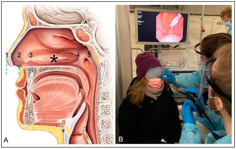
lateral wall of nasal cavity turbinate meatus anatomy diagram
Posterior Wall of Nose (Nasal Cavity Anatomy) - 15 Marks
1. Introduction
2. Walls of the Nasal Cavity
(a) Roof
- Frontonasal part - sloping upward (nasal bones + frontal process of maxilla)
- Ethmoidal part - horizontal, formed by the cribriform plate of ethmoid (site of olfactory nerve filaments passing through)
- Sphenoidal part - sloping downward (body of sphenoid/anterior wall of sphenoid sinus)
(b) Floor
- Palatine process of maxilla (anterior 3/4)
- Horizontal plate of palatine bone (posterior 1/4)
(c) Medial Wall (Nasal Septum)
- Perpendicular plate of ethmoid (posterosuperior)
- Vomer (posteroinferior)
- Septal cartilage (anterosuperior)
- Columella (anteroinferior, membranous/mobile septum)
- Keystone area = junction of nasal bones, perpendicular plate of ethmoid, upper lateral cartilage (ULC), and cartilaginous septum
- Posterior septal branch of sphenopalatine artery (SPA)
- Anterior ethmoidal artery (AEA)
- Greater palatine artery
- Alar branch of superior labial artery
3. Lateral Wall of Nose (Most Important / High-Yield)
Bones forming the lateral wall:
- Nasal bone (anterosuperior)
- Frontal process of maxilla (anterior)
- Lacrimal bone
- Ethmoid (labyrinth - main contributor)
- Inferior turbinate bone (separate bone)
- Perpendicular plate of palatine bone (posterior)
- Medial pterygoid plate of sphenoid (most posterior)
Turbinates (Conchae):
| Turbinate | Origin | Meatus Below It |
|---|---|---|
| Inferior | Separate bone (largest) | Inferior meatus |
| Middle | Ethmoid bone | Middle meatus |
| Superior | Ethmoid bone | Superior meatus |
| Supreme (Santorini) | Ethmoid (inconsistent) | Supreme meatus |
Meati and Their Openings:
- Between inferior turbinate and nasal floor
- Opening of nasolacrimal duct (under the anterior end - Hasner's valve)
- Preferred route for nasal airway devices (widest meatus)
- Between middle and inferior turbinates
- Contains the ostiomeatal complex (OMC)
- Structures seen here:
- Uncinate process (UP) - sickle-shaped bony projection
- Ethmoid bulla (EB) - most prominent anterior ethmoid cell
- Hiatus semilunaris - 2D crescent-shaped gap between UP (anteriorly) and EB (posteriorly)
- Ethmoid infundibulum - 3D trough bounded by UP medially, lamina papyracea laterally, EB posterosuperiorly
- Sinus drainage into middle meatus:
- Maxillary sinus
- Frontal sinus (via frontonasal recess)
- Anterior ethmoidal sinuses
- Between superior and middle turbinates
- Drains: posterior ethmoidal sinuses
- Above and behind the superior turbinate
- Drains: sphenoid sinus
Important Structures on Lateral Wall:
- Sphenopalatine foramen - within superior meatus, between basal lamella of middle turbinate and superior turbinate; transmits the sphenopalatine artery and nerve
- Eustachian (pharyngotympanic) tube orifice - on lateral wall of nasopharynx
- Fossa of Rosenmüller (pharyngeal recess) - posterosuperior to ET orifice; common site of nasopharyngeal carcinoma
4. Posterior Wall of Nose - Choana
Bony Margins of the Choana:
| Border | Bone |
|---|---|
| Superior | Body of sphenoid (+ root of medial pterygoid plate) |
| Medial | Vomer (+ ala of vomer superiorly) |
| Inferior | Horizontal plate of palatine bone (+ posterior nasal spine) |
| Lateral | Perpendicular plate of palatine bone + medial pterygoid plate of sphenoid |
- Inferior nasal concha
- Middle nasal concha
- Ethmoidal bulla
- Uncinate process
Mucosal Landmarks Behind the Choana (Nasopharynx):
- Posterior pharyngeal wall
- Uvula
- Soft palate (inferior boundary between nasopharynx and oropharynx)
- Pharyngeal tonsil (adenoids) - in roof/posterior wall of nasopharynx
5. Blood Supply of Nasal Cavity
Arterial Supply (two main sources):
- Anterior ethmoidal artery (AEA) - exits orbit via anterior ethmoidal foramen, supplies anterosuperior septum and lateral wall
- Posterior ethmoidal artery (PEA) - exits ~12 mm posterior to AEA, 6 mm anterior to optic canal; supplies posterior nasal cavity
- Sphenopalatine artery (SPA) - main artery of the nasal cavity; enters via sphenopalatine foramen in superior meatus
- Posterior septal branch - courses along sphenoid rostrum; supplies nasal septum (forms pedicle of nasoseptal flap)
- Posterior lateral nasal branch - supplies middle and inferior turbinates
- Descending palatine → greater palatine artery - supplies anteroinferior nasal cavity and septum (via incisive foramen)
- Pharyngeal branch of IMA (palatovaginal artery) - courses through pharyngeal canal from posterior wall of pterygopalatine fossa; supplies posterosuperior nasal cavity and nasopharynx
Vascular Plexuses:
- Kiesselbach's plexus (Little's area) - anteroinferior septum; most common site of anterior epistaxis
- Woodruff's plexus - posteroinferior meatus (posterior lateral nasal + pharyngeal branch of IMA); previously thought arterial, now known to be venous; site of posterior epistaxis
6. Venous Drainage
- Anterior/posterior ethmoidal veins → superior ophthalmic vein → cavernous sinus
- Sphenopalatine vein → pterygoid plexus
- Diploic veins of Breschet → communicate with cavernous and superior sagittal sinuses
- Valveless system - allows retrograde intracranial spread of infection
7. Nerve Supply
Sensory Innervation:
- Internal nasal branch of anterior ethmoidal nerve - anterosuperior septum and lateral wall
- Posterior ethmoidal nerve - posterior nasal cavity
- Sphenopalatine (nasopalatine) nerve - posterior nasal cavity and septum
- Anterior superior alveolar nerve - anterior nasal cavity and floor
Autonomic Innervation (via Vidian nerve - nerve of pterygoid canal):
- Sympathetic (vasoconstriction): preganglionic from sympathetic chain → superior cervical ganglion → deep petrosal nerve → joins GSPN → Vidian nerve → nasal mucosa (without synapsing in SPG)
- Parasympathetic (vasodilation + secretion): preganglionic from superior salivary nucleus (CN VII) → nervus intermedius → GSPN → Vidian nerve → sphenopalatine ganglion (synapse) → postganglionic fibers with branches of V2 to nasal mucosa
Olfactory:
- Olfactory nerve (CN I) - filaments pass through cribriform plate of ethmoid to upper nasal cavity (superior turbinate and upper third of septum)
Summary Table - Quick Revision
| Feature | Details |
|---|---|
| Choana borders | Superior: sphenoid body; Medial: vomer; Inferior: palatine bone; Lateral: palatine + medial pterygoid plate |
| Main artery | Sphenopalatine artery (branch of IMA) |
| Anterior epistaxis site | Kiesselbach's plexus / Little's area |
| Posterior epistaxis site | Woodruff's plexus |
| NLD opens into | Inferior meatus (Hasner's valve) |
| Sphenoid sinus drains into | Sphenoethmoidal recess |
| Posterior ethmoid drains into | Superior meatus |
| Middle meatus drains | Maxillary, frontal, anterior ethmoid sinuses |
| Olfactory area | Superior concha + upper 1/3 septum |
| Vidian nerve formed by | Greater superficial petrosal nerve (GSPN) + deep petrosal nerve |
Gimme a diagram of posterior wall of nose
posterior wall of nose choana anatomy diagram bones
nasal cavity posterior aperture choana vomer sphenoid palatine bone
Detailed anatomical diagram of the posterior wall of the nose (posterior nasal apertures / choanae), as seen from a posterior view. The diagram should clearly label all bony borders: Superior border - body of sphenoid bone and root of medial pterygoid plate; Medial border - vomer (vertical midline bone) and ala of vomer; Inferior border - horizontal plate of palatine bone and posterior nasal spine; Lateral border - perpendicular plate of palatine bone and medial pterygoid plate of sphenoid. Also label the two choanae (left and right posterior nasal apertures), the pterygoid canal (Vidian canal), the inferior nasal concha, the middle nasal concha, the nasal septum, and the soft palate below. Use clean medical illustration style with clear labels and arrows, white background, professional ENT anatomy diagram quality.
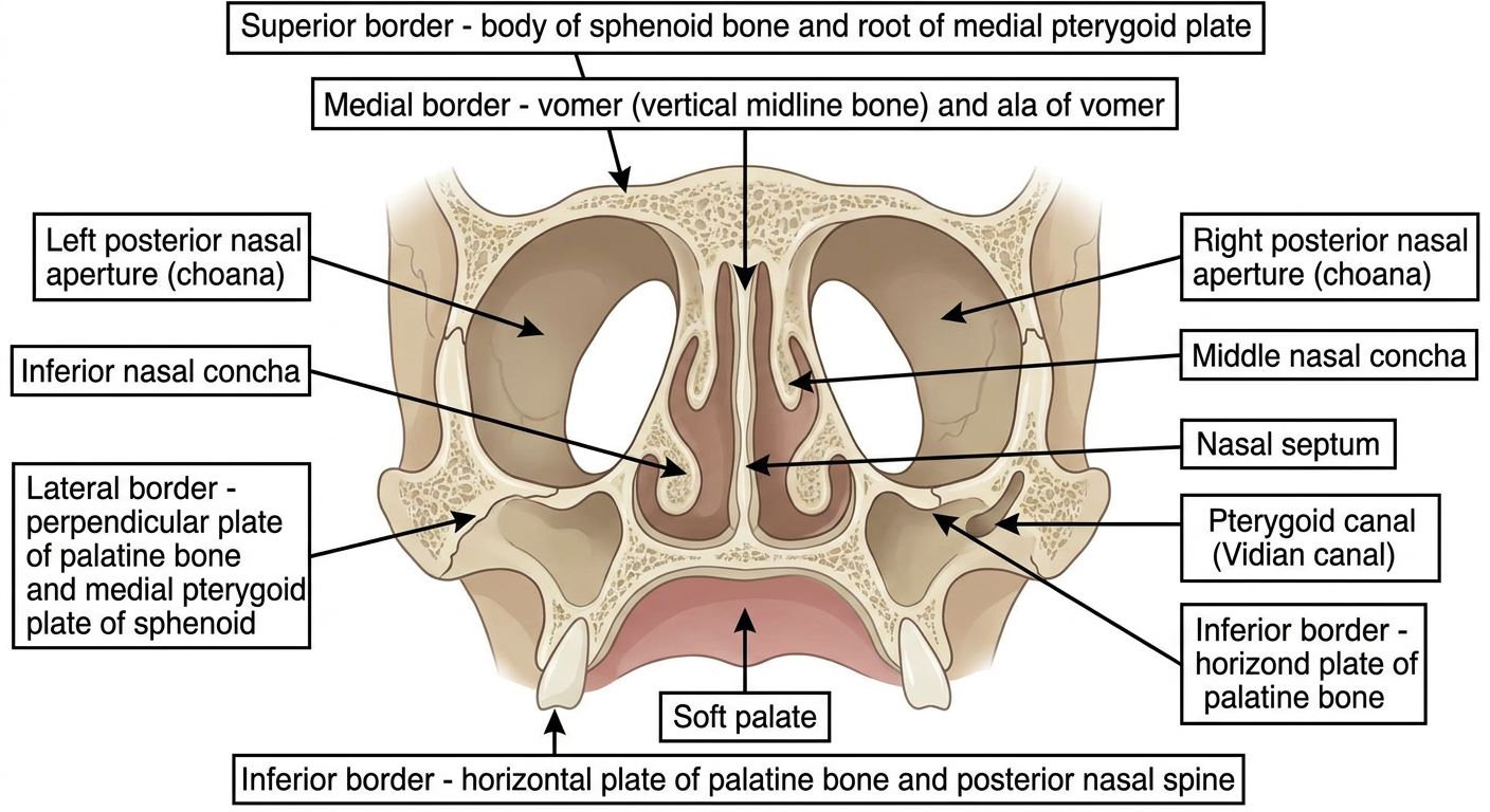
- Superior - body of sphenoid + root of medial pterygoid plate (with pterygoid/Vidian canal)
- Medial - vomer (the vertical midline divider between the two choanae)
- Inferior - horizontal plate of palatine bone + posterior nasal spine
- Lateral - perpendicular plate of palatine bone + medial pterygoid plate of sphenoid
- Inferior nasal concha
- Middle nasal concha
- Soft palate (below)
- Sphenoid (superior) | Vomer (medial) | Palatine - horizontal plate (inferior) | Lateral - palatine perpendicular plate + Medial Pterygoid plate