
Auditory pathway flowchart for mbbs 1st year prof
Reading File
Finding Sources
Finding Sources
Reading File
Reading File
Reading File
Reading File
Loading Image
Loading Image
Here is a complete, exam-ready auditory pathway flowchart for MBBS 1st year, built from Ganong's Physiology and Localization in Clinical Neurology.
Auditory Pathway - MBBS 1st Year
Pathway Diagram (from textbook)
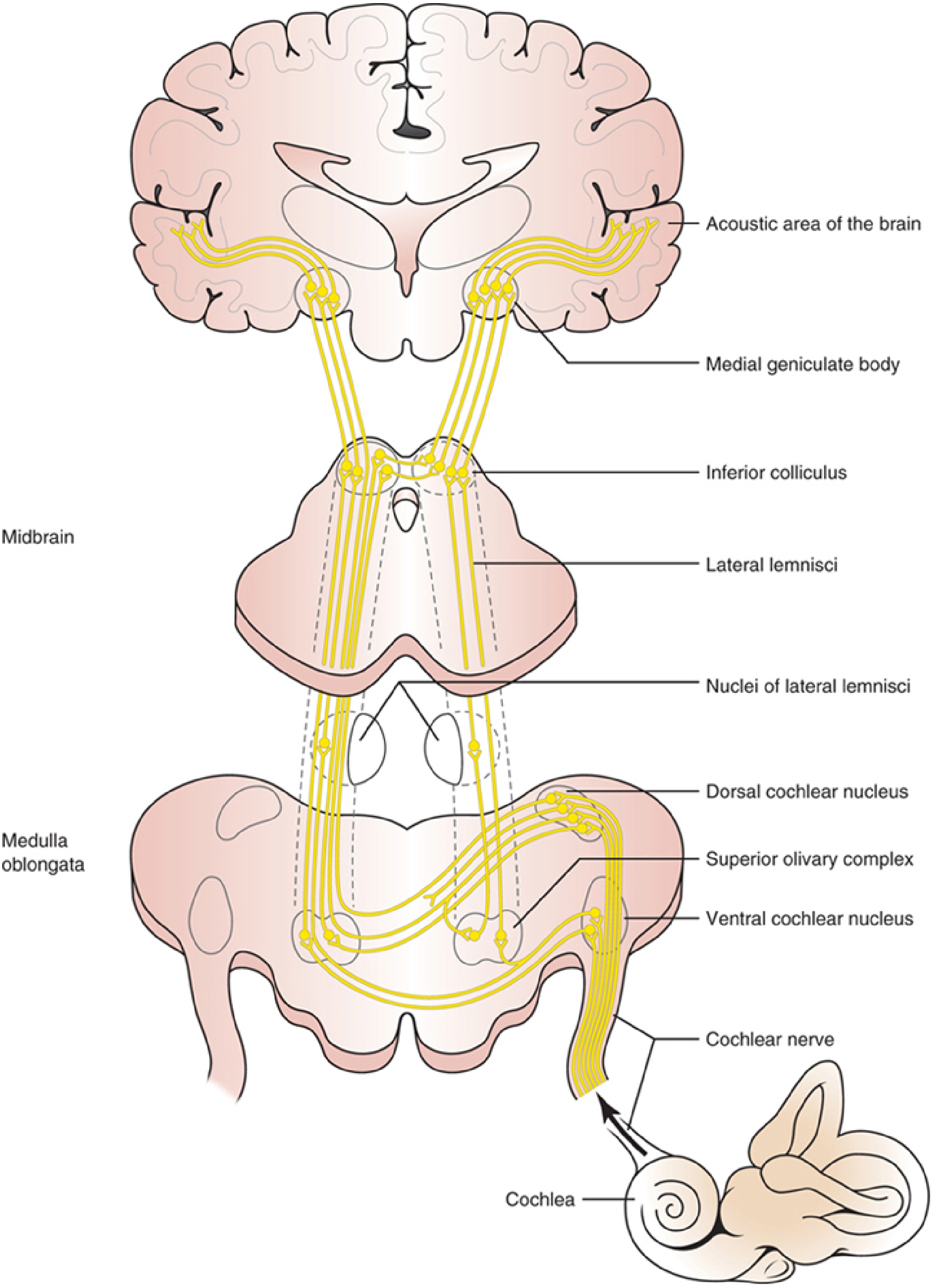
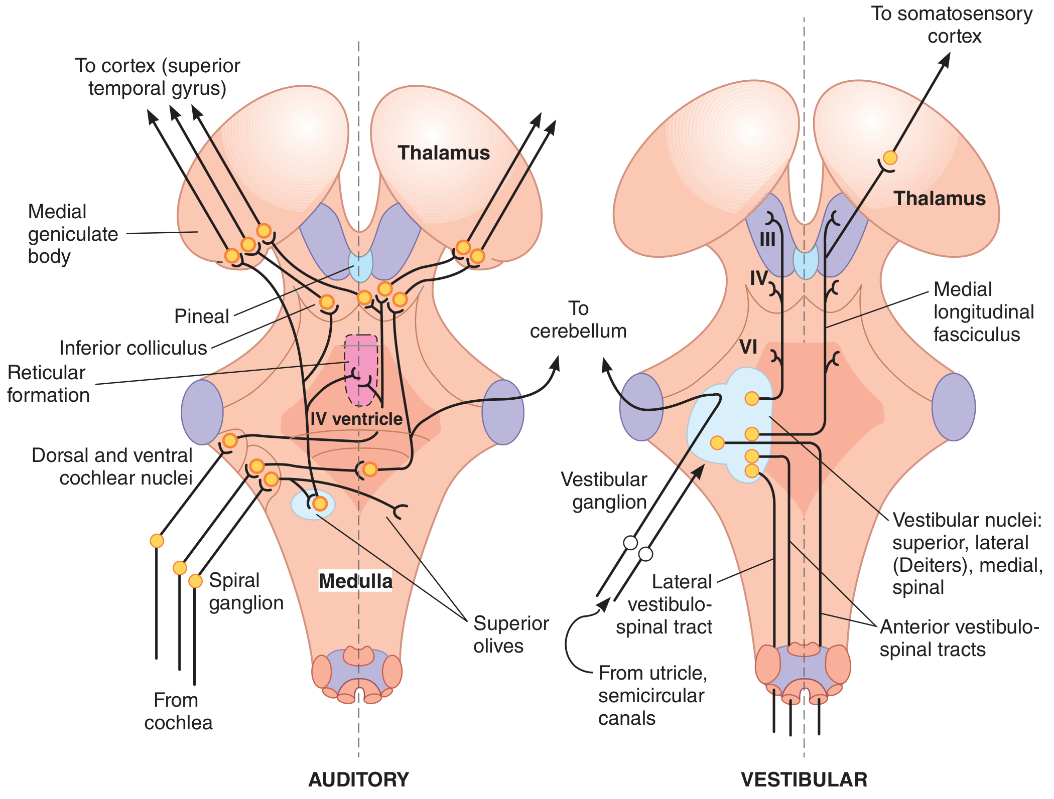
Flowchart
SOUND STIMULUS (External Ear → Middle Ear → Inner Ear)
                        ↓
         ORGAN OF CORTI (Hair cells on Basilar Membrane)
         • Inner hair cells → main receptors
         • Tonotopic: apex = low freq | base = high freq
                        ↓
              1st ORDER NEURONS
         Cell body: Spiral Ganglion (in Canal of Rosenthal)
                        ↓
              COCHLEAR NERVE (CN VIII)
                        ↓
         Enters brainstem at medullopontine junction
                        ↓
              2nd ORDER NEURONS
     ┌─────────────────────────────────────┐
     ↓                                     ↓
Dorsal Cochlear Nucleus           Ventral Cochlear Nucleus
(in medulla)                   (Anteroventral + Posteroventral)
     ↓                                     ↓
Dorsal Acoustic Striae      Intermediate + Ventral Acoustic Striae
     ↓                                     ↓
     └──────────── Most fibers CROSS ──────┘
                   (Trapezoid Body - in caudal pons)
                        ↓
              SUPERIOR OLIVARY COMPLEX
         (First site of BINAURAL convergence)
         (Both ipsilateral + contralateral input)
                        ↓
              LATERAL LEMNISCUS
         (Runs up through pons and midbrain tegmentum)
                        ↓
              3rd ORDER NEURONS
         INFERIOR COLLICULUS (Midbrain tectum)
         • Central relay nucleus
         • ALL ascending auditory pathways end here
         • Auditory reflex center
                        ↓
         Brachium of inferior colliculus
                        ↓
              4th ORDER NEURONS
    MEDIAL GENICULATE BODY / NUCLEUS (MGB) - Thalamus
         • Thalamic auditory relay nucleus
         • Generates auditory radiations (geniculotemporal fibers)
                        ↓
         Auditory Radiations (in internal capsule)
                        ↓
         PRIMARY AUDITORY CORTEX
         Heschl's Gyrus (Transverse Temporal Gyrus)
         Brodmann Area 41 - Superior Temporal Gyrus
         + Auditory Association Cortex (BA 42)
         • High freq → medial | Low freq → lateral
Summary Table
| Order | Neuron Location | Key Structure | Level |
|---|---|---|---|
| 1st | Spiral ganglion | Cochlear nerve (CN VIII) | Peripheral |
| 2nd | Cochlear nuclei (D + V) | Trapezoid body / Lateral lemniscus | Medulla |
| 3rd | Inferior colliculus | Brachium of IC | Midbrain |
| 4th | Medial geniculate body | Auditory radiations | Thalamus |
| End | Primary auditory cortex | Heschl's gyrus | Temporal lobe |
High-Yield Points for Exams
-
Crossing (decussation): Most fibers cross at the trapezoid body in the caudal pons - so each auditory cortex receives input from BOTH ears (bilateral representation).
-
First binaural site: Superior olivary complex is where signals from both ears first converge - this is the key nucleus for sound localization.
-
Auditory reflex center: Inferior colliculus (midbrain) - mediates the acoustic startle reflex.
-
Thalamic relay: Medial geniculate body (NOT lateral geniculate, which is for vision).
-
Cortical area: Brodmann area 41 (primary) + BA 42 (association) on the superior temporal gyrus.
-
Tonotopy: Preserved throughout the entire pathway (cochlea → cortex).
-
Unilateral cortical lesion: Does NOT cause complete deafness because of bilateral representation - only causes subtle deficits in sound localization.
-
Commissure of Probst: Connects the two nuclei of the lateral lemniscus (an additional crossing point).
Sources: Ganong's Review of Medical Physiology, 26th Ed., p. 225 | Localization in Clinical Neurology, 8th Ed., pp. 818-821
This is a shared conversation. Sign in to Orris to start your own chat.