
Hanau quint
- Condylar guidance - determined by the patient's temporomandibular joint anatomy
- Incisal guidance - the angle of the anterior teeth
- Occlusal plane - orientation of the teeth relative to the condyles
- Cusp height - steepness of posterior tooth cusps
- Compensating curves - curves built into the denture arch (Curve of Spee, Curve of Wilson)
Hanau quint five factors balanced occlusion complete dentures
Hanau quint five factors balanced occlusion complete denture
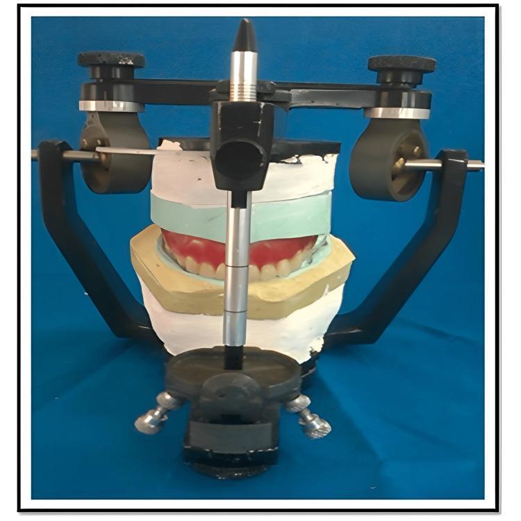
This procedural image illustrates a semi-adjustable dental articulator (Hanau type) used in the fabrication of a removable prosthesis. The device houses a stone core assembly consisting of multiple dental plaster and stone layers. A trial denture, featuring white artificial teeth set in a red wax-up gingiva, is sandwiched between these layers. The layered structure comprises a white maxillary mounting stone, a light-green stone layer, and a yellow Type III dental stone base. A vertically oriented metal incisal pin with adjustable markings is visible at the midline to maintain and verify the established vertical dimension of occlusion. The articulator's mechanical components, including the condylar elements and U-shaped frame, facilitate the replication of mandibular movements such as centric, protrusive, and lateral excursions. This setup is specifically used for the functionally generated path technique, where the stone core represents the recorded movements of the opposing arch to ensure balanced occlusion and eliminate interferences during the try-in phase.
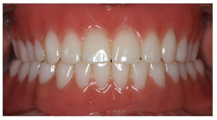
This intraoral clinical photograph presents an anterior view of complete maxillary and mandibular dentures in centric occlusion. The image illustrates a successful prosthetic restoration using a CAD/CAM workflow. The artificial teeth exhibit natural-appearing morphology, with central incisors, lateral incisors, and canines arranged in a balanced dental arch. The maxillary incisors show appropriate dominance and size relative to the mandibular teeth. The pink-pigmented denture base material (polymethyl methacrylate) mimics natural gingival tissue color and texture, showing smooth adaptation to the cervical margins of the teeth. The occlusion appears bilaterally balanced with stable intercuspation. This image serves as an educational example of prosthetic dentistry, demonstrating proper tooth arrangement, gingival esthetics, and the achievement of a stable vertical dimension in the rehabilitation of an edentulous patient using implant-supported mandibular overdentures and a tissue-supported maxillary denture.
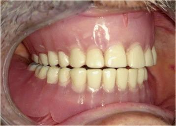
This intraoral clinical photograph showcases a set of complete maxillary and mandibular dentures in a state of bilateral balanced occlusion. The visual demonstrates the prosthetic alignment of off-white artificial teeth, which mimic natural tooth morphology including incisors, canines, and premolars. The teeth are set into a pink acrylic denture base designed to simulate natural gingival tissue. Key visual features include the simultaneous, even contact between the upper and lower arches across both the anterior and posterior segments. This occlusal scheme is a fundamental concept in prosthetic dentistry, intended to provide stability to the denture bases during functional movements and parafunctional habits. The photograph highlights the relationship between the prosthetic teeth and the supporting soft tissue, emphasizing the aesthetic and functional integration of the dental prostheses within the oral cavity.
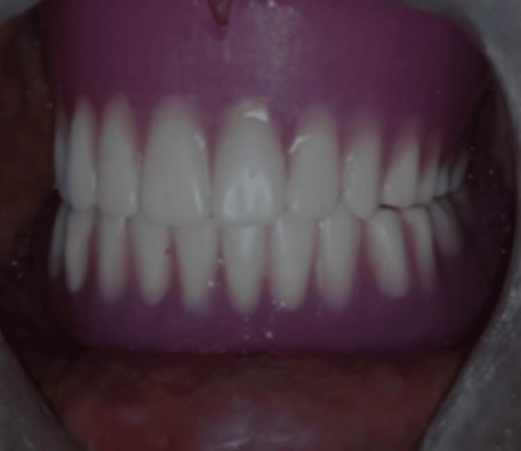
This intraoral clinical photograph depicts a set of maxillary and mandibular complete dentures in situ, demonstrating the final prosthetic outcome for an edentulous patient. The visual focus is on the occlusion and aesthetic rehabilitation. The artificial teeth are characterized by a uniform, opaque white shade and are arranged in a balanced arch form. The maxillary anterior teeth exhibit a slight vertical overlap (overbite) and horizontal overlap (overjet) relative to the mandibular teeth. The denture base is constructed from a pink heat-polymerized acrylic resin, designed to simulate natural gingival tissue and provide support via adaptation to the underlying alveolar ridges. Clinical significance includes the demonstration of proper occlusal contact and intercuspation, which are essential for masticatory efficiency and stability in prosthodontic rehabilitation, particularly in cases involving specialized impression techniques for flabby ridges.
Hanau's Quint - The Five Factors of Balanced Occlusion
The Five Factors
| # | Factor | Description |
|---|---|---|
| 1 | Condylar Guidance (CG) | The path the condyle travels along the articular eminence during protrusive/lateral movements. It is anatomically fixed and non-adjustable by the clinician. It is the primary, fixed determinant - all other factors are adjusted relative to it. Steeper condylar inclination requires steeper compensating curves or higher cusps to maintain balance. |
| 2 | Incisal Guidance (IG) | The angle formed between the occlusal plane and the path of the lower incisors as they move along the lingual surfaces of the upper incisors during protrusion. Influenced by anterior tooth position and aesthetics. In complete dentures, it is recommended to minimize incisal guidance (keep it as flat as possible) to reduce tipping/destabilizing forces on the denture base. |
| 3 | Occlusal Plane (OP) | The orientation of the plane of the posterior teeth relative to the condyles and incisal edges. A more anteriorly inclined (tipped-down) occlusal plane increases the tendency for posterior contact in protrusion. Adjusting this plane is one of the main ways to compensate for a fixed condylar path. |
| 4 | Compensating Curve (CC) | The anteroposterior and lateral curvature built into the alignment of the artificial teeth (a combination of the Curve of Spee and Curve of Wilson). It is unique to complete denture occlusion. The more prominent the curve, the easier it is to maintain posterior contact as the mandible moves forward/laterally. |
| 5 | Cuspal Inclination / Cusp Height (CI) | The steepness of the cuspal inclines of posterior teeth. Higher cusps favor balanced contact but also generate more lateral forces on the ridges. Lower cusps (or even cuspless/flat teeth) reduce lateral forces but sacrifice balanced occlusion. |
Thielmann's Formula (the mathematical expression of Hanau's Quint)
C = (OP × IG × CI × CC)
Clinical Significance
- Condylar guidance and incisal guidance are the two fixed/semi-fixed factors in clinical practice. They guide the adjustment of the other three.
- Goal: Achieve simultaneous contact of anterior and posterior teeth in protrusive and lateral excursions - this stabilizes the denture base and prevents tipping/rocking.
- Flat-cusped (monoplane) occlusion sacrifices some balanced occlusion in exchange for reduced horizontal forces on the edentulous ridge - acceptable in certain clinical situations (e.g., severe ridge resorption, Class II/III skeletal relationships).
- A 2025 PMC study validated Hanau's Quint digitally, confirming the classic interrelationships hold true in CAD/CAM-assisted denture fabrication.
A Helpful Mnemonic
Objective of complete DENTURE fabrication
objectives of complete denture fabrication prosthodontics
https://www.thejpd.org/article/0022-3913(54)90018-9/abstract
complete denture objectives restoration function esthetics phonetics tissue health prosthodontics textbook
https://dokumen.pub/textbook-of-complete-denture-prosthodont…
Objectives of Complete Denture Fabrication
1. Restoration of Masticatory Function (Chewing Efficiency)
- Replace the lost natural dentition so the patient can chew food adequately.
- Achieve bilateral balanced occlusion - simultaneous contact of teeth in centric occlusion and during all excursive movements to stabilize the denture base.
- Restore proper vertical dimension of occlusion (VDO) - the correct jaw height at which the teeth occlude, preventing over-closure or over-opening.
- Restore proper centric relation - the most retruded, unstrained position of the mandibular condyles in the glenoid fossae, used as the reproducible jaw reference point.
2. Restoration of Phonetics (Speech)
- Replace missing teeth and lost alveolar bone volume so air is directed correctly for sound production.
- Proper placement of anterior teeth is especially important for sibilant sounds ("s", "sh"), labial sounds ("f", "v"), and linguodental sounds ("th").
- Incorrect tooth position or a poor palatal contour of the upper denture distorts speech.
3. Restoration of Aesthetics (Appearance)
- Restore a natural, youthful facial appearance - lost teeth and bone cause inward collapse of the lips and cheeks, giving a "sunken" look.
- Select and arrange artificial teeth to match the patient's age, sex, facial form, and complexion.
- Restore proper lip support - the upper anterior teeth support the upper lip and determine lip fullness.
- Restore correct vertical dimension to prevent the over-closed appearance (deepened nasolabial folds, sunken chin, "witch's chin" deformity).
4. Preservation and Maintenance of Oral and General Health
a. Preservation of the Residual Ridges
- Distribute occlusal loads evenly over the edentulous ridge to minimize resorption of the alveolar bone (one of the most important long-term objectives).
- Avoid excessive pressure on any one area; use an appropriate impression technique to record the tissues under functional load.
b. Protection of the Oral Mucosa
- The denture base must fit accurately to avoid trauma, ulceration, and chronic irritation of the mucosa.
- Proper extension of flanges (borders) to achieve peripheral seal without overextension that impinges on muscle attachments.
c. Preservation of the Temporomandibular Joint (TMJ)
- Correct VDO and centric relation protect the TMJ from excessive strain.
- Poorly made dentures with incorrect VDO can contribute to TMJ disorders.
d. Nutritional Health
- Edentulous patients without dentures (or with poorly fitting ones) restrict their diet to soft, processed foods, leading to nutritional deficiencies.
- Adequate chewing efficiency promotes a more varied, nutritious diet and supports systemic health.
5. Psychological and Social Rehabilitation
- Restoration of appearance and speech has a profound impact on the patient's self-confidence, social interaction, and quality of life.
- Young (1954) in the Journal of Prosthetic Dentistry listed rehabilitation as the overriding objective, with comfort, esthetics, and function as sub-objectives - recognizing that the ultimate goal is restoring the whole patient, not just replacing teeth.
6. Retention, Stability, and Support (The Mechanical Triad)
| Property | Definition | How Achieved |
|---|---|---|
| Retention | Resistance to displacement away from the tissue (dislodgement) | Accurate border seal, adhesion, atmospheric pressure, correct denture base extension |
| Stability | Resistance to horizontal/rotational displacement during function | Balanced occlusion, accurate fit, correct ridge form coverage |
| Support | Resistance to vertical occlusal forces driving the denture into the tissues | Maximum coverage of the primary stress-bearing areas (hard palate, buccal shelf, posterior palatal seal) |
7. Comfort
- The denture must be comfortable at rest and during function.
- No pressure spots, no areas of tissue impingement, smooth polished surfaces, and correct occlusion all contribute to patient comfort.
- Comfortable dentures lead to patient compliance and long-term use.
Summary (H.A. Young's Classic Classification, 1954)
- Rehabilitation - the overriding objective
- Comfort - physical and mental
- Esthetics - natural, youthful appearance
- Masticatory efficiency - adequate chewing
- Preservation of remaining structures - ridges, TMJ, mucosa
Explain the difference between centric occlusion and balanced occlusion.
centric occlusion vs balanced occlusion difference complete denture prosthodontics
https://pmc.ncbi.nlm.nih.gov/articles/PMC4762337
balanced occlusion centric excursive protrusive lateral complete denture diagram
This intraoral clinical photograph presents an anterior view of complete maxillary and mandibular dentures in centric occlusion. The image illustrates a successful prosthetic restoration using a CAD/CAM workflow. The artificial teeth exhibit natural-appearing morphology, with central incisors, lateral incisors, and canines arranged in a balanced dental arch. The maxillary incisors show appropriate dominance and size relative to the mandibular teeth. The pink-pigmented denture base material (polymethyl methacrylate) mimics natural gingival tissue color and texture, showing smooth adaptation to the cervical margins of the teeth. The occlusion appears bilaterally balanced with stable intercuspation. This image serves as an educational example of prosthetic dentistry, demonstrating proper tooth arrangement, gingival esthetics, and the achievement of a stable vertical dimension in the rehabilitation of an edentulous patient using implant-supported mandibular overdentures and a tissue-supported maxillary denture.
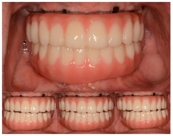
This composite intraoral clinical photograph displays maxillary and mandibular definitive complete-arch implant-supported prostheses in a patient with Sjögren's syndrome. The large top panel illustrates the prostheses in centric occlusion, showing a balanced arrangement of off-white artificial teeth and pink gingiva-colored prosthetic bases that mimic natural soft tissue morphology. The three smaller bottom panels demonstrate functional occlusal dynamics: lateral excursions (left and right) and protrusive excursion (center). The lateral views show working-side contacts without posterior interferences, while the protrusive view demonstrates anterior guidance, where the mandibular incisors slide along the palatal surfaces of the maxillary incisors. This visual documentation serves as an educational example of successful prosthetic rehabilitation using titanium-zirconia and monolithic zirconia materials to restore aesthetics and function in complex restorative cases.
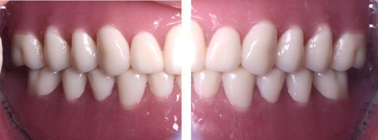
This composite intra-oral photograph shows two lateral views of newly placed CAD/CAM (Computer-Aided Design/Computer-Aided Manufacturing) complete dentures in a patient with edentulism. The image illustrates the maxillary and mandibular prostheses in centric occlusion. The artificial teeth exhibit a light shade, anatomical ovoid morphology, and natural-looking interdigitation with a stable occlusal relationship. The denture bases are fabricated from pink-pigmented acrylic resin designed to mimic natural gingival tissue and show smooth contours with appropriate adaptation to the vestibular mucosa. Key educational features include the demonstration of dental esthetics, tooth arrangement, and the integration of digital prosthetic technology in managing cases with significant alveolar ridge resorption. The clinical focus is on the final functional and esthetic outcome of digital denture fabrication.
Centric Occlusion vs. Balanced Occlusion
Definitions
| Term | Definition (GPT - Glossary of Prosthodontic Terms) |
|---|---|
| Centric Occlusion (CO) | The occlusion of opposing teeth when the mandible is in centric relation - the maximum intercuspation of the teeth with the condyles in their most superior, anterior, unstrained position in the glenoid fossae. |
| Balanced Occlusion | "The simultaneous contacting of the maxillary and mandibular teeth on the right and left, and in the posterior and anterior occlusal areas, in centric and eccentric positions, developed to lessen or limit tipping or rotating of the denture bases in relation to the supporting structures." |
The Core Difference
- Centric occlusion = tooth contact at ONE specific jaw position (maximum intercuspation / the "home" position)
- Balanced occlusion = tooth contact maintained THROUGHOUT all jaw movements (centric + eccentric positions)
Centric occlusion is a point. Balanced occlusion is a range.
Detailed Comparison
| Feature | Centric Occlusion | Balanced Occlusion |
|---|---|---|
| Jaw position | One fixed position - maximum intercuspation | All positions - centric, protrusive, left lateral, right lateral |
| Tooth contacts | Simultaneous contact of all opposing teeth at rest/closure | Simultaneous anterior + posterior contacts during all excursive movements |
| Applicable to | Natural dentition AND complete dentures | Primarily complete dentures (rarely seen in natural dentition) |
| In natural teeth | The normal "bite" position | Considered an interference in natural dentition (unwanted) |
| In complete dentures | The starting/reference position of occlusion | The ideal occlusal scheme - essential for denture stability |
| Purpose | Establishes the reference jaw closing position | Prevents denture tipping/rocking/displacement during jaw movements |
| Number of contacts | Maximum tooth contacts (intercuspation) | Contacts distributed bilaterally front and back at all times |
| Movement involved | Static - no jaw movement | Dynamic - involves all mandibular excursions |
Why Centric Occlusion Alone is Insufficient for Dentures
- Denture dislodgement
- Mucosal ulceration and soreness
- Accelerated alveolar bone resorption
- Loss of retention
The Relationship Between the Two
Balanced Occlusion = Centric Occlusion + Eccentric Balance
                     (tooth contact    (tooth contact during
                      at rest/closure)  all movements)
- Proper centric occlusion - maximum intercuspation when the jaw closes
- Bilateral balance - maintained contacts as the jaw moves in all directions
The "Enter Bolus, Exit Balance" Problem
"Enter bolus, exit balance"
- Swallowing (occurs ~2,000 times/day with tooth contact)
- Bruxism and parafunction
- Repositioning the denture after each chewing stroke
- Stability at rest
Eccentric Positions Covered by Balanced Occlusion
- Protrusive - mandible moves straight forward; posterior teeth on both sides contact simultaneously with anterior teeth
- Right lateral - mandible moves right; both the right (working) and left (balancing) sides maintain contact
- Left lateral - mandible moves left; both the left (working) and right (balancing) sides maintain contact
Quick Summary
| Centric Occlusion | Balanced Occlusion | |
|---|---|---|
| What it is | The "bite" position | A dynamic occlusal scheme |
| When it matters | At jaw closure | During all jaw movements |
| In natural teeth | Normal and desired | Unwanted interference |
| In complete dentures | Necessary but not sufficient | The gold standard occlusal goal |
| Clinical benefit | Establishes vertical dimension, jaw reference | Prevents denture displacement, protects ridges |
Parts of CD
parts of complete denture anatomy denture base flange land area border seal prosthodontics
parts of complete denture labeled diagram anatomy flange base
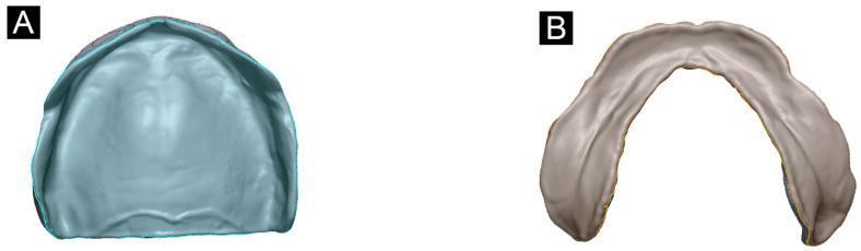
This clinical diagram illustrates the anatomical design of complete denture bases. Figure A shows a maxillary denture base, characterized by a broad horseshoe arch form with full palatal coverage. The internal fitting surface is smooth, demonstrating the contours of the hard palate, maxillary tuberosities, and alveolar ridge. Figure B depicts a mandibular denture base, which features a narrower U-shaped design to accommodate the tongue. It lacks palatal coverage and exhibits a distinct lingual flange and labial/buccal borders that follow the residual alveolar ridge. Both models represent Class I, type A residual ridge morphology used for evaluating the accuracy and dimensional stability of 3D-printed prosthodontics. The digital renderings highlight the surface texture and peripheral border extensions necessary for optimal retention and stability within an edentulous oral cavity. This comparison is essential for dental students and prosthodontists in understanding the morphological differences between upper and lower dental arch rehabilitations.
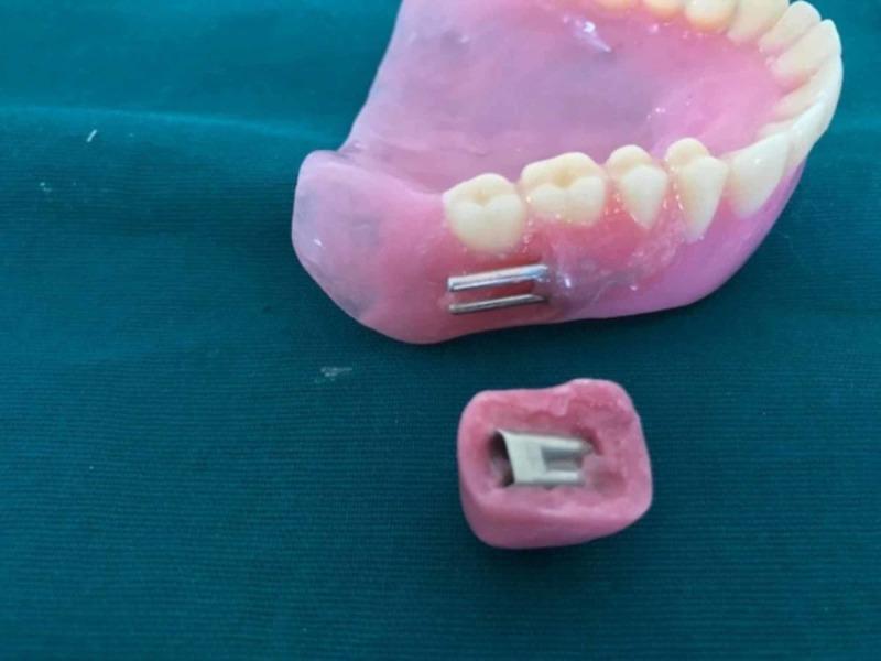
A clinical photograph of a maxillary complete denture and a separate detachable cheek plumper, illustrating a mechanical attachment system for prosthodontic rehabilitation. The denture base is constructed from pink heat-polymerized acrylic resin with anatomical artificial teeth. Positioned on the buccal flange in the molar region are two parallel, cylindrical metallic die pins that project outwards. Below the denture lies a small, matching pink acrylic cheek plumper containing an embedded metallic sleeve designed to receive the die pins. This configuration demonstrates a friction-fit, interlocking mechanism used to provide extraoral support for slumped facial musculature in patients with severe resorption or loss of dental esthetics. The image highlights the laboratory phase of prosthodontic fabrication, focusing on the precise alignment and housing of retentive elements to ensure a stable, yet removable, connection between the primary prosthesis and the cosmetic accessory.
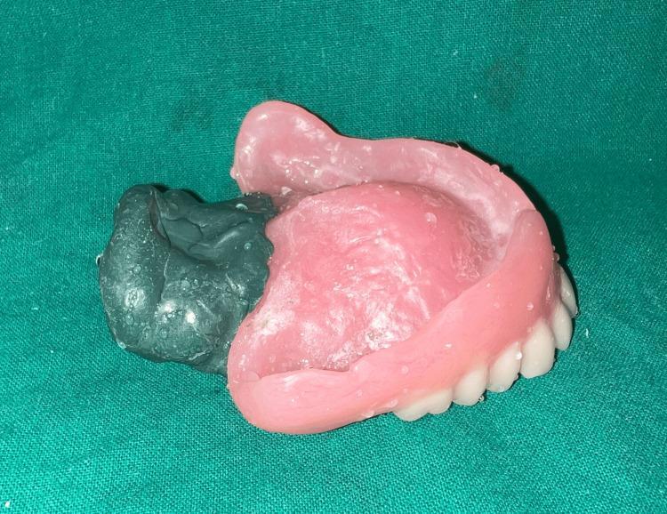
Clinical photograph of a maxillary complete denture being used for a functional impression of a velopharyngeal defect. The image shows a pink acrylic denture base with artificial white teeth set into the flange. Attached to the posterior aspect of the denture is a dark gray, high-viscosity impression compound. This material has been molded to capture the morphology and dimensions of a velopharyngeal defect, facilitating the fabrication of a speech bulb obturator. The impression material exhibits a matte, irregular, and functionally contoured surface, contrasting with the smooth, polished texture of the pink denture base. The prosthesis is resting on a green surgical drape. This visual demonstrates a critical step in maxillofacial prosthodontics for rehabilitating patients with hypernasal speech and palatal insufficiency, showing the spatial relationship between the dental arch support and the posterior extension required for velopharyngeal closure.
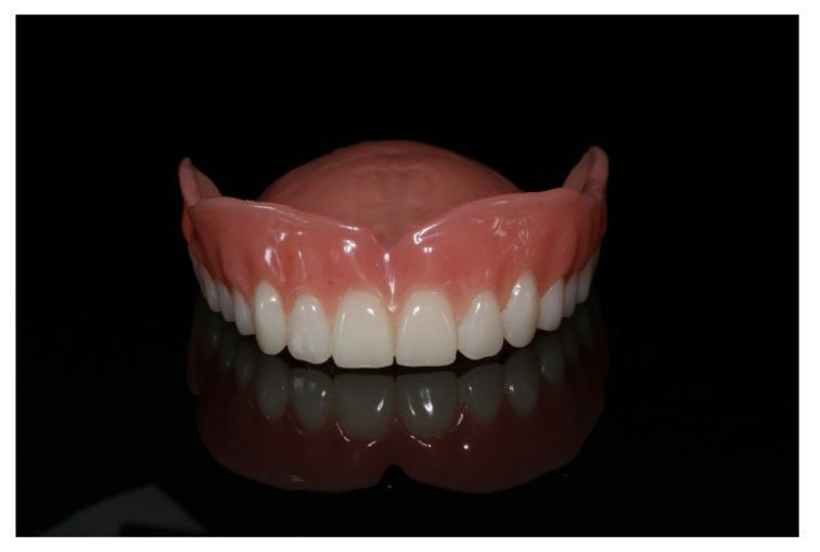
A clinical photograph of a final, polished maxillary (upper jaw) complete denture, shown against a dark, reflective background before insertion. The prosthesis features a pink, gingiva-colored base made of smooth, polished polymeric material (typically PMMA or milled CAD/CAM resin). The denture base includes a well-defined labial flange and a full palatal plate for suction-based retention. The artificial teeth are arranged in a symmetrical, curved dental arch, exhibiting a uniform bright white color and anatomical morphology consistent with natural dentition. The surface of both the base and teeth is highly reflective, indicating a high-quality finish aimed at reducing plaque accumulation and improving patient comfort. This image demonstrates the outcome of a prosthetic workflow, likely involving CAD/CAM fabrication for the restoration of an edentulous upper arch.
https://www.scribd.com/document/622861780/Complete-Denture-P…
Parts of a Complete Denture (CD)
- Denture Base (with its sub-parts)
- Artificial Teeth
I. DENTURE BASE
A. Three Surfaces of the Denture Base
| Surface | Also Called | Description |
|---|---|---|
| Impression Surface (Tissue Surface / Intaglio Surface) | Basal surface, fitting surface | The concave inner surface that directly contacts and rests on the oral mucosa. It must be an accurate replica of the denture-bearing tissues. |
| Polished Surface (Outer Surface) | Non-fitting surface | The outer surface that contacts the cheeks, lips, and tongue. Must be smoothly polished to allow muscle action to help seat and retain the denture (neutral zone). |
| Occlusal Surface | - | The surface that carries the artificial teeth and contacts the opposing denture or natural teeth. |
B. Parts / Regions of the Denture Base
1. Denture Flange
- Labial flange - extends into the labial (lip) vestibule, anteriorly
- Buccal flange - extends into the buccal (cheek) vestibule, posteriorly
- Lingual flange (mandibular denture only) - extends into the lingual sulcus between the ridge and the tongue
- Maxillary denture has labial and buccal flanges only
- Mandibular denture has labial, buccal, and lingual flanges
2. Denture Border (Periphery / Border Roll)
- Peripheral seal - to maintain retention through atmospheric pressure
- Avoiding impingement on muscle attachments (overextension causes dislodgement)
3. Post-Dam Area (Posterior Palatal Seal) - Maxillary Denture Only
- Pterygomaxillary seal - extends across the hamular notch bilaterally
- Postpalatal seal - runs between the two pterygomaxillary seals across the midline
4. Denture Heel
5. Land Area (Ledge)
6. Palatal Vault / Plate - Maxillary Denture Only
- Large surface area for support and retention
- Contributes to atmospheric pressure retention
- Provides palatal contour for phonetics
7. Retromolar Pad Coverage - Mandibular Denture Only
II. ARTIFICIAL TEETH
Classified by Position:
| Type | Description |
|---|---|
| Anterior teeth | Incisors (central and lateral) and canines - 6 per arch. Primarily for aesthetics, phonetics, and cutting food. |
| Posterior teeth | Premolars and molars - 8 per arch (4 on each side). Primarily for mastication. |
Classified by Material:
| Material | Advantages | Disadvantages |
|---|---|---|
| Acrylic resin (PMMA) | Easy to adjust, bond well to base, lightweight, aesthetic | Wear faster, less durable |
| Porcelain | Highly aesthetic, wear-resistant, natural feel | Brittle, click sound, harder to adjust, can damage opposing ridges |
Classified by Cusp Form:
| Type | Features | Use |
|---|---|---|
| Anatomic (cusped) - 30°-33° | Natural cusp form, good chewing efficiency | Balanced, well-aligned ridges |
| Semi-anatomic - 20° | Moderate cusps | Moderate ridge conditions |
| Non-anatomic (0°/flat/monoplane) | No cusps, flat occlusal surface | Resorbed ridges, poor ridge relations, Class II/III |
III. OTHER NOTABLE COMPONENTS
| Part | Description |
|---|---|
| Denture teeth-base junction | Where artificial teeth are embedded into the acrylic base - must be an intimate bond to prevent fracture or detachment |
| Gingival festooning | Simulated gingival contour carved into the polished surface to mimic natural gum tissue - improves aesthetics and food shedding |
| Root eminences | Slight convexities on the labial surface of the denture base mimicking natural root prominences - enhances natural appearance |
| Interdental papilla | Simulated gingival tissue between teeth on the polished surface |
Visual Summary
COMPLETE DENTURE
│
├── DENTURE BASE
│   ├── Impression surface (contacts oral tissues)
│   ├── Polished surface (contacts cheeks/lips/tongue)
│   ├── Occlusal surface (carries the teeth)
│   ├── Flanges: Labial / Buccal / Lingual
│   ├── Denture border (peripheral seal)
│   ├── Post-dam (maxillary only)
│   ├── Palatal plate (maxillary only)
│   └── Retromolar pad coverage (mandibular only)
│
└── ARTIFICIAL TEETH
    ├── Anterior (incisors + canines) → aesthetics, phonetics
    └── Posterior (premolars + molars) → mastication, occlusion