
Explain Heart
Reading File
Reading File
Finding Sources
Searching PubMed
"heart"[MeSH Terms] AND anatomy
Searching the Web
heart anatomy chambers valves circulation NIH
Searching Images
labeled anatomical diagram of human heart chambers valves blood flow
Reading File
Heart: basic explanation
The heart is a muscular pump, about the size of a clenched fist, located slightly left of the middle of the chest. Its job is to keep blood moving continuously:
- To the lungs to collect oxygen
- To the body to deliver oxygen and nutrients
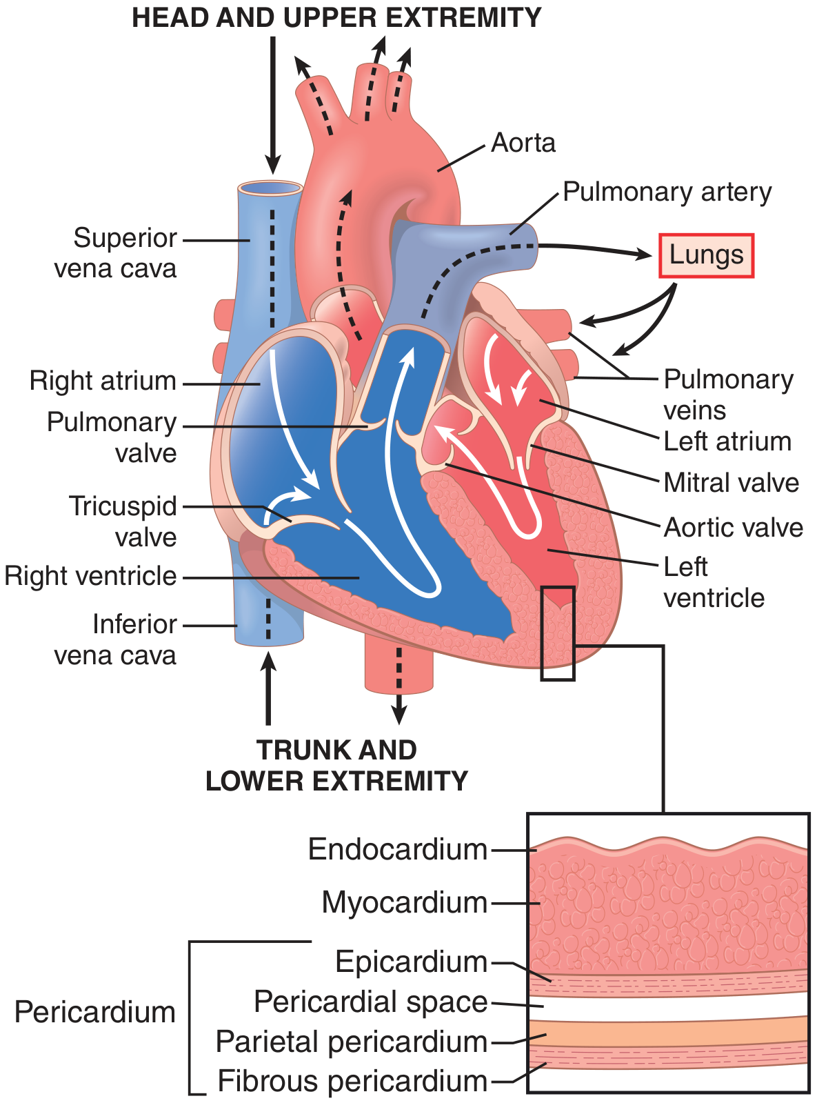
Four chambers
The heart has four hollow chambers:
| Side | Upper chamber | Lower chamber | Main role |
|---|---|---|---|
| Right | Right atrium | Right ventricle | Sends oxygen-poor blood to the lungs |
| Left | Left atrium | Left ventricle | Sends oxygen-rich blood to the body |
A wall called the septum separates the right and left sides.
Blood flow pathway
- Oxygen-poor blood returns from the body through the superior and inferior vena cava into the right atrium.
- It passes through the tricuspid valve into the right ventricle.
- The right ventricle pumps it through the pulmonary valve and pulmonary artery to the lungs.
- In the lungs, blood releases carbon dioxide and picks up oxygen.
- Oxygen-rich blood returns through the pulmonary veins to the left atrium.
- It crosses the mitral valve into the left ventricle.
- The left ventricle, the strongest chamber, pumps it through the aortic valve into the aorta and then to the entire body.
In short:
Body → right heart → lungs → left heart → body
This matches the NHLBI description of normal heart anatomy and blood flow.
Valves
The four valves act like one-way doors, opening and closing due to pressure differences to prevent backward blood flow:
- Tricuspid valve: right atrium → right ventricle
- Pulmonary valve: right ventricle → pulmonary artery
- Mitral valve: left atrium → left ventricle
- Aortic valve: left ventricle → aorta
How the heart beats
The heart has its own electrical system:
- The SA node is the natural pacemaker. It starts an electrical signal.
- This makes the atria contract.
- The signal travels through the AV node and conducting fibers to the ventricles.
- The ventricles then contract and pump blood out.
One heartbeat has two main phases:
- Diastole: the heart relaxes and fills with blood.
- Systole: the ventricles contract and eject blood.
Cardiac muscle cells connect through intercalated discs and gap junctions, allowing electrical signals to spread quickly so that the heart contracts in an organized rhythm. Guyton and Hall Textbook of Medical Physiology, cardiac muscle anatomy section.
Key point
The right heart handles blood going to the lungs, while the left heart handles blood going to the body. Because it pumps blood around the whole body, the left ventricle has the thickest muscle wall.
Explain Urinary system detail
Reading File
The urinary system can be studied in these connected parts:
Asking for Preferences
Which part would you like explained in detail first?
Heart: basic explanation
The heart is a muscular pump, about the size of a clenched fist, located slightly left of the middle of the chest. Its job is to keep blood moving continuously:
- To the lungs to collect oxygen
- To the body to deliver oxygen and nutrients
Four chambers
The heart has four hollow chambers:
| Side | Upper chamber | Lower chamber | Main role |
|---|---|---|---|
| Right | Right atrium | Right ventricle | Sends oxygen-poor blood to the lungs |
| Left | Left atrium | Left ventricle | Sends oxygen-rich blood to the body |
A wall called the septum separates the right and left sides.
Blood flow pathway
- Oxygen-poor blood returns from the body through the superior and inferior vena cava into the right atrium.
- It passes through the tricuspid valve into the right ventricle.
- The right ventricle pumps it through the pulmonary valve and pulmonary artery to the lungs.
- In the lungs, blood releases carbon dioxide and picks up oxygen.
- Oxygen-rich blood returns through the pulmonary veins to the left atrium.
- It crosses the mitral valve into the left ventricle.
- The left ventricle, the strongest chamber, pumps it through the aortic valve into the aorta and then to the entire body.
In short:
Body → right heart → lungs → left heart → body
This matches the NHLBI description of normal heart anatomy and blood flow.
Valves
The four valves act like one-way doors, opening and closing due to pressure differences to prevent backward blood flow:
- Tricuspid valve: right atrium → right ventricle
- Pulmonary valve: right ventricle → pulmonary artery
- Mitral valve: left atrium → left ventricle
- Aortic valve: left ventricle → aorta
How the heart beats
The heart has its own electrical system:
- The SA node is the natural pacemaker. It starts an electrical signal.
- This makes the atria contract.
- The signal travels through the AV node and conducting fibers to the ventricles.
- The ventricles then contract and pump blood out.
One heartbeat has two main phases:
- Diastole: the heart relaxes and fills with blood.
- Systole: the ventricles contract and eject blood.
Cardiac muscle cells connect through intercalated discs and gap junctions, allowing electrical signals to spread quickly so that the heart contracts in an organized rhythm. Guyton and Hall Textbook of Medical Physiology, cardiac muscle anatomy section.
Key point
The right heart handles blood going to the lungs, while the left heart handles blood going to the body. Because it pumps blood around the whole body, the left ventricle has the thickest muscle wall.The urinary system can be studied in these connected parts:The urinary system has five main areas:
- Kidneys: their location, gross structure, nephron, and blood supply
- Urine formation: filtration, reabsorption, secretion, and concentration
- Urine drainage pathway: renal pelvis, ureters, bladder, and urethra
- Micturition (urination): bladder filling, reflexes, sphincters, and nervous control
- Regulation and clinical relevance: water, electrolytes, acid-base balance, blood pressure, and common disorders
Brief overview
The urinary system consists mainly of two kidneys, two ureters, the urinary bladder, and the urethra. Its visible role is producing and excreting urine, but it also maintains the internal environment of the body.
The kidneys filter blood, remove metabolic wastes such as urea and creatinine, regulate body water and electrolytes such as sodium and potassium, help control blood pressure, maintain acid-base balance, produce erythropoietin for red blood cell formation, and activate vitamin D.
Urine is produced in microscopic functional units called nephrons. It moves from the kidneys into the ureters, is stored in the bladder, and leaves the body through the urethra during urination.
Which section would you like first: 1) kidney anatomy, 2) urine formation, 3) bladder and urination, or 4) full system explanation in exam-note style?
Explain nephron structure and urine formation step by step
Reading File
Finding Sources
Finding Sources
Searching PubMed
"Renal Tubules"[MeSH Terms] AND "glomerular filtration"
Searching the Web
NIDDK kidneys nephron urine formation filtration reabsorption secretion
Searching Images
labeled nephron diagram glomerulus proximal tubule loop of Henle distal tubule collecting duct
Reading File
Finding Sources
query_images_db
Reading File
Reading File
Loading Image
Quick answer
Nephron: structure and urine formation
A nephron is the microscopic structural and functional unit of the kidney. Each kidney contains roughly one million nephrons. Its work is to filter blood, retain substances the body needs, add selected wastes to the tubule, and finally make urine.
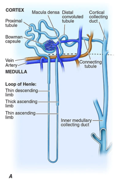
1. Main parts of a nephron
A nephron has two major components:
- Renal corpuscle: filters blood
- Renal tubule: modifies the filtered fluid into urine
A. Renal corpuscle
Located in the renal cortex, it consists of:
- Glomerulus: a tuft of capillaries supplied by the afferent arteriole
- Bowman's capsule: a cup-shaped structure surrounding the glomerulus that receives the filtrate
- Efferent arteriole: carries blood away from the glomerulus
The efferent arteriole then forms capillaries around the tubule:
- Peritubular capillaries around cortical nephrons
- Vasa recta alongside the loops of Henle in juxtamedullary nephrons
These vessels allow exchange between blood and tubular fluid.
B. Renal tubule
Fluid flows through the following sequence:
Bowman's capsule → proximal convoluted tubule → loop of Henle → distal convoluted tubule → collecting duct → renal pelvis
| Segment | Location | Main function |
|---|---|---|
| Proximal convoluted tubule, PCT | Cortex | Bulk reabsorption of water, sodium, glucose, amino acids, bicarbonate |
| Descending limb of loop of Henle | Medulla | Water reabsorption |
| Ascending limb of loop of Henle | Medulla to cortex | Sodium, potassium, and chloride reabsorption; does not allow water out |
| Distal convoluted tubule, DCT | Cortex | Fine control of sodium, potassium, calcium, and acid-base balance |
| Collecting duct | Cortex through medulla | Final adjustment of water, sodium, potassium, hydrogen ions, and urea; forms final urine |
Types of nephrons
- Cortical nephrons: most nephrons; short loops of Henle and mainly located in the cortex.
- Juxtamedullary nephrons: have long loops extending deep into the medulla. They are especially important for producing concentrated urine.
Urine formation: step by step
Urine formation depends on three processes:
[
\textbf{Urinary excretion = Filtration - Reabsorption + Secretion}
]
Guyton and Hall Textbook of Medical Physiology, p. 328 and p. 347.
Step 1: Glomerular filtration
What happens?
Blood enters the glomerulus through the afferent arteriole. The pressure inside the glomerular capillaries forces water and small dissolved substances out of blood and into Bowman's capsule.
The resulting fluid is called glomerular filtrate.
What is filtered?
Normally filtered substances include:
- Water
- Sodium, potassium, chloride, bicarbonate
- Glucose
- Amino acids
- Urea
- Creatinine
- Small molecules and some drugs
What normally remains in the blood?
The filtration barrier normally prevents passage of:
- Blood cells
- Platelets
- Most plasma proteins, especially albumin
- Large molecules
Thus, normal filtrate is essentially protein-free and initially resembles plasma in its small-solute composition. Guyton and Hall Textbook of Medical Physiology, p. 328.
Filtration barrier
The barrier has three layers:
- Fenestrated capillary endothelium: keeps blood cells inside capillaries
- Glomerular basement membrane: restricts many large and negatively charged proteins
- Podocytes with filtration slits: provide the final selective barrier
GFR
Glomerular filtration rate, GFR, is the total filtrate formed by both kidneys per minute.
- Approximately 125 mL/min
- Approximately 180 L/day of filtrate
Yet only about 1 to 2 L of urine is normally passed daily because most filtrate is reabsorbed.
Step 2: Tubular reabsorption
What is reabsorption?
Reabsorption means movement of substances from the tubule back into the blood, mainly through the peritubular capillaries.
This is the major reason the body does not lose enormous amounts of water and useful nutrients in urine.
A. Proximal convoluted tubule
The PCT performs the largest amount of reabsorption.
It reabsorbs approximately:
- 65% to 70% of filtered sodium and water
- Most chloride and potassium
- Most bicarbonate
- Nearly all glucose and amino acids under normal conditions
- Some urea, phosphate, calcium, and other solutes
Water follows sodium osmotically, so fluid leaving the PCT remains roughly isotonic to plasma.
Clinical link: If blood glucose is very high, glucose transporters become saturated. Glucose then remains in tubular fluid and appears in urine, drawing water with it. This causes increased urination in uncontrolled diabetes mellitus.
B. Descending limb of the loop of Henle
The descending limb is highly permeable to water but relatively less permeable to solutes.
- Water leaves the tubule into the hyperosmotic medulla.
- Tubular fluid becomes more concentrated as it descends.
C. Ascending limb of the loop of Henle
The ascending limb, especially the thick ascending limb:
- Reabsorbs sodium, potassium, and chloride.
- Is relatively impermeable to water.
- Dilutes the tubular fluid.
This segment helps create the high solute concentration in the renal medulla, which is needed for later water reabsorption.
D. Distal convoluted tubule
The DCT fine-tunes ion composition.
Important actions include:
- Sodium chloride reabsorption
- Calcium reabsorption, stimulated by parathyroid hormone
- Further regulation of acid-base balance and potassium handling
E. Collecting duct
The collecting duct makes the final decisions about the urine's water content and acidity.
It is regulated strongly by hormones:
- ADH, vasopressin: increases water reabsorption by inserting aquaporin water channels. Urine becomes concentrated and lower in volume.
- Aldosterone: increases sodium reabsorption and promotes potassium secretion.
- Atrial natriuretic peptide, ANP: promotes sodium and water excretion in settings of volume expansion.
When ADH is low, the collecting duct stays relatively impermeable to water. More water remains in the urine, producing a larger volume of dilute urine.
Step 3: Tubular secretion
What is secretion?
Tubular secretion is movement of substances from the peritubular blood into the tubular fluid.
It allows the kidneys to eliminate substances that were not sufficiently filtered or that need precise regulation.
Important secreted substances include:
- Hydrogen ions, H⁺: helps maintain acid-base balance
- Potassium ions, K⁺: regulated especially by aldosterone
- Ammonium, NH₄⁺
- Creatinine, in small amount
- Organic acids and bases
- Some medications, including penicillin and other drugs
The final amount excreted depends not only on what is filtered, but also on how much is reabsorbed or secreted. Potassium and hydrogen ion secretion are particularly important in the final composition of urine. Guyton and Hall Textbook of Medical Physiology, p. 347.
How concentrated urine is produced
The kidney can conserve water by making concentrated urine through the countercurrent mechanism.
1. Countercurrent multiplier
This occurs in the loop of Henle:
- Descending limb loses water.
- Ascending limb removes salt but does not lose water.
- This establishes a progressively hyperosmotic medulla.
2. Vasa recta
The vasa recta act as a countercurrent exchanger. They preserve the medullary concentration gradient without washing away the accumulated solutes.
3. Role of ADH
When the body is dehydrated:
- ADH secretion rises.
- The collecting ducts become more permeable to water.
- Water moves from the collecting ducts into the hyperosmotic medulla and then back to blood.
- Urine volume decreases and urine becomes concentrated.
When water intake is high, ADH falls, less water is reabsorbed, and urine becomes dilute.
Final pathway of urine
Once final urine leaves the collecting ducts, it travels through:
Papillary ducts → minor calyces → major calyces → renal pelvis → ureter → urinary bladder → urethra
High-yield summary
Blood route:
Renal artery → afferent arteriole → glomerulus → efferent arteriole → peritubular capillaries/vasa recta → renal vein
Tubular fluid route:
Bowman's capsule → PCT → descending loop → ascending loop → DCT → collecting duct → renal pelvis
Core processes:
- Filtration: blood → Bowman's capsule
- Reabsorption: tubule → blood
- Secretion: blood → tubule
- Excretion: tubule → outside the body as urine
The nephron therefore filters a large volume of plasma, selectively returns most water and useful solutes to blood, removes excess ions and wastes, and produces urine that matches the body's needs.
This is a shared conversation. Sign in to Orris to start your own chat.