
Introduction to the Urinary System Anatomy of the Kidney Location Size and shape External structure Internal structure (cortex, medulla, renal pyramids, calyces, renal pelvis)
Reading File
Now let me search the library for kidney anatomy content in parallel:
Finding Sources
Finding Sources
Finding Sources
Finding Sources
Finding Sources
Finding Sources
Excellent - found great sources. Let me now read the most comprehensive sections in parallel:
Reading File
Reading File
Reading File
I found a kidney anatomy diagram. Let me fetch that image and also read the histology section for more detail:
Loading Image
Reading File
Finding Sources
I found another diagram. Let me fetch that too and get additional detail on the external structure and hilus:
Loading Image
Reading File
I now have rich, comprehensive content from multiple authoritative textbooks. Let me also fetch the hemisection diagram of the kidney:
Loading Image
I now have everything needed for a thorough, well-illustrated answer. Here is the complete, textbook-sourced overview:
Introduction to the Urinary System & Anatomy of the Kidney
Introduction to the Urinary System
The urinary system consists of two kidneys, two ureters, the urinary bladder, and the urethra. The kidneys are the primary organs and serve three essential roles:
- Filtration and excretion - they remove metabolic waste products, toxins, and foreign substances from the blood, excreting them in urine.
- Homeostatic regulation - they regulate fluid volume, electrolyte balance (Na⁺, K⁺, Cl⁻, HCO₃⁻), and acid-base balance.
- Endocrine functions - they produce or activate hormones including erythropoietin (stimulates red blood cell production), renin (blood pressure regulation), and 1,25-dihydroxyvitamin D₃ (calcitriol, active vitamin D for calcium metabolism). The kidneys also perform gluconeogenesis during prolonged fasting.
- Medical Physiology, p. 3064; Guyton and Hall Textbook of Medical Physiology, p. 3177
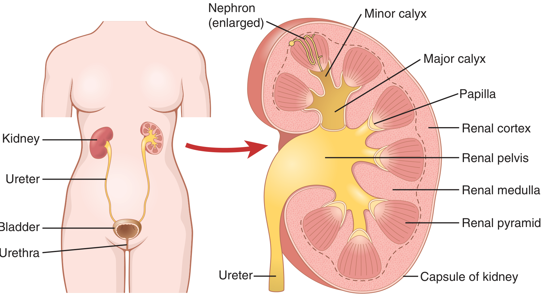
Anatomy of the Kidney
Location
The kidneys are paired, bean-shaped organs located in the retroperitoneal space of the posterior abdominal wall, on either side of the vertebral column. They extend from the 12th thoracic vertebra (T12) to the 3rd lumbar vertebra (L3). The right kidney sits slightly lower than the left because of the large right lobe of the liver above it. On the upper pole of each kidney, nestled within the renal fascia and perirenal fat, lies an adrenal (suprarenal) gland.
- Histology: A Text and Atlas, p. 1852; Medical Physiology, p. 3074
Size and Shape
Each adult kidney is:
-
Shape: bean-shaped (reniform), with a convex lateral border and a concave medial border
-
Dimensions: approximately 10 cm long × 6.5 cm wide × 3 cm thick
-
Weight: approximately 150 g (range: 125-170 g in men; 115-155 g in women)
-
Together, both kidneys account for slightly less than 0.5% of total body weight
-
Guyton and Hall, p. 3186; Medical Physiology, p. 3074; Histology: A Text and Atlas, p. 1852
External Structure
The external features of the kidney include:
| Structure | Description |
|---|---|
| Fibrous capsule | A tough, almost nondistensible layer of connective tissue surrounding the entire kidney. It has an outer layer of fibroblasts/collagen and an inner layer of myofibroblasts. It protects the delicate internal structures. |
| Convex (lateral) border | The smooth, rounded outer edge |
| Concave (medial) border | Contains the hilum - a deep vertical fissure through which structures pass |
| Hilum | The entry/exit port on the medial border. Structures passing through: renal artery and nerves enter; renal vein, lymphatics, and ureter exit |
| Renal sinus | A shallow space just inside the hilum, completely surrounded by renal parenchyma. It contains the renal pelvis, calyces, blood vessels, nerves, and fat. The capsule reflects inward at the hilum to line the sinus walls. |
The capsule passes inward at the hilum, where it becomes continuous with the connective tissue forming the walls of the calyces and renal pelvis.
- Histology: A Text and Atlas, p. 1852-1853; Medical Physiology, p. 3076
Internal Structure
A coronal (frontal) section through the kidney reveals two major zones and a collecting system:
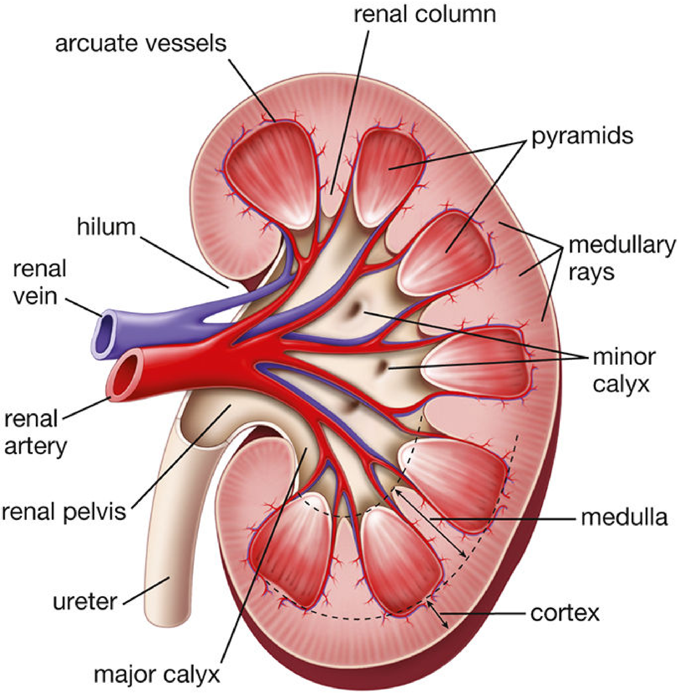
1. Renal Cortex
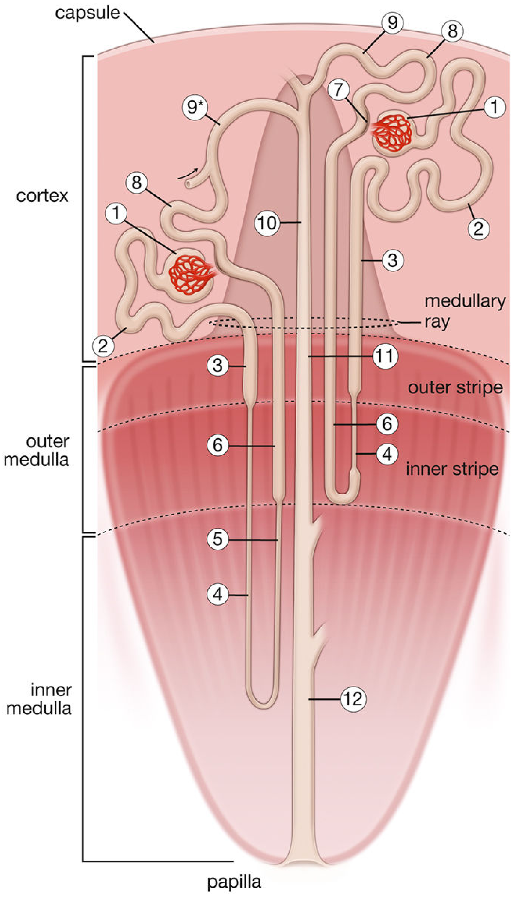
The cortex is the outer, granular layer of the kidney. Its granular appearance (on a cut surface) results from the presence of millions of glomeruli (microscopic capillary tufts) and highly convoluted tubules.
-
Contains: renal corpuscles (glomerulus + Bowman's capsule), proximal convoluted tubules, distal convoluted tubules, connecting tubules, and cortical collecting ducts
-
Receives approximately 90-95% of total renal blood flow
-
Projects downward between the pyramids as renal columns (of Bertin) - extensions of cortical tissue that dip into the medulla, separating adjacent pyramids
-
Also contains medullary rays (of Ferrein): approximately 400-500 vertical striations visible in the cortex, each being an aggregation of straight tubules and collecting ducts projecting upward from the medulla
-
Histology: A Text and Atlas, p. 1854-1855; Campbell Walsh Wein Urology, p. 2163
2. Renal Medulla
The medulla is the inner, darker region, which lacks glomeruli. It consists of a parallel arrangement of straight tubules (loops of Henle, collecting ducts) and the specialized vasa recta capillary network. The medulla is further divided into an outer medulla (outer stripe + inner stripe) and inner medulla.
3. Renal Pyramids
The medulla is divided into 8-18 conical renal pyramids (typically 8-12 in humans):
-
Base: faces the corticomedullary border (toward the cortex)
-
Apex (papilla): points toward the renal sinus; the tip is called the area cribrosa, perforated by the openings of collecting ducts through which urine drains
-
Each pyramid + its associated cortical tissue = one renal lobe
-
The tissue between pyramids (the renal columns) is cortical in nature
-
Histology: A Text and Atlas, p. 1855-1856; Guyton and Hall, p. 3188; Medical Physiology, p. 3082
4. Calyces (Minor and Major)
The calyces are cup-shaped structures that form the initial part of the urine-collecting system:
-
Minor calyces: Each is a cup-shaped extension that cups around the papilla of one renal pyramid, receiving urine directly from the collecting ducts at the area cribrosa. There are typically 8-18 minor calyces (one per papilla).
-
Major calyces: Several (2-3) minor calyces merge to form each major calyx. There are typically 2-3 major calyces per kidney.
-
The walls of the calyces contain smooth muscle that contracts to propel urine toward the renal pelvis and bladder.
-
Histology: A Text and Atlas, p. 1855; Junqueira's Basic Histology, p. 3846; Guyton and Hall, p. 3188
5. Renal Pelvis
The renal pelvis is the funnel-shaped, expanded upper end of the ureter. It is located within the renal sinus and receives urine from the major calyces. It narrows at the ureteropelvic junction (UPJ) to become the ureter, which then exits the hilum and carries urine down to the bladder. Like the calyces, its walls contain smooth muscle for peristaltic urine transport.
- Medical Physiology, p. 3076; Guyton and Hall, p. 3188; Histology: A Text and Atlas, p. 1852
Summary Table: Internal Structures
| Structure | Location | Key Feature |
|---|---|---|
| Cortex | Outer zone | Contains glomeruli, convoluted tubules; granular appearance |
| Medulla | Inner zone | Contains pyramids, straight tubules, vasa recta; striated appearance |
| Renal pyramids | Medulla | 8-18 cones; base at corticomedullary border, apex = papilla |
| Renal columns | Between pyramids | Extensions of cortex penetrating into medulla |
| Minor calyces | Renal sinus | Cup around each papilla; collect urine from ducts |
| Major calyces | Renal sinus | 2-3 per kidney; formed by merging minor calyces |
| Renal pelvis | Renal sinus/hilum | Funnel-shaped; narrows to become ureter |
This is a shared conversation. Sign in to Orris to start your own chat.