

IMG-20260707-WA0003.jpg
Answer
Answer: b. Borderline tuberculoid leprosy
Reasoning:
The key features in this case are:
- Multiple hypoesthetic, mildly erythematous large plaques with elevated margins - this points away from pure tuberculoid (which has very few, well-defined lesions) and away from lepromatous (which has many small, poorly defined, symmetric lesions)
- Bilateral enlarged peripheral nerves (ulnar + lateral popliteal) - nerve enlargement is prominent in the tuberculoid spectrum
- Hypoesthesia - indicates significant immune-mediated nerve damage, typical of the tuberculoid end of the spectrum
Why the other options are wrong:
| Option | Why excluded |
|---|---|
| a. Lepromatous leprosy | Lesions are numerous, small, poorly defined, symmetric macules/nodules; nerves enlarged but less dramatically; sensation often preserved early |
| c. Borderline leprosy (BB) | Most unstable form; lesions are "Swiss cheese" shaped with punched-out centers; nerve involvement less prominent |
| d. Borderline lepromatous (BL) | Many small lesions, less defined margins, less hypoesthesia, more bacilli; nerve thickening less pronounced |
Borderline tuberculoid (BT) is characterized by:
- Few to several plaques (more than pure TT, but not numerous)
- Large plaques with raised, well-defined outer edges and sloping inner edges
- Marked hypoesthesia
- Peripheral nerve thickening (classically ulnar and lateral popliteal nerves)
- Paucibacillary on slit-skin smear
The combination of large plaques with elevated margins + bilateral nerve enlargement + hypoesthesia is classic for BT leprosy.
Help me learn about different types of leprosy in simple terms first and then in detail for a final year mbbs student
Reading File
Now let me search the library for comprehensive leprosy content in parallel:
Finding Sources
Finding Sources
Excellent sources found. Let me read the most relevant textbooks in parallel - Harrison's, Dermatology, Park's, and Robbins:
Reading File
Reading File
Reading File
Reading File
Good data from Robbins and Park's. Now let me get the Harrison's classification section and Dermatology clinical features in depth:
Reading File
Reading File
Excellent content from Harrison's. Now let me get the Harrison's clinical features of each type and the treatment section:
Reading File
Reading File
Now let me get the Harrison's clinical features section and reactions/treatment section:
Reading File
I now have comprehensive content from Harrison's covering all types of leprosy in great detail. I have enough to write a thorough, well-structured answer. Let me also fetch the key histology image from Robbins:
Loading Image
Loading Image
I now have comprehensive, authoritative content from Harrison's 22e, Robbins Pathology, and Park's PSM to write an outstanding answer. Here it is:
Leprosy (Hansen's Disease) - Complete Guide
PART 1: SIMPLE EXPLANATION (Beginner Level)
What is leprosy?
Leprosy is a slow, chronic infection caused by the bacterium Mycobacterium leprae. It mainly attacks two things: skin and peripheral nerves (the nerves outside the brain and spinal cord). It is NOT highly contagious - most people (about 95%) who are exposed never get the disease.
The One Big Concept: It's a SPECTRUM
Think of leprosy like a slider between two extremes:
STRONG IMMUNITY ←————————————————————→ WEAK IMMUNITY

      TT ——— BT ——— BB ——— BL ——— LL

   Few lesions,            Many lesions,
   few bacteria,           many bacteria,
   lots of nerve damage    less nerve damage early
- Left side (TT): Your immune system fights hard. It kills most bacteria. But in doing so, it destroys the nerves too. Few skin lesions, lots of nerve damage.
- Right side (LL): Your immune system barely responds. Bacteria multiply everywhere. Many skin lesions all over, but nerve damage comes later.
- Middle (Borderline types): Mix of both features.
Simple Memory Trick
| Feature | Tuberculoid (TT) | Lepromatous (LL) |
|---|---|---|
| Skin lesions | Few (1-3) | Many (dozens) |
| Bacteria in lesions | Almost none | Packed full |
| Nerve thickening | Prominent, asymmetric | Symmetric, less prominent early |
| Sensation loss | Severe | Less severe early |
| Immunity | Strong | Weak |
| Lepromin test | Positive | Negative |
PART 2: DETAILED GUIDE (Final Year MBBS Level)
The Organism
Mycobacterium leprae is an obligate intracellular pathogen that:
- Cannot be cultured in vitro (grown in labs)
- Grows best at 27-33°C (prefers cool body areas: skin, peripheral nerves, testes, nasal mucosa, ear lobes)
- Its virulence is partly due to PGL-1 (phenolic glycolipid-1), which helps it invade host cells
- Primarily infects Schwann cells and dermal macrophages
- A newer species, M. lepromatosis (discovered 2008), causes diffuse lepromatous leprosy mainly in Mexico/Central America
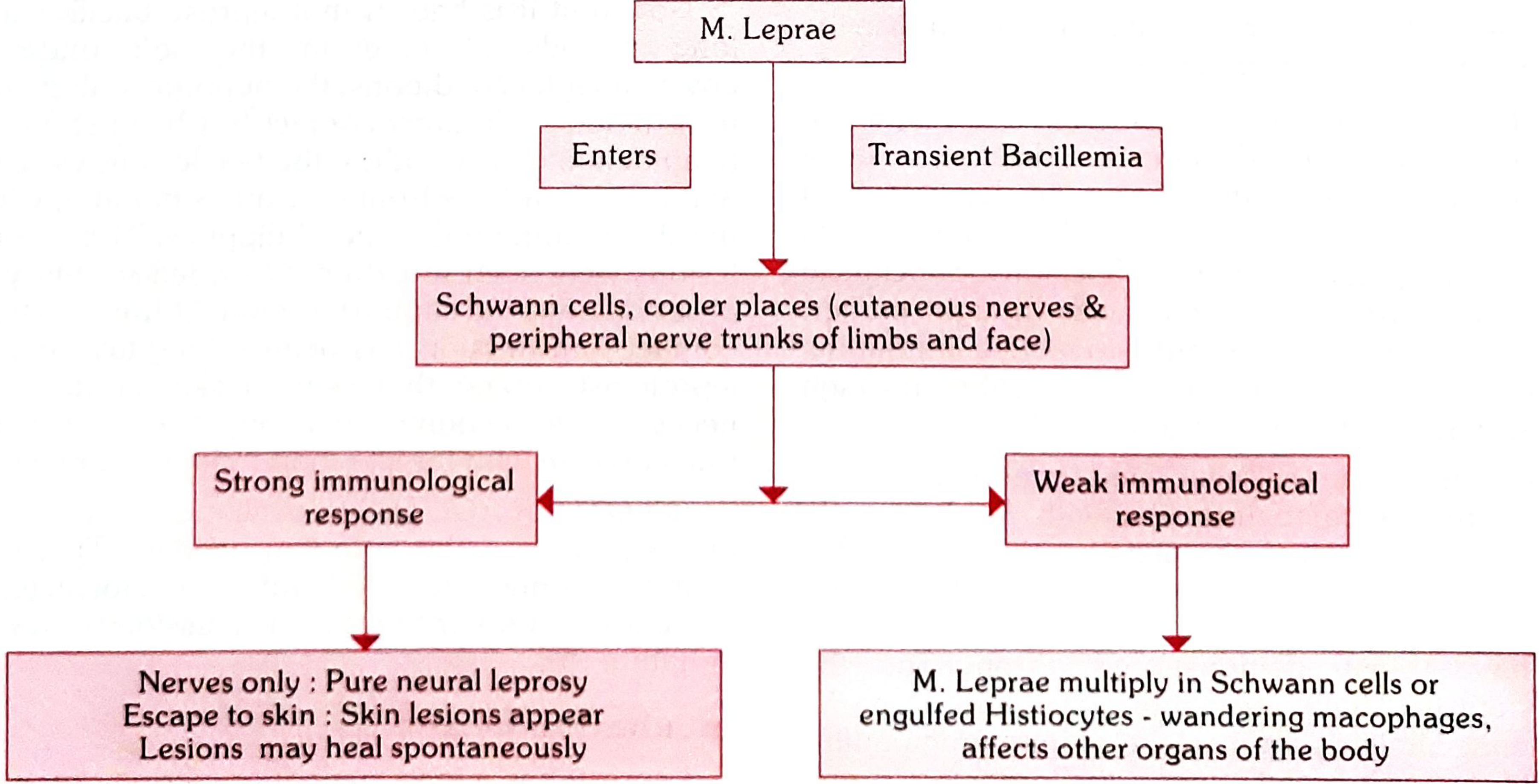
Park's Textbook of Preventive and Social Medicine - Pathogenesis of Leprosy
Transmission
- Primarily via respiratory secretions (nasal droplets) from untreated multibacillary patients
- Rare zoonotic transmission from armadillos
- Prolonged close contact required; the disease is NOT highly contagious
- Incubation period: 2-12 years (average ~5 years)
Immunology - The Key to Understanding the Spectrum
The type of leprosy that develops depends entirely on the cell-mediated immune (CMI) response:
| Immune response | Th1 (IFN-γ, IL-2) | Th2/Tregs | CMI | Result |
|---|---|---|---|---|
| Tuberculoid end | Strong | Weak | Strong | Few lesions, paucibacillary |
| Lepromatous end | Weak | Dominant | Weak | Many lesions, multibacillary |
HLA associations:
- HLA-DR2 and HLA-DR3 → tuberculoid form
- HLA-DQ1 → lepromatous form
In TT leprosy: Macrophages + lymphocytes → epithelioid cells + Langerhans giant cells → tight granuloma → kills bacteria but destroys nerves
In LL leprosy: Macrophages engulf bacteria but cannot kill them → histiocytes become "lepra cells" (Virchow cells) packed with bacteria → "globi" = clumps of acid-fast bacilli inside macrophages
Classifications of Leprosy
1. Ridley-Jopling Classification (Most important for exams)
| Type | Code | Key Features |
|---|---|---|
| Indeterminate | IL | Earliest form; may self-heal or progress |
| Tuberculoid | TT | Polar; few lesions, strong immunity |
| Borderline Tuberculoid | BT | Several lesions, asymmetric nerve involvement |
| Mid-Borderline | BB | Unstable; "Swiss cheese" lesions |
| Borderline Lepromatous | BL | Many lesions, less defined |
| Lepromatous | LL | Polar; many lesions, weak immunity |
2. WHO Classification (Used in field/control programs)
| Type | Criteria | Treatment |
|---|---|---|
| Paucibacillary (PB) | 1-5 lesions, ≤1 nerve, smear negative | 6 months MDT |
| Multibacillary (MB) | ≥6 lesions, >1 nerve, OR smear positive | 12 months MDT |
3. Indian Classification (Used in India)
- Indeterminate, Tuberculoid, Borderline, Lepromatous, Pure Neuritic (no skin lesions - unique to Indian classification)
Cardinal Features of Leprosy (Diagnose if ANY ONE present)
- Hypopigmented/erythematous skin patch with loss of sensation
- Thickened peripheral nerve trunk
- AFB positive on slit-skin smear or biopsy
Each Type in Detail
1. Indeterminate Leprosy (IL)
- Earliest stage - may be the only manifestation
- 1-2 ill-defined hypopigmented or faintly erythematous macules, 1-5 cm
- Occurs on limbs, buttocks, face
- Mild to moderate loss of touch/thermal sensation
- No nerve thickening
- Either self-heals or progresses to any determinate type depending on immunity
- Biopsy shows only lymphocytic infiltration (no granuloma yet)
2. Tuberculoid Leprosy (TT)
Skin:
- Single or very few (up to 3) well-defined lesions
- Raised, erythematous/copper-colored plaque with clearly defined outer edge sloping inward to a hypopigmented, depressed center (central healing)
- Can be of any size; surface is dry, hairless, anhidrotic, and completely anesthetic
- Complete loss of fine touch and temperature sensation over the lesion
Nerves:
- One peripheral nerve near the lesion may be thickened
- Asymmetric nerve involvement
- Nerve thickening with sensory loss and possible motor deficit
Bacteriology: AFB absent on slit-skin smear (paucibacillary)
Lepromin test: Strongly positive (Mitsuda reaction ≥5mm)
Histology: Tight, well-formed epithelioid cell granulomas with Langerhans giant cells and lymphocytes; nerves may be completely destroyed; no AFB seen
Prognosis: Relatively stable; may self-heal
3. Borderline Tuberculoid (BT) Leprosy
- 3 to 9+ lesions, asymmetrically distributed
- Plaques with edges sloping outward (opposite to TT)
- Smaller "satellite" lesions around the main lesion
- Variable loss of sensation (less than TT)
- Several peripheral nerves enlarged asymmetrically (ulnar, lateral popliteal commonly)
- Highly prone to Type 1 (reversal) reactions
- Bacteriologic Index (BI): 0 to 1+
- Lepromin test: Positive (weakly)
- If untreated, may downgrade toward BB or BL
This is the most common type seen in Indian clinical exams (as in the MCQ from your image)
4. Mid-Borderline (BB) Leprosy
- Most immunologically unstable form - can upgrade or downgrade
- Multiple lesions of varying sizes and shapes
- Characteristic "Swiss cheese" (punched-out) lesions - central clearing with a sharp inner edge but sloping outer edge
- Lesions may look like annular plaques with a "punched-out" pale center
- Sensation impaired but not totally lost
- Nerves involved but less severely than BT
- BI: 2+ to 3+
- Lepromin test: Negative or weakly positive
- Most likely to undergo lepra reactions
5. Borderline Lepromatous (BL) Leprosy
- Many lesions (more numerous and smaller than BB)
- Less-defined margins, more symmetric distribution than BT/BB
- Macules, papules, plaques, nodules
- Reduced sensation but not absent
- Nerve involvement: multiple nerves, more symmetric than BT
- BI: 3+ to 5+
- Lepromin test: Negative
- Can undergo Type 2 reactions (ENL - Erythema Nodosum Leprosum)
6. Lepromatous Leprosy (LL)
Skin:
- Many (dozens to hundreds) of lesions, symmetric distribution
- Early: poorly defined hypopigmented macules
- Late: papules, nodules, and plaques - especially on face, ears, wrists, elbows, knees
- Leonine facies = thickened, corrugated skin of face due to nodule coalescence
- Madarosis = loss of lateral eyebrows (pathognomonic)
- Saddle-nose deformity (nasal collapse from septal destruction)
- Ear lobes thickened and infiltrated
- Sensation relatively preserved early
Nerves:
- Symmetric peripheral nerve involvement
- Nerves invaded with bacteria but minimal inflammation
- Commonly affected: ulnar, median, lateral popliteal, posterior tibial, radial cutaneous, facial, greater auricular
- "Glove and stocking" sensory loss pattern
Systemic features:
- Orchitis → testicular atrophy → sterility and gynaecomastia
- Eye involvement: iridocyclitis, corneal anesthesia, lagophthalmos
- Laryngeal involvement → hoarseness
- Lymphadenopathy
- Hepatosplenomegaly
- In advanced disease: bacilli in blood and sputum
Bacteriology: AFB 4+ to 6+ (massive numbers)
- "Globi" = clumps of AFB in macrophages
- Virchow cells (lepra cells) = lipid-laden macrophages packed with AFB
Lepromin test: Negative (no CMI)
Histology: Sheets of foamy macrophages (Virchow cells); very few lymphocytes; nerves invaded but architecture preserved; AFB easily found in clumps ("globi")
Histology at a Glance
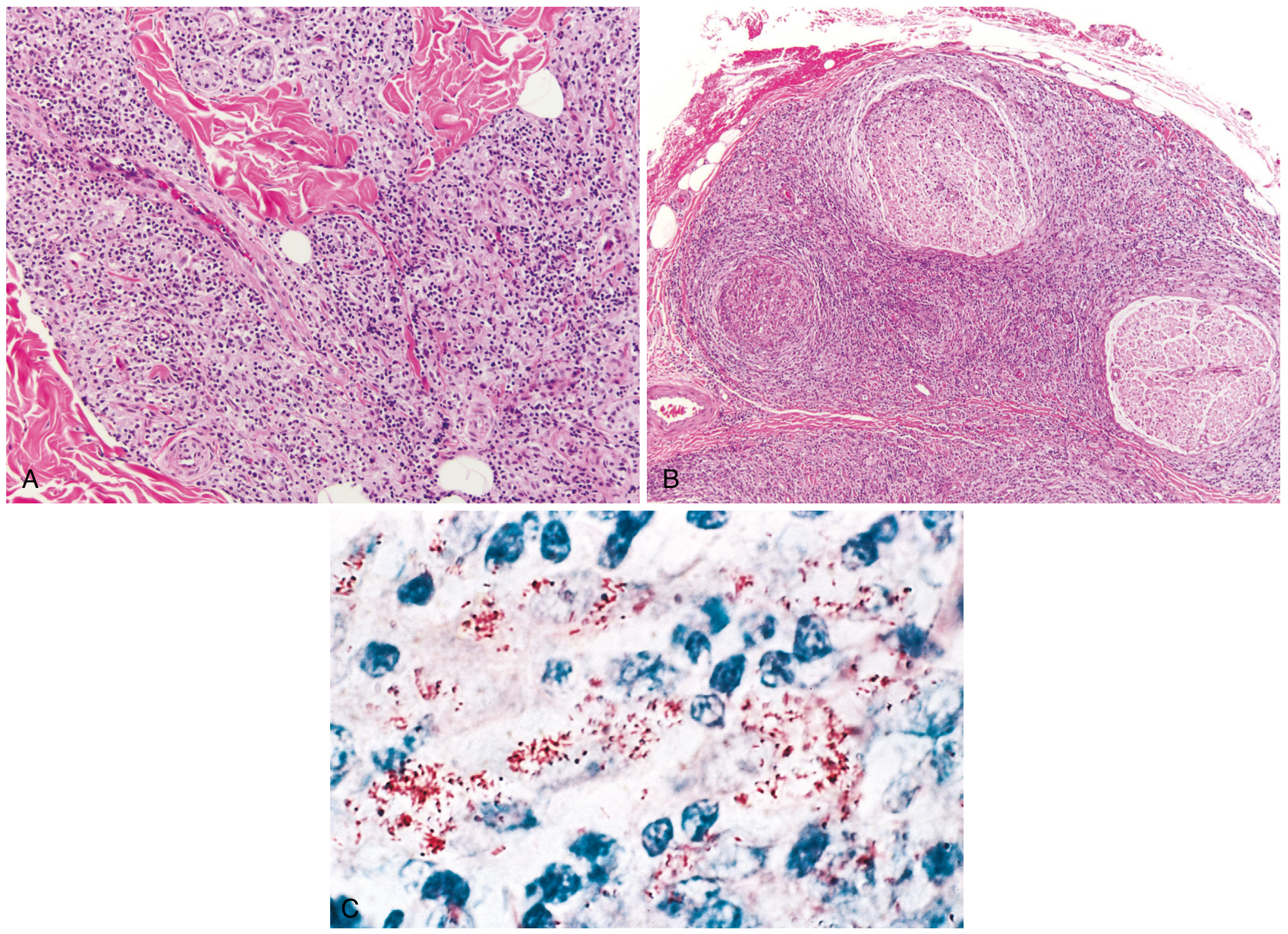
Robbins Pathology - (A) Tuberculoid leprosy: macrophage infiltration surrounding nerves and adnexa; (B) Lepromatous leprosy: dense lymphocytic infiltration into large nerve bundles; (C) AFB (red) within macrophages in lepromatous form
| Feature | Tuberculoid (TT) | Lepromatous (LL) |
|---|---|---|
| Granuloma type | Tight epithelioid + Langerhans giant cells | Loose; foamy macrophages (Virchow/lepra cells) |
| Lymphocytes | Many | Very few |
| AFB | Absent | Abundant (globi) |
| Nerve architecture | Destroyed | Preserved but infiltrated |
| Subepidermal clear zone | Absent | Present (Grenz zone) |
Nerves Commonly Affected (Must Know)
| Nerve | Site of thickening | Consequence |
|---|---|---|
| Ulnar nerve | Behind medial epicondyle | Claw hand (4th, 5th fingers) |
| Median nerve | At wrist | Claw hand (2nd, 3rd fingers), ape hand |
| Radial nerve | Lateral arm | Wrist drop |
| Lateral popliteal (common peroneal) | Neck of fibula | Foot drop |
| Posterior tibial | Behind medial malleolus | Plantar anesthesia, trophic ulcers |
| Facial nerve | Zygomatic branch | Lagophthalmos |
| Greater auricular nerve | Neck | Visible/palpable thickening |
| Supra-orbital nerve | Forehead |
Leprosy Reactions (Acute Episodes)
Type 1 Reaction (Reversal Reaction)
- Who: BT, BB, BL patients (borderline types only)
- Mechanism: Sudden increase in CMI (delayed hypersensitivity) - upgrade reaction
- Features: Existing lesions become acutely inflamed (red, hot, edematous, tender); new lesions; acute nerve palsies (very important!)
- Treatment: Prednisolone 40-60 mg/day, tapered over months
Type 2 Reaction - Erythema Nodosum Leprosum (ENL)
- Who: BL and LL patients only (multibacillary end)
- Mechanism: Immune complex deposition (Type III hypersensitivity) - NOT a change in CMI
- Features: Painful, tender, erythematous nodules on skin; fever, malaise; can have uveitis, orchitis, neuritis, arthritis, lymphadenopathy
- Treatment: Thalidomide (drug of choice), or clofazimine, or prednisolone
Lucio's Phenomenon
- Only in diffuse lepromatous leprosy (M. lepromatosis)
- Necrotizing vasculitis → skin necrosis and ulceration
Diagnosis
| Test | Finding |
|---|---|
| Slit-skin smear (SSS) | AFB on Ziehl-Neelsen stain; Bacteriologic Index (BI) 0-6+ |
| Skin biopsy | Histopathology (key for classification) |
| Lepromin test (Mitsuda) | Tests CMI; positive in TT, negative in LL (not a diagnostic test - measures resistance) |
| PCR | Confirms M. leprae DNA |
| Nerve conduction studies | Assess nerve damage |
WHO MDT Treatment (Current Regimen)
Paucibacillary (PB) - 6 months
| Drug | Dose | Frequency |
|---|---|---|
| Rifampicin | 600 mg | Monthly (supervised) |
| Dapsone | 100 mg | Daily (self-administered) |
Multibacillary (MB) - 12 months
| Drug | Dose | Frequency |
|---|---|---|
| Rifampicin | 600 mg | Monthly (supervised) |
| Clofazimine | 300 mg monthly + 50 mg daily | Monthly + daily |
| Dapsone | 100 mg | Daily |
Side effects to remember:
- Rifampicin: hepatotoxicity, orange urine
- Dapsone: hemolytic anemia (especially in G6PD deficiency), methemoglobinemia, dapsone hypersensitivity syndrome
- Clofazimine: skin pigmentation (orange-brown discoloration), ichthyosis, GI symptoms
High-Yield Points for Exams
- Most common nerve affected in leprosy overall: Ulnar nerve
- Most common cause of foot drop in leprosy: Lateral popliteal (common peroneal) nerve
- Pathognomonic skin finding: Madarosis (loss of lateral eyebrow)
- Only nerve visible AND palpable: Greater auricular nerve
- Lepromin test = test of immunity, NOT a diagnostic test
- Most unstable type: BB (mid-borderline)
- Type 1 reaction only in borderline; Type 2 (ENL) only in BL and LL
- Drug of choice for ENL: Thalidomide (contraindicated in pregnancy - teratogenic)
- BCG vaccination gives partial protection (~50%) against leprosy
- Deformity of nose in LL = Saddle nose deformity (not "button nose")
- "Leonine facies" = LL leprosy
- Most common cause of blindness in leprosy = Lagophthalmos (facial nerve palsy → corneal exposure)
- Pure neuritic leprosy (no skin lesions) - recognized in Indian classification only
- Armadillos are the only non-human animal reservoir
Sources
- Harrison's Principles of Internal Medicine, 22nd Edition, 2025 - Chapter 184
- Robbins, Cotran & Kumar - Pathologic Basis of Disease - Chapter 8
- Park's Textbook of Preventive and Social Medicine - Chapter on Leprosy
Now tell me everything about the Anatomy and physiology of Gastrointestinal System in details from basics to clinicals. Also include the appropriate diagrams and pictures wherever required.
Reading File
Finding Sources
Finding Sources
Finding Sources
Finding Sources
Reading File
Reading File
Reading File
Now let me read from Guyton's GI physiology (the most authoritative for physiology) and fetch key images in parallel:
Reading File
Reading File
Loading Image
Reading File
Reading File
Reading File
Loading Image
Now let me read key GI secretion and absorption content from Guyton and images for the gut wall structure:
Reading File
Finding Sources
Loading Image
Reading File
Reading File
Loading Image
Reading File
Reading File
Loading Image
I now have all the content needed to write a comprehensive, well-illustrated GI anatomy and physiology guide. Let me compile the full response:# Gastrointestinal System - Complete Guide (Basics to Clinicals)
PART 1: SIMPLE ORIENTATION (The Big Picture)
What is the GI System?
The gastrointestinal (GI) system is essentially a long tube - about 9 meters from mouth to anus - whose primary job is:
- Take in food (ingestion)
- Break it down (digestion - mechanical + chemical)
- Absorb nutrients into the bloodstream
- Expel waste (defecation)
It also has massive immune, endocrine, and neurological functions that make it one of the most complex systems in the body.
The GI Tract at a Glance
Mouth → Pharynx → Esophagus → Stomach → Small Intestine (Duodenum → Jejunum → Ileum)
→ Large Intestine (Cecum → Ascending → Transverse → Descending → Sigmoid Colon)
→ Rectum → Anal Canal → Anus
Accessory organs that pour secretions into the gut: Salivary glands, Liver (bile), Gallbladder (bile storage), Pancreas (enzymes + bicarbonate)
PART 2: ANATOMY - LAYER BY LAYER
General Wall Structure (Same Throughout the GI Tract)
The gut wall has 4 fundamental layers from inside out:
| Layer | Structure | Key Contents | Nerve Plexus |
|---|---|---|---|
| Mucosa | Epithelium + Lamina propria + Muscularis mucosae | Glands, villi, goblet cells | - |
| Submucosa | Dense connective tissue | Meissner's (submucosal) plexus, blood vessels, Brunner's glands (duodenum) | Meissner's plexus |
| Muscularis propria | Inner circular + Outer longitudinal smooth muscle | Controls peristalsis | Auerbach's (myenteric) plexus |
| Serosa/Adventitia | Peritoneum or fibrous coat | Outer covering | - |
Key clinical point: Auerbach's plexus (between circular and longitudinal muscle layers) controls motility. Loss of these ganglia = Hirschsprung's disease (aganglionic megacolon). - Yamada's Textbook of Gastroenterology, 7th edition
The Enteric Nervous System (ENS) - "The Second Brain":
- Over 100 million neurons in the gut wall
- Functions largely independently of the CNS
- Two main plexuses: Meissner's (secretion/absorption) and Auerbach's (motility)
- Can coordinate entire peristaltic reflexes without input from the brain
PART 3: SEGMENT-BY-SEGMENT ANATOMY AND PHYSIOLOGY
1. MOUTH (Oral Cavity)
Anatomy:
- Teeth: incisors (cutting), molars (grinding)
- Tongue: mixes food, contains taste buds (CN VII, IX, X)
- Salivary glands: parotid (largest, Stensen's duct → opposite upper 2nd molar), submandibular (Wharton's duct → floor of mouth), sublingual
Physiology - Mastication (Chewing):
- Jaw muscles innervated by CN V (trigeminal) motor branch
- Chewing controlled by reticular areas in brain stem, hypothalamus, amygdala
- Incisors generate ~55 lbs force; molars ~200 lbs
- Chewing reflex: bolus → jaw inhibition → stretch → rebound contraction (automatic rhythmic cycle)
- Function: increases surface area for enzyme access; especially important for plant foods with cellulose membranes
Salivary Secretion:
- Volume: ~1000-1500 mL/day
- Contents:
- Ptyalin (salivary amylase): begins starch → maltose hydrolysis (α-1,4 bonds); continues 1-2 hours in stomach
- Mucus: lubrication
- IgA: antibacterial defense
- Lysozyme: antibacterial
- Controlled by parasympathetic (CN VII, IX) stimulation → copious watery saliva; sympathetic → thick mucous saliva
- pH ~6.0-7.0
2. PHARYNX AND ESOPHAGUS
Anatomy of Esophagus:
- Length: ~25 cm; begins at C6 (cricopharyngeus) → ends at T10 (gastroesophageal junction, GEJ)
- Three anatomic narrowings (important for impaction/carcinoma):
- At cricopharyngeus (C6) - 15 cm from incisor
- At aortic arch/left main bronchus (T4) - 25 cm from incisor
- At GEJ/diaphragmatic hiatus (T10) - 40 cm from incisor
- Upper 1/3: striated muscle; Lower 2/3: smooth muscle; Middle: mixed
- Upper Esophageal Sphincter (UES): cricopharyngeus muscle (striated, normally CLOSED, ~60-80 mmHg)
- Lower Esophageal Sphincter (LES): smooth muscle, 3-4 cm above diaphragm (normally CLOSED, ~10-25 mmHg)
Physiology - Swallowing (Deglutition):
3 stages:
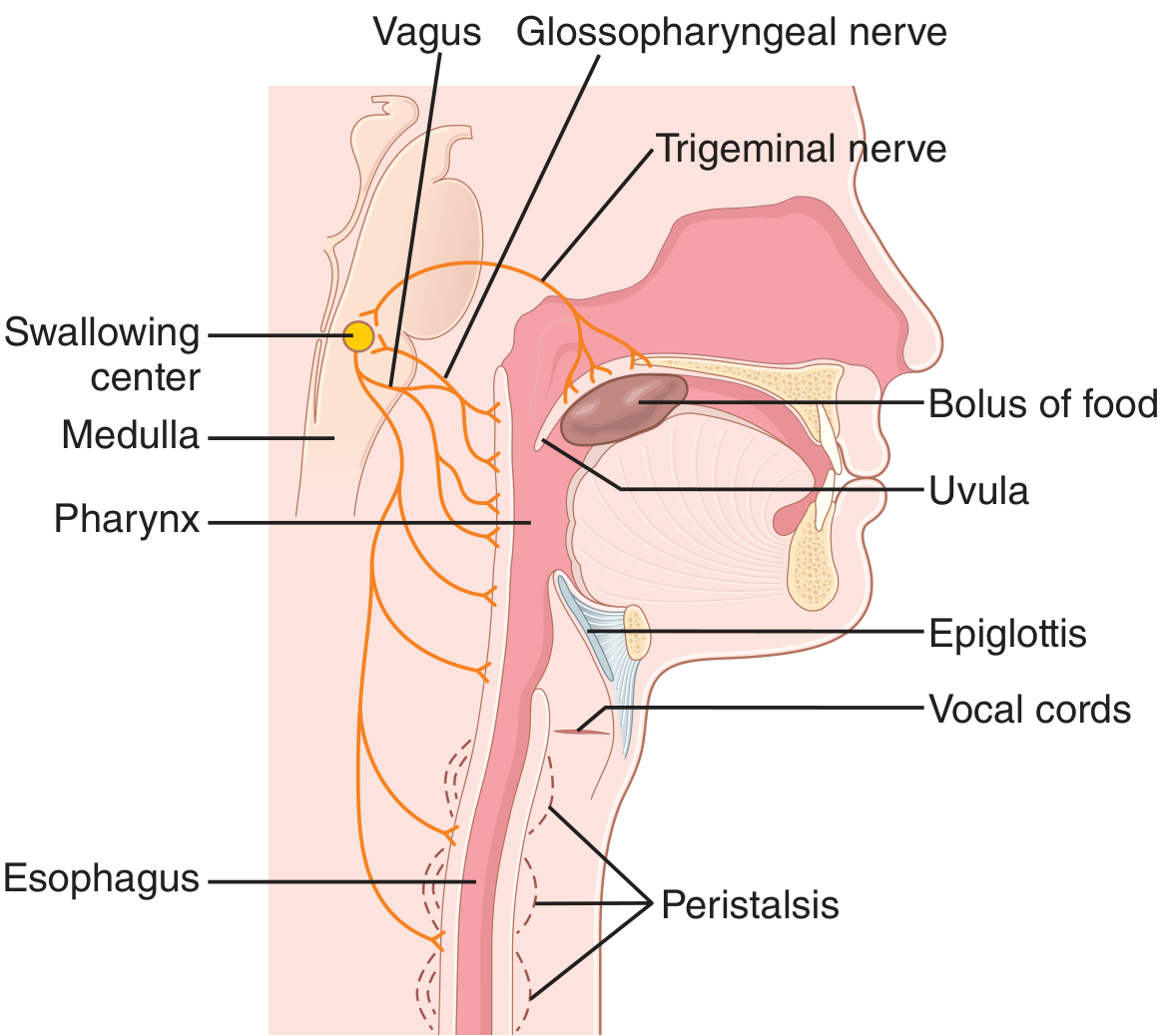
Guyton & Hall - Swallowing mechanism showing neural control via Vagus, Glossopharyngeal, and Trigeminal nerves
Stage 1 - Voluntary: Tongue pushes bolus posteriorly into pharynx
Stage 2 - Pharyngeal (Involuntary, <1 second):
- Soft palate elevates → seals nasopharynx
- Palatopharyngeal folds approximate medially → form selective slit
- Vocal cords approximate; larynx pulled up + forward → epiglottis tips back over larynx (airway protected)
- UES relaxes → bolus enters esophagus
- Swallowing center: medulla oblongata
- Nerves involved: CN V, VII, IX, X, XII
Stage 3 - Esophageal (Involuntary, 8-10 seconds):
- Primary peristalsis: initiated by swallowing reflex; progressive wave from UES to LES
- Secondary peristalsis: initiated by esophageal distension (even without swallowing); clears remaining bolus
- LES relaxes just before peristaltic wave arrives → bolus enters stomach
- LES is maintained closed by: intrinsic muscle tone, abdominal pressure, angle of His (cardiac angle), diaphragmatic crura
Clinical relevance: LES incompetence → GERD (gastroesophageal reflux disease). Failure of LES relaxation → Achalasia (absent peristalsis + incomplete LES relaxation). Loss of LES tone in pregnancy (progesterone) → heartburn in 80% of pregnancies.
3. STOMACH
Anatomy:
- J-shaped; volume ranges from ~30 mL (neonate) to 1.5-2 L (adult)
- Located at T10-L1; esophagogastric junction lies left of T10, gastroduodenal junction at L1
- Rotation during embryology: stomach rotates 90° around its longitudinal axis → explains why the left vagus innervates the anterior wall and right vagus innervates the posterior wall
4 Anatomical Regions:
| Region | Location | Key Feature |
|---|---|---|
| Cardia | Near GEJ | Transition zone; mucous glands |
| Fundus | Superior dome | Gas bubble on X-ray; relaxes to accommodate food |
| Body (Corpus) | Largest part | Oxyntic glands (acid + pepsinogen) |
| Antrum/Pylorus | Distal 20% | Pyloric glands (gastrin, mucus); pyloric sphincter |
Curvatures:
- Greater curvature (left, inferior) - site of gastric vessels (gastroepiploic)
- Lesser curvature (right, superior) - "incisura angularis" is an important landmark; site of gastric ulcer
Relations:
- Anterior: liver (right), anterior abdominal wall (left)
- Posterior: lesser sac, pancreas, transverse colon, spleen, left kidney, left adrenal (the "stomach bed")
Blood Supply:
- Right gastric + Left gastric (lesser curvature)
- Right gastroepiploic + Left gastroepiploic (greater curvature)
- Short gastric arteries (fundus, from splenic)
- All ultimately from celiac axis
Lymph Drainage:
- Follows blood supply to celiac nodes
- Clinical significance: gastric cancer spreads via lymphatics to Virchow's node (left supraclavicular), Troisier's sign
Histology - Gastric Glands:
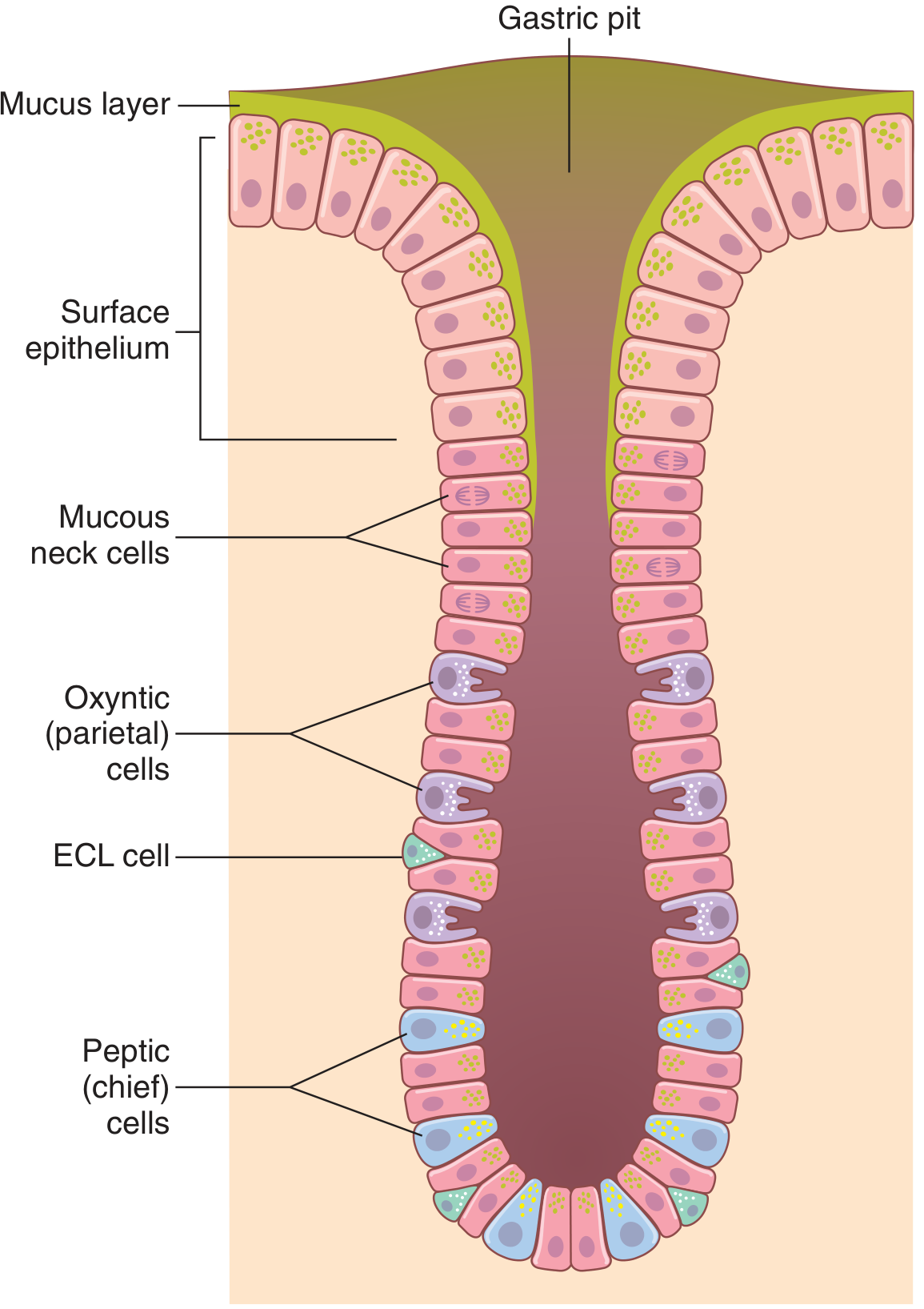
Guyton & Hall - Gastric (oxyntic) gland from body of stomach showing: Mucous neck cells, Parietal (oxyntic) cells, ECL cells, and Peptic (chief) cells
| Cell Type | Location | Secretion | Function |
|---|---|---|---|
| Surface mucous cells | Entire stomach | Mucus (HCO3 rich) | Mucosal protection (alkaline mucus barrier) |
| Mucous neck cells | Neck of oxyntic glands | Mucus (watery) | Protection |
| Parietal (oxyntic) cells | Body/fundus | HCl + Intrinsic Factor | Acid secretion; IF for B12 absorption |
| Chief (peptic) cells | Base of glands | Pepsinogen | Protein digestion (converts to pepsin in acid pH) |
| ECL cells | Oxyntic glands | Histamine | Stimulates parietal cells (via H2 receptors) |
| G cells | Antrum/pylorus | Gastrin | Stimulates parietal cells + ECL cells |
| D cells | Antrum + oxyntic glands | Somatostatin | Inhibits gastrin (paracrine) + HCl |
Gastric Acid Secretion - In Detail
Mechanism of HCl secretion by parietal cell:
- Driven by H+/K+-ATPase (proton pump) on the canalicular membrane
- H+ pumped OUT into lumen; K+ pumped IN; Cl- follows H+ via channels
- Results in ~160 mmol/L HCl, pH ~0.8 (3 million times more acidic than arterial blood)
- "Alkaline tide": as H+ is secreted, HCO3- enters blood → gastric venous blood more alkaline than arterial
Three stimulants of acid secretion (the "magic triangle"):
        GASTRIN (G cells)
              ↓
        ECL CELLS → HISTAMINE
              ↓
ACETYLCHOLINE (vagus) → PARIETAL CELL → HCl
| Stimulant | Receptor on Parietal Cell | Source |
|---|---|---|
| Gastrin | CCK-B receptor | G cells (antrum) |
| Histamine | H2 receptor | ECL cells |
| Acetylcholine | M3 muscarinic receptor | Vagus nerve |
Clinical relevance: Proton pump inhibitors (PPIs) - e.g., omeprazole - block the final common pathway (H+/K+-ATPase). H2 blockers - e.g., ranitidine - block histamine receptor. Vagotomy reduces acid by removing ACh stimulus - historically used for peptic ulcers.
Three phases of gastric secretion:
| Phase | Trigger | Mediator | % of Total Acid |
|---|---|---|---|
| Cephalic | Sight, smell, taste, thought of food | Vagus → ACh + gastrin | 30% |
| Gastric | Food in stomach → distension + protein | Gastrin, local reflexes | 60% |
| Intestinal | Chyme enters duodenum | Gastrin from duodenum (small) | 10% |
Inhibition of gastric acid:
- Acid itself (pH <3): inhibits G cells (feedback)
- Secretin (from S cells of duodenum): when duodenal pH <4.5 → inhibits gastrin and HCl
- CCK: inhibits gastric emptying
- GIP (Glucose-dependent Insulinotropic Peptide): from K cells → inhibits acid
- Somatostatin (D cells): paracrine inhibition
Gastric Motility
Reservoir function (receptive relaxation):
- As food enters, fundus relaxes (vagally mediated, via VIP and NO) to accommodate 1.5-2L with minimal pressure rise
- Absent in post-vagotomy patients → early satiety
Gastric mixing and propulsion:
- Antrum acts as a pump (pyloric pump)
- Peristaltic waves originate from a pacemaker (gastric pacemaker) at mid-corpus, rate ~3/min
- Antrum contracts → chyme propelled toward pylorus → pylorus closes → chyme rebounds back = retropulsion and mixing
Gastric emptying:
- Liquids: emptied rapidly (half-time ~20 minutes)
- Solids: require grinding into particles <1 mm before pylorus allows passage; half-time ~2-4 hours
- Fats empty most slowly
Factors inhibiting gastric emptying (enterogastric reflexes):
- Duodenal distension
- Duodenal acidity (pH <3.5-4)
- Hyper/hypoosmolarity of chyme
- Fat (fatty acids) → secretin + CCK → inhibit pyloric pump
- Protein breakdown products
Clinical relevance: Gastroparesis (delayed gastric emptying) is common in diabetes (autonomic neuropathy) → early satiety, nausea, vomiting; treated with metoclopramide. Dumping syndrome post-gastrectomy: too-rapid gastric emptying → hyperosmolar load enters jejunum → massive fluid shift → early dizziness, late hypoglycemia.
4. SMALL INTESTINE
Anatomy:
| Segment | Length | Key Features |
|---|---|---|
| Duodenum | 25-30 cm (12 inches, "duodenum" = 12) | C-shaped loop around head of pancreas; retroperitoneal (except bulb) |
| Jejunum | ~2.5 m | Proximal 2/5 of jejuno-ileum; thicker wall, prominent circular folds (plicae circulares), wider lumen, more vascular |
| Ileum | ~3.5 m | Distal 3/5; thinner wall, fewer folds, Peyer's patches, terminates at ileocecal valve |
Duodenum - 4 parts:
- 1st part (D1/duodenal bulb): 5 cm; intraperitoneal; posterior relations: gastroduodenal artery, bile duct, portal vein; site of >90% duodenal ulcers
- 2nd part (D2/descending): 7-10 cm; retroperitoneal; Ampulla of Vater on posteromedial wall (bile + pancreatic duct entry); minor papilla 2 cm proximal
- 3rd part (D3/horizontal): crosses vertebral column, IVC, aorta; crossed anteriorly by superior mesenteric vessels (SMA/SMV)
- 4th part (D4/ascending): ends at duodenojejunal flexure (ligament of Treitz) = marker for upper vs. lower GI bleeding
Clinical relevance: SMA syndrome = 3rd part of duodenum compressed between aorta (posterior) and SMA (anterior) → vomiting after meals; occurs in severe weight loss.
Jejunum vs. Ileum - how to tell apart:
| Feature | Jejunum | Ileum |
|---|---|---|
| Wall thickness | Thicker | Thinner |
| Lumen diameter | Wider (~4 cm) | Narrower (~3 cm) |
| Plicae circulares | Tall, prominent | Fewer, shorter |
| Vascularity (arcades) | 1-2 arcades, long vasa recta | 3-5 arcades, short vasa recta |
| Peyer's patches | Rare | Prominent (anti-mesenteric border) |
| Fat in mesentery | Less | More ("creeping fat") |
Surface Area Amplification - The Key to Absorption:
The small intestine maximizes absorptive area ~1000-fold by three mechanisms:
- Plicae circulares (Folds of Kerckring): permanent circular folds → 3x surface area
- Villi: finger-like projections 0.5-1.5 mm tall → further 10x surface area
- Microvilli (brush border): 1000 microvilli per cell, 1 μm long → another 20x
Total absorptive area = ~250 m² (size of a tennis court!) - Guyton & Hall, Medical Physiology
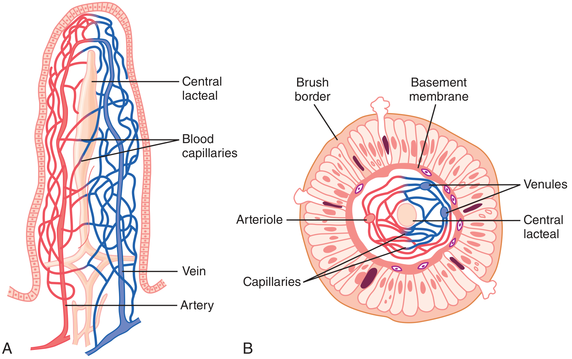
Guyton & Hall - Intestinal villus: (A) Longitudinal section showing blood capillary network and central lacteal for lymphatic absorption; (B) Cross section showing brush border, basement membrane, arteriole, venules, capillaries, and central lacteal
Villus structure (from outside in):
- Single layer of columnar epithelium (enterocytes) with tight junctions
- Central lacteal: lymphatic capillary that absorbs fat (chylomicrons)
- Blood capillaries: absorb water-soluble nutrients → portal vein → liver (first-pass metabolism)
- Goblet cells (mucus)
- Enteroendocrine cells
Crypts of Lieberkühn: tubular glands at base of villi
- Site of stem cells → cell renewal every 3-5 days
- Contain Paneth cells (antimicrobial peptides - defensins, lysozyme)
Special features:
- Brunner's glands (in submucosa of duodenum only): secrete alkaline HCO3-rich mucus → neutralize gastric acid entering duodenum; Brunner's glands are stimulated by secretin and CCK
- Peyer's patches (in submucosa of ileum): lymphoid aggregates for immune surveillance; M cells overlying them sample luminal antigens
5. LARGE INTESTINE
Anatomy:
Cecum → Ascending colon → Hepatic flexure → Transverse colon → Splenic flexure
→ Descending colon → Sigmoid colon → Rectum → Anal canal → Anus
Total length: ~1.5 m
Distinguishing features of colon (vs. small intestine):
- Teniae coli: 3 longitudinal bands of outer longitudinal muscle (instead of continuous coat) → cause colon to gather into haustra (sacculations)
- Haustra: pouches between teniae coli
- Appendices epiploicae: fat-filled peritoneal tags
- Wider caliber, no villi
Cecum and Appendix:
- Cecum: blind pouch at ileocecal junction; intraperitoneal
- Ileocecal valve: prevents backflow from colon; opens transiently with each peristaltic wave
- Appendix: arises from posteromedial cecum, 2-3 cm below ileocecal valve; McBurney's point = 1/3 of the way from ASIS to umbilicus; all layers of gut present; contains lymphoid tissue (immune function); base projection of all 3 teniae coli = landmark to find appendix
Rectum and Anal Canal:
- Rectum: 12-15 cm; follows sacral curve; 3 lateral curves (Houston's valves)
- Anorectal junction (at level of levator ani = puborectalis sling)
- Anal canal: 4 cm; has dentate (pectinate) line at midpoint
- Above dentate line: columnar epithelium, internal hemorrhoids; innervation visceral (autonomic) - painless
- Below dentate line: squamous epithelium, external hemorrhoids; innervation somatic (pudendal nerve) - painful
- Internal anal sphincter (IAS): smooth muscle, involuntary, tonically contracted (sympathetic)
- External anal sphincter (EAS): striated muscle, voluntary (pudendal nerve/somatic)
Clinical relevance: Hemorrhoids above dentate = internal (painless, bleed on defecation); below dentate = external (painful, thrombose). Fistula in ano - Park's classification based on relation to sphincters. Hirschsprung's disease - aganglionic segment always includes internal sphincter → absence of recto-anal inhibitory reflex (RAIR).
PART 4: GI PHYSIOLOGY - DIGESTION AND ABSORPTION
GI Hormones - The Master Controllers
| Hormone | Site of Secretion | Stimulus | Actions |
|---|---|---|---|
| Gastrin | G cells (antrum) | Protein, distension, vagus, hypoglycemia | ↑ HCl, ↑ pepsinogen, ↑ gastric motility, trophic to GI mucosa |
| Secretin | S cells (duodenum) | Duodenal acid (pH <4.5) | ↑ pancreatic HCO3-, ↓ HCl, ↑ bile flow, ↑ pepsinogen |
| CCK (Cholecystokinin) | I cells (duodenum + jejunum) | Fats + proteins (amino acids) | ↑ pancreatic enzymes, gallbladder contraction, ↓ gastric emptying, satiety |
| GIP | K cells (duodenum + jejunum) | Glucose + fat | ↑ insulin (incretin effect), ↓ gastric acid |
| Motilin | M cells (duodenum + jejunum) | Fasting (every 90 min) | Initiates MMC (migrating motor complex), ↑ LES tone |
| VIP | Neurons (ENS) | Distension | ↑ intestinal secretion, ↓ LES tone, vasodilation |
| Somatostatin | D cells (stomach + pancreas) | Acid, fat, glucose | Inhibits gastrin, secretin, CCK, GIP, motilin (universal OFF switch) |
| GLP-1 | L cells (ileum + colon) | Glucose, fat | ↑ insulin, ↓ glucagon, ↓ gastric emptying (incretin) |
Mnemonic for CCK functions: "CCK - Contracts gallbladder, Curtails gastric emptying, Kicks out pancreatic enzymes"
Pancreatic Secretion
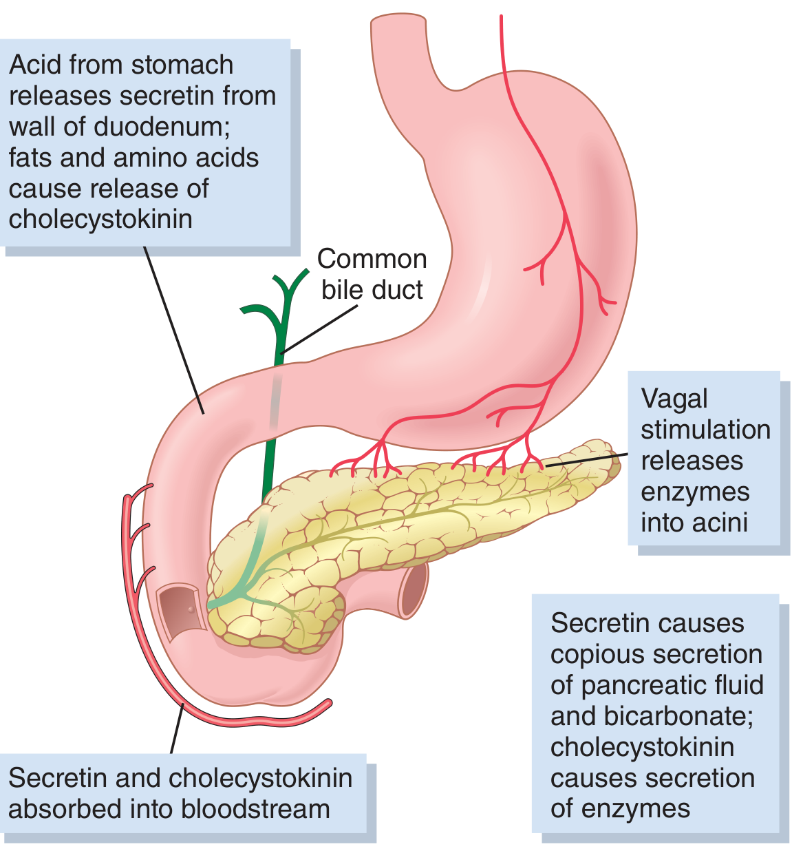
Guyton & Hall - Regulation of pancreatic secretion: Secretin (from duodenal acid) → HCO3- + water; CCK (from fats/amino acids) → enzymes; Vagal stimulation → enzyme secretion
Volume: ~1.5-2 L/day; pH ~8.0-8.3 (alkaline)
Two components:
- Aqueous component (from duct cells): NaHCO3-rich fluid → neutralizes gastric acid in duodenum; stimulated by secretin
- Enzymatic component (from acinar cells): digestive enzymes; stimulated by CCK + vagus
Pancreatic enzymes:
| Enzyme | Precursor | Activator | Substrate |
|---|---|---|---|
| Trypsin | Trypsinogen | Enteropeptidase (duodenal brush border) then trypsin (autocatalytic) | Proteins |
| Chymotrypsin | Chymotrypsinogen | Trypsin | Proteins |
| Elastase | Proelastase | Trypsin | Proteins |
| Carboxypeptidase | Procarboxypeptidase | Trypsin | Proteins |
| Pancreatic amylase | (active) | - | Starch → maltose |
| Pancreatic lipase | (active) | Colipase (needed to displace bile salts) | Triglycerides → fatty acids + monoglycerides |
| Phospholipase A2 | Prophospholipase | Trypsin | Phospholipids |
Clinical relevance: Acute pancreatitis = premature activation of trypsinogen inside acinar cells → autodigestion. Trypsin then activates all other zymogens. Enterokinase (enteropeptidase) deficiency → failure to activate trypsinogen → malabsorption + failure to thrive in infants.
Digestion of the Three Macronutrients
Carbohydrate Digestion
| Site | Enzyme | Substrate → Product |
|---|---|---|
| Mouth | Salivary amylase (ptyalin) | Starch → maltose + oligosaccharides |
| Stomach | Salivary amylase continues (until acid inactivates it) | Continues |
| Small intestine (lumen) | Pancreatic amylase | Starch → maltose, maltotriose, α-limit dextrins |
| Small intestine (brush border) | Maltase, Sucrase, Lactase, Isomaltase | Disaccharides → monosaccharides (glucose, galactose, fructose) |
Absorption:
- Glucose + Galactose: SGLT-1 (Na+/glucose cotransporter, secondary active transport) on apical membrane → basolateral exit via GLUT-2
- Fructose: GLUT-5 (facilitated diffusion) on apical → GLUT-2 basolateral
Clinical relevance: Lactase deficiency (most common enzyme deficiency worldwide, especially in East Asians and Africans) → undigested lactose enters colon → fermented by bacteria → gas, bloating, osmotic diarrhea. Glucose-galactose malabsorption = mutation in SGLT-1 → severe neonatal diarrhea.
Protein Digestion
| Site | Enzyme | Action |
|---|---|---|
| Stomach | Pepsin (from pepsinogen, pH<3) | Proteins → large peptides (10-15% of total protein digestion) |
| Small intestine (lumen) | Trypsin, chymotrypsin, elastase (endopeptidases) | Peptide bonds at specific sites |
| Small intestine (lumen) | Carboxypeptidase A & B (exopeptidases) | Remove C-terminal amino acids |
| Brush border | Aminopeptidases, dipeptidases | Oligopeptides → amino acids + di/tripeptides |
Absorption:
- Amino acids: active transport with Na+ cotransporters (multiple, specific for groups)
- Di- and tripeptides: PepT1 (H+-coupled cotransporter) on apical membrane → hydrolyzed intracellularly
- Neonates: endocytosis of intact proteins (immunoglobulins from breast milk)
Fat Digestion
| Step | Location | Process |
|---|---|---|
| 1. Emulsification | Stomach + duodenum | Bile salts + lecithin break fat droplets into small emulsion droplets |
| 2. Hydrolysis | Small intestine | Pancreatic lipase (+ colipase) → monoglycerides + fatty acids |
| 3. Micelle formation | Small intestine | Bile salts form mixed micelles with digestion products → transport to brush border |
| 4. Absorption | Jejunum brush border | Fatty acids + monoglycerides diffuse into enterocytes |
| 5. Re-synthesis | Enterocyte SER | Triglycerides re-formed; packaged as chylomicrons with apolipoprotein B-48 |
| 6. Exocytosis | Basolateral membrane | Chylomicrons → central lacteal (lymphatics) → thoracic duct → bloodstream |
Key points:
- Short-chain fatty acids (< C12): water-soluble → portal blood directly (NOT lymphatics)
- Long-chain fatty acids (> C12): require micelles + chylomicron route via lymphatics
- Bile salt reabsorption: 94-96% reabsorbed in terminal ileum (active transport) → portal vein → liver → enterohepatic circulation; pool recycles 2-3x per meal
- Fat-soluble vitamins (A, D, E, K): absorbed with fat via micelle/chylomicron route
Clinical relevance: Steatorrhea (fat malabsorption) occurs in: pancreatic exocrine insufficiency (no lipase), bile salt deficiency (cholestasis, terminal ileal disease/resection), celiac disease (villous atrophy), short bowel syndrome. Characterized by bulky, greasy, foul-smelling, floating stools.
Absorption - Site Specificity (High-Yield Table)
| Nutrient/Substance | Primary Site of Absorption |
|---|---|
| Iron | Duodenum (ferrous Fe2+ via DMT-1) |
| Calcium | Duodenum (vitamin D-dependent) |
| Folate | Duodenum + Jejunum |
| Most amino acids, sugars, fats | Jejunum (proximal) |
| Vitamin B12 (cobalamin) | Terminal ileum (requires Intrinsic Factor from parietal cells) |
| Bile salts | Terminal ileum (active) |
| Water and electrolytes | Small intestine + colon |
| Vitamin B12 | Terminal ileum |
| Fat-soluble vitamins (A,D,E,K) | Small intestine (with fat) |
| Water-soluble vitamins (C, B1, B2, etc.) | Small intestine |
| Cholesterol | Proximal jejunum (via NPC1L1) |
Clinical relevance: Terminal ileal disease or resection (Crohn's disease, right hemicolectomy) → B12 deficiency (megaloblastic anemia, subacute combined degeneration of spinal cord) + bile salt malabsorption (cholerheic diarrhea or steatorrhea if extensive).
PART 5: BILE AND THE LIVER
Bile composition:
- Water (97%), bile salts (conjugated = glyco- or tauro-cholic acid), cholesterol, phospholipids (lecithin), bilirubin, electrolytes
- Volume: 600-1000 mL/day
- Stored + concentrated (10-20x) in gallbladder
Two functions of bile:
- Fat digestion: bile salts emulsify fat + form mixed micelles
- Excretion: bilirubin, cholesterol, drugs, metabolites
Gallbladder:
- Stores and concentrates bile between meals
- CCK (released by fat + protein in duodenum) → gallbladder contraction + Sphincter of Oddi relaxation → bile into duodenum
- Fasting → gallbladder fills; eating → gallbladder empties
Clinical relevance: Gallstones (cholelithiasis): cholesterol stones (80%, from supersaturated bile) or pigment stones (hemolysis, cirrhosis). Courvoisier's law: palpable non-tender gallbladder + jaundice = malignant obstruction (NOT gallstones, which cause contracted fibrotic gallbladder). Murphy's sign: tenderness at gallbladder point on inspiration = acute cholecystitis.
PART 6: COLONIC PHYSIOLOGY
Main functions of the colon:
- Water and electrolyte absorption: colon can absorb up to 4-5 L/day; normally ~1-1.5 L arrives, 100-200 mL excreted
- Na+ absorption: aldosterone-regulated in distal colon (same as kidney)
- Short-chain fatty acid absorption: bacterial fermentation of undigested carbohydrates → butyrate (colonocyte fuel) + propionate + acetate
- Storage of feces
Colonic motility:
- Segmentation contractions: haustra mix contents (slow)
- Mass movements: 1-3 times/day; propel large amounts of feces toward rectum
- Initiated by gastrocolic reflex (eating → distension of stomach/duodenum → mass movements in colon) - basis of post-meal urge to defecate
- Also by duodenocolic reflex
Defecation:
- Rectum normally empty; feces held in sigmoid by anorectal angle + sphincters
- Defecation reflex:
- Mass movement fills rectum → rectal distension → urge to defecate
- Intrinsic reflex (myenteric): peristalsis in sigmoid + rectum, IAS relaxes (RAIR - Recto-Anal Inhibitory Reflex)
- Extrinsic reflex (spinal, via sacral parasympathetics S2-S4): amplifies intrinsic reflex
- Voluntary decision: if appropriate → EAS relaxes + Valsalva maneuver → defecation; if inappropriate → EAS contracts → rectum accommodates → urge subsides
- Valsalva maneuver: closes glottis, contracts abdominal muscles → ↑ intra-abdominal pressure
Clinical relevance: Hirschsprung's: no RAIR; Fecal incontinence: EAS weakness (obstetric injury, MS, cauda equina); Constipation: slow transit, pelvic floor dysfunction, or anorectal.
PART 7: GI MOTILITY PATTERNS
Migrating Motor Complex (MMC)
The "housekeeper" of the gut:
- Active during fasting (not after meals)
- Cycle every 90 minutes; initiated by motilin from M cells
- 3 phases:
- Phase I: quiescence (~45 min)
- Phase II: irregular contractions (~30 min)
- Phase III: intense "burst" of contractions sweeping from stomach to ileum (~5-10 min) - clears residual food, bacteria, secretions
- Disappears after eating (replaced by fed pattern)
- Absent MMC → Small Intestinal Bacterial Overgrowth (SIBO) → malabsorption, bloating, diarrhea
Peristalsis
- Ascending excitation (proximal contraction) + Descending inhibition (distal relaxation)
- Mediated by ENS: serotonin (5-HT) from enterochromaffin cells initiates peristaltic reflex
- "Law of the gut" (Bayliss-Starling): contraction above, relaxation below the bolus
Clinical relevance: Serotonin (5-HT3) antagonists (ondansetron) block the peristaltic reflex in the gut → used as antiemetics. Metoclopramide is a dopamine antagonist that enhances GI motility (prokinetic).
PART 8: ENTERIC NERVOUS SYSTEM (ENS)
- Contains ~100 million neurons (more than spinal cord)
- Two ganglionated plexuses:
- Submucosal (Meissner's plexus): controls secretion and blood flow
- Myenteric (Auerbach's plexus): controls motility (between circular and longitudinal muscle)
- Can function independently of CNS
- Receives modulating input from:
- Parasympathetic (vagus + S2-S4): generally stimulatory (via ACh, VIP)
- Sympathetic (T5-L2): generally inhibitory (via NA) - decreases motility, constricts sphincters
- Neurotransmitters: ACh (excitatory), VIP (inhibitory/relaxant), NO (relaxant), substance P, 5-HT (serotonin), neuropeptide Y
Gut-Brain Axis: 90% of vagal fibers are AFFERENT (gut → brain), not the other way. The gut senses its environment constantly and reports to the brain. IBS is partly a gut-brain axis dysregulation disorder.
PART 9: HIGH-YIELD CLINICAL CORRELATIONS
Key Clinical Points for Exams
| Condition | Key Anatomy/Physiology | Clinical Implication |
|---|---|---|
| GERD | LES incompetence; reflux of acid into esophagus | Barrett's esophagus (squamous → columnar metaplasia) → risk of adenocarcinoma |
| Achalasia | Failure of LES relaxation + absent peristalsis; loss of inhibitory neurons (VIP, NO) in LES | "Bird-beak" on barium swallow; dysphagia to both solids AND liquids |
| Peptic ulcer | H. pylori destroys mucous layer; NSAIDs inhibit COX-1 (prostaglandins protect mucosa) | Duodenal ulcer: high acid, relieved by food; Gastric ulcer: may worsen with food |
| Zollinger-Ellison | Gastrinoma (non-beta islet cell tumor) → massive gastrin → massive HCl → multiple peptic ulcers | Elevated fasting gastrin; secretin stimulation test → paradoxical rise; treat with PPIs + resect tumor |
| Celiac disease | Gluten (gliadin) → immune reaction → villous atrophy in proximal small intestine | Malabsorption of iron (duodenum), folate, fat; Anti-tTG antibodies |
| Crohn's disease | Transmural inflammation, skip lesions, ANY part of GI tract (mouth to anus), cobblestone mucosa | Terminal ileum most common site; B12 + bile salt malabsorption; risk of fistulae, strictures |
| Ulcerative colitis | Mucosal inflammation only; starts at rectum, continuous proximal spread; colon only | Bloody diarrhea; toxic megacolon; risk of colorectal carcinoma (after 10 years) |
| Hirschsprung's | Absence of Auerbach's + Meissner's ganglia; always includes internal sphincter | No RAIR; presents as neonatal obstruction; aganglionic segment on biopsy (absent ganglion cells) |
| Meckel's diverticulum | Remnant of vitello-intestinal duct; 2 feet from ileocecal valve; 2 inches long; 2% of population; 2:1 male; presents by age 2 | 50% contain ectopic gastric mucosa → acid secretion → bleeding/ulceration; "Rule of 2s" |
| Carcinoid tumor | From enterochromaffin (Kulchitsky) cells; ileum most common; secretes serotonin | Carcinoid syndrome (flushing, diarrhea, wheezing, tricuspid stenosis) only with liver mets (5-HIAA in urine) |
| SMA syndrome | 3rd part of duodenum compressed between aorta + SMA | Occurs in rapid weight loss (loss of fat pad around SMA); vomiting, epigastric pain relieved by prone position |
| Short gut syndrome | After extensive small bowel resection | Malabsorption, B12 deficiency, diarrhea; TPN dependence |
| VIPoma | VIP-secreting tumor | WDHA syndrome: Watery Diarrhea, Hypokalemia, Achlorhydria (VIP inhibits acid) |
QUICK REVISION SUMMARY TABLE
| Parameter | Mouth | Esophagus | Stomach | Small Intestine | Large Intestine |
|---|---|---|---|---|---|
| Length | - | 25 cm | Capacity 1.5-2L | ~6 m (duod 30cm, jej 2.5m, ileum 3.5m) | ~1.5 m |
| Epithelium | Stratified squamous | Stratified squamous | Simple columnar | Simple columnar | Simple columnar |
| pH | 6-7 | 7.0 | 1.5-3.5 | 6-7.5 (rises) | 5.5-7 |
| Key secretion | Saliva (amylase) | Mucus | HCl + pepsinogen | Succus entericus | Mucus |
| Transit time | Seconds | 8-10 sec | 2-4 hrs | 3-5 hrs | 10-60 hrs |
| Main function | Mastication, salivary digestion | Transport | Storage, acid digestion, mixing | Digestion + absorption | Water/electrolyte absorption, storage |
| Key sphincter | - | UES + LES | Pyloric sphincter | Ileocecal valve | IAS + EAS |
Sources
- Guyton and Hall Textbook of Medical Physiology, 14th Edition - Units XII-XIII (GI Physiology)
- Ganong's Review of Medical Physiology, 26th Edition - GI Section
- Sleisenger and Fordtran's GI and Liver Disease, 11th Edition - Stomach + Duodenum Anatomy chapters
- Yamada's Textbook of Gastroenterology, 7th Edition - GI wall layers and histopathology
- Harrison's Principles of Internal Medicine, 22nd Edition - GI clinical correlations
Create a comprehensive study guide PDF on GI pathophysiology and common GI disorders
Your credits are exhausted. You can purchase a top-up or subscribe to a plan to continue using Orris. If you're already subscribed, your daily credits will reset tomorrow.
Hepatobiliary system
Your credits are exhausted. You can purchase a top-up or subscribe to a plan to continue using Orris. If you're already subscribed, your daily credits will reset tomorrow.
This is a shared conversation. Sign in to Orris to start your own chat.