Scrambled cell carcinoma. Observe a clinical cancer, show keratinization or not.

Reading File
Finding Sources
Searching PubMed

squamous cell carcinoma AND keratinization

Searching the Web

squamous cell carcinoma histology keratinization keratin pearls pathology

Searching Images

histology squamous cell carcinoma keratin pearls well differentiated
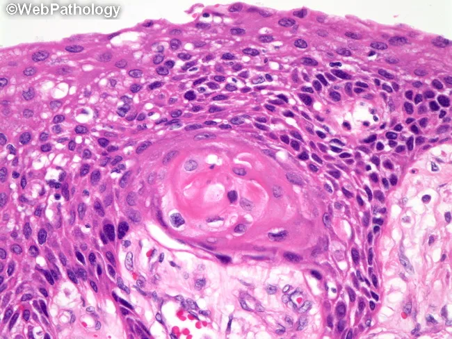
Imaging modality: Light microscopy of a hematoxylin and eosin stained tissue section, often obtained by biopsy. The slide demonstrates conventional histology of a keratinizing squamous cell carcinoma with well-differentiated epithelial nests infiltrating the surrounding stroma. Tumor cells are polygonal with abundant eosinophilic cytoplasm and intercellular bridges; keratinization is evident as keratin pearls within tumor nests. The basal layer shows increased mitotic activity and nuclear atypia, with occasional keratinizing halos surrounding nests. In focal areas, intracellular mucin may be present in scattered tumor cells, adding diagnostic nuance. The overall architecture includes cohesive nests of malignant squamous cells, keratin formation, and a fibrous stromal response. Immunohistochemistry would support squamous lineage if keratinization is less prominent or ambiguous, with p40, p63, CK5/6 positivity favoring squamous differentiation and TTF-1 negativity helping exclude pulmonary adenocarcinoma or other TTF-1 expressing tumors. Clinically, this pattern correlates with a well-differentiated squamous carcinoma that tends to show better differentiation than non-keratinizing variants but still requires complete excision or staged therapy based on invasion depth and anatomic site. This image is representative for pathology education, tumor grading, and diagnostic pathology workflow, illustrating classic keratinization, keratin pearls, and intercellular bridges characteristic of well-differentiated keratinizing squamous cell carcinoma.
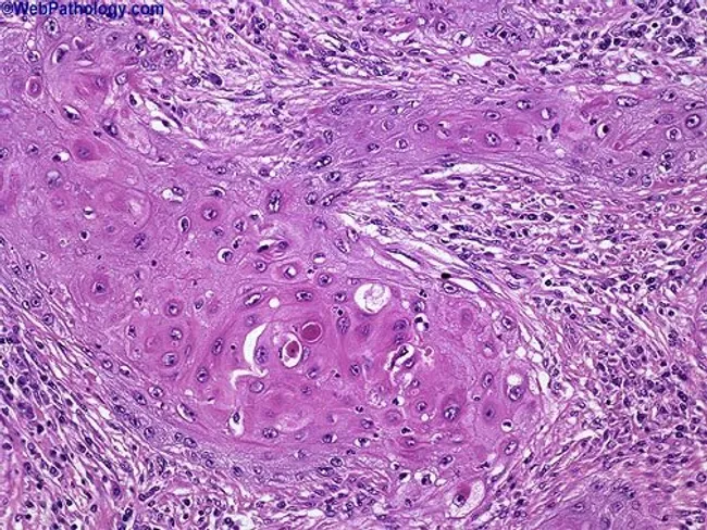
Imaging modality: Light microscopy of a hematoxylin and eosin stained tissue section, often obtained by biopsy. The slide demonstrates conventional histology of a keratinizing squamous cell carcinoma with well-differentiated epithelial nests infiltrating the surrounding stroma. Tumor cells are polygonal with abundant eosinophilic cytoplasm and intercellular bridges; keratinization is evident as keratin pearls within tumor nests. The basal layer shows increased mitotic activity and nuclear atypia, with occasional keratinizing halos surrounding nests. In focal areas, intracellular mucin may be present in scattered tumor cells, adding diagnostic nuance. The overall architecture includes cohesive nests of malignant squamous cells, keratin formation, and a fibrous stromal response. Immunohistochemistry would support squamous lineage if keratinization is less prominent or ambiguous, with p40, p63, CK5/6 positivity favoring squamous differentiation and TTF-1 negativity helping exclude pulmonary adenocarcinoma or other TTF-1 expressing tumors. Clinically, this pattern correlates with a well-differentiated squamous carcinoma that tends to show better differentiation than non-keratinizing variants but still requires complete excision or staged therapy based on invasion depth and anatomic site. This image is representative for pathology education, tumor grading, and diagnostic pathology workflow, illustrating classic keratinization, keratin pearls, and intercellular bridges characteristic of well-differentiated keratinizing squamous cell carcinoma.

This histopathology image depicts a cutaneous squamous cell carcinoma on routine hematoxylin and eosin stained section. The biopsy shows nests of malignant squamous cells with keratinization and abundant keratin pearls, indicating squamous differentiation. The tumor exhibits moderate differentiation, evidenced by intercellular bridges, eosinophilic cytoplasm, nuclear pleomorphism, hyperchromasia, and mitotic activity. Invasion into the surrounding dermal stroma is evident, with desmoplastic-type fibrous response in some areas. The epidermis overlying the lesion shows dysplastic changes, and keratin pearl formation within tumor nests is a hallmark of keratinizing SCC. The architecture includes irregular cords and nests of polygonal cells, occasional central keratinization, and keratinized pearls of varying sizes. Clinically, these features suggest a malignant cutaneous neoplasm with potential for local invasion; staging would consider depth of invasion and perineural or lymphovascular involvement. This image is relevant for diagnostic pathology, histology education, and tumor biology research, illustrating characteristic squamous differentiation, keratin production, and invasion patterns. Differential considerations include well-differentiated keratinizing SCC versus moderately-to-poorly differentiated forms, verrucous carcinoma, and basal cell carcinoma with squamous features. The visual cues—keratin pearls, keratinization, and cohesive epidermal-derived tumor islands—assist in confirming diagnosis and guiding surgical management. Correlation with clinical data will inform prognosis and adjuvant therapy decisions and multidisciplinary care planning.
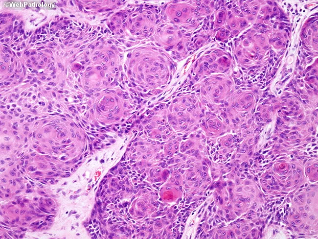
This histopathology image depicts a cutaneous squamous cell carcinoma on routine hematoxylin and eosin stained section. The biopsy shows nests of malignant squamous cells with keratinization and abundant keratin pearls, indicating squamous differentiation. The tumor exhibits moderate differentiation, evidenced by intercellular bridges, eosinophilic cytoplasm, nuclear pleomorphism, hyperchromasia, and mitotic activity. Invasion into the surrounding dermal stroma is evident, with desmoplastic-type fibrous response in some areas. The epidermis overlying the lesion shows dysplastic changes, and keratin pearl formation within tumor nests is a hallmark of keratinizing SCC. The architecture includes irregular cords and nests of polygonal cells, occasional central keratinization, and keratinized pearls of varying sizes. Clinically, these features suggest a malignant cutaneous neoplasm with potential for local invasion; staging would consider depth of invasion and perineural or lymphovascular involvement. This image is relevant for diagnostic pathology, histology education, and tumor biology research, illustrating characteristic squamous differentiation, keratin production, and invasion patterns. Differential considerations include well-differentiated keratinizing SCC versus moderately-to-poorly differentiated forms, verrucous carcinoma, and basal cell carcinoma with squamous features. The visual cues—keratin pearls, keratinization, and cohesive epidermal-derived tumor islands—assist in confirming diagnosis and guiding surgical management. Correlation with clinical data will inform prognosis and adjuvant therapy decisions and multidisciplinary care planning.

This is a high-fidelity light microscopy image of a formalin-fixed, paraffin-embedded skin biopsy stained with Hematoxylin and Eosin, viewed under bright-field illumination at 400x total magnification (40x objective). The epidermal squamous cells show cohesive nests and finger-like islands with pronounced keratinization. The most striking feature is a striking whorling pattern composed of concentric lamellations of epithelial cells, forming keratin pearls in a well-differentiated squamous phenotype. The cells are polygonal with abundant eosinophilic cytoplasm, distinct intercellular bridges, and prominent, often irregular nuclei with mild-to-moderate atypia. Mitotic figures are present in some areas, but the overall architecture demonstrates orderly differentiation with lamellated keratin deposition. The surrounding dermis exhibits a mild inflammatory infiltrate and scattered fibrovascular stroma, with no obvious overt invasion in this field; however, invasion may be present in deeper sections. Pathologic interpretation centers on squamous differentiation with keratinization; keratin pearls and whorling patterns support a squamous lineage and are characteristic of well-differentiated squamous cell carcinoma or keratoacanthoma depending on context. Clinically, this histology informs diagnostic considerations, helps distinguish from basal cell carcinoma and benign hyperkeratosis, and guides management decisions such as surgical excision with clear margins and staging assessment. Correlation with clinical history and ancillary studies improves accuracy and prognostication.

This is a high-fidelity light microscopy image of a formalin-fixed, paraffin-embedded skin biopsy stained with Hematoxylin and Eosin, viewed under bright-field illumination at 400x total magnification (40x objective). The epidermal squamous cells show cohesive nests and finger-like islands with pronounced keratinization. The most striking feature is a striking whorling pattern composed of concentric lamellations of epithelial cells, forming keratin pearls in a well-differentiated squamous phenotype. The cells are polygonal with abundant eosinophilic cytoplasm, distinct intercellular bridges, and prominent, often irregular nuclei with mild-to-moderate atypia. Mitotic figures are present in some areas, but the overall architecture demonstrates orderly differentiation with lamellated keratin deposition. The surrounding dermis exhibits a mild inflammatory infiltrate and scattered fibrovascular stroma, with no obvious overt invasion in this field; however, invasion may be present in deeper sections. Pathologic interpretation centers on squamous differentiation with keratinization; keratin pearls and whorling patterns support a squamous lineage and are characteristic of well-differentiated squamous cell carcinoma or keratoacanthoma depending on context. Clinically, this histology informs diagnostic considerations, helps distinguish from basal cell carcinoma and benign hyperkeratosis, and guides management decisions such as surgical excision with clear margins and staging assessment. Correlation with clinical history and ancillary studies improves accuracy and prognostication.

Reading File
You likely mean squamous cell carcinoma (SCC).
Keratinization may be present or absent, depending on differentiation:
  • Well-differentiated SCC: prominent keratinization, often forming keratin pearls, with intercellular bridges.
  • Moderately differentiated SCC: less keratinization and greater nuclear atypia.
  • Poorly differentiated / non-keratinizing SCC: little or no obvious keratinization; may need immunohistochemistry such as p40/p63 and CK5/6 to confirm squamous differentiation.
So, the classic histologic sign of a keratinizing SCC is a keratin pearl: concentric eosinophilic keratin in a nest of invasive malignant squamous cells. Robbins, Cotran & Kumar Pathologic Basis of Disease describes SCC as characterized by keratinization and/or intercellular bridges, most evident in well-differentiated tumors.
Keratinizing squamous cell carcinoma showing keratin pearls
This distinction also appears in the pulmonary SCC classification: keratinizing, non-keratinizing, and basaloid types.
This is a shared conversation. Sign in to Orris to start your own chat.