An interno and ab externo subconjunctival migs steps
ab interno ab externo subconjunctival MIGS XEN Preserflo surgical steps
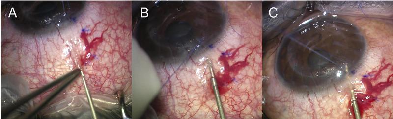
This clinical photographic sequence illustrates the three critical surgical steps of an ab-externo XEN gel stent implantation, a minimally invasive glaucoma surgery (MIGS). Panel A shows the initial conjunctival insertion, where the injector needle is positioned approximately 7 mm posterior to the limbus, stabilized by conjunctival forceps. Panel B depicts the scleral entry phase, with the needle advancing through the scleral tissue approximately 2.5 mm behind the limbus at a shallow angle. Panel C demonstrates the final intraocular positioning, where the injector tip has penetrated into the anterior chamber, visible through the clear cornea. Throughout the procedure, the bulbar conjunctiva shows significant vascular engorgement and localized subconjunctival hemorrhage at the entry sites, which are typical findings. Blue surgical markings on the sclera indicate the measured distances for precise entry. This sequence serves as an educational guide for ophthalmology trainees on the external-to-internal approach for glaucoma drainage device placement.
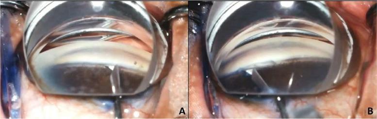
This clinical photograph displays two panels (A and B) showcasing the surgical steps of Bent Ab interno Needle Goniotomy (BANG), a minimally invasive glaucoma surgery (MIGS). The images are captured through a surgical gonioscopy lens, which provides a clear, magnified view of the anterior chamber angle of the eye. In Panel A, a 25-gauge angled needle tip is shown making contact with and penetrating the nasal trabecular meshwork. The needle is positioned to access Schlemm's canal. In Panel B, the post-incisional result is visible, demonstrating a 90-degree sectoral excision of the trabecular meshwork across the superonasal, nasal, and inferonasal quadrants. The surgical lens appears as a transparent, dome-shaped interface on the corneal surface. This educational visual illustrates the precise anatomical targeting required to enhance aqueous humor outflow in the management of intraocular pressure. The imagery is intended for advanced ophthalmology education, specifically focusing on surgical techniques for glaucoma treatment.
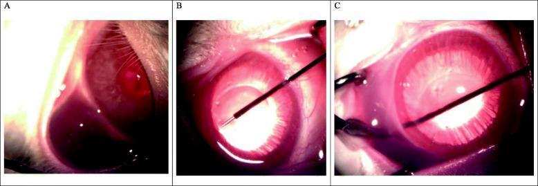
This sequence of three clinical intraoperative photographs (labeled A, B, and C) demonstrates the stages of an ab-interno laser glaucoma procedure in a lab setting. Panel A shows the initial subconjunctival injection of mitomycin C (MMC), resulting in a localized, dark, fluid-filled bleb intended to inhibit postoperative fibrosis. Panels B and C illustrate the surgical approach using a 2940nm Er:YAG thermal-ablating laser system. In Panel B, a thin laser probe is inserted into the anterior chamber through a 1mm clear corneal incision, positioned across the iris plane. Panel C depicts the advancement of the laser probe to perform tissue ablation at the trabecular meshwork, creating a permanent drainage channel (scleral tunnel) from the anterior chamber to the subconjunctival space. The images highlight key landmarks including the cornea, iris, and the surgical instrumentation. This material is designed for advanced ophthalmic surgical training, focusing on minimally invasive glaucoma surgery (MIGS) techniques and the use of adjunct antimetabolites in filtration procedures.
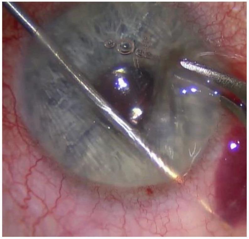
This clinical photograph is an intraoperative view of an ophthalmic surgical procedure, specifically a XEN Gel Stent implantation for glaucoma management. The image displays the anterior segment of the eye with a thin, metallic injector inserted through a temporal-inferior side port incision. The injector traverses the anterior chamber, with its distal tip positioned approximately 2 mm posterior to the corneal limbus, residing underneath the conjunctiva to facilitate the ab interno creation of a subconjunctival drainage bleb. The conjunctiva shows localized vascular congestion and mild erythema, consistent with surgical manipulation and the administration of mitomycin C. The cornea remains relatively transparent but exhibits surface reflections and minor irregularities likely due to the application of viscoelastic substances. Small air bubbles are visible within the fluid-filled space above the cornea. This visual demonstrates the precise needle-passing technique required to bridge the anterior chamber and the subconjunctival space to ensure proper stent placement for intraocular pressure reduction.
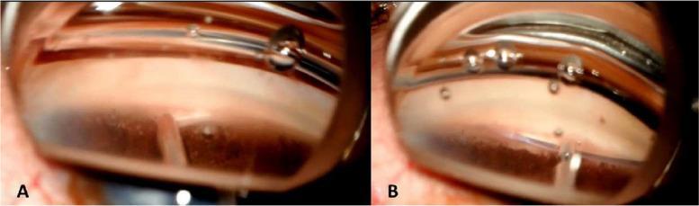
This clinical procedural image consists of two panels (A and B) demonstrating the intraoperative steps of Gonioscopy-Assisted Transluminal Trabeculotomy (GATT), a minimally invasive glaucoma surgery (MIGS). The view is obtained via an ab interno approach through a surgical goniolens, visualizing the nasal anterior chamber angle. Panel A shows the initial step where a 25-gauge needle is used to create a focal goniotomy, incising the trabecular meshwork to expose the underlying Schlemm's canal. Panel B illustrates the subsequent cannulation phase, where a 5-0 Prolene (polypropylene) suture wire is inserted through the goniotomy and advanced 360 degrees into the lumen of Schlemm's canal, guided by 23-gauge microforceps. Key anatomical landmarks visible include the clear cornea with surgical air bubbles, the pigmented trabecular meshwork band, and the iris periphery. This procedure is performed to reduce intraocular pressure by bypassing the primary site of aqueous outflow resistance.
Ab Interno vs Ab Externo Subconjunctival MIGS - Surgical Steps
Classification
| Feature | Ab Interno | Ab Externo |
|---|---|---|
| Entry point | Clear corneal incision (inside out) | Conjunctival/scleral surface (outside in) |
| Gonioscopy needed | Yes (for some) | No |
| Conjunctival incision | Not required | Required |
| Representative device | XEN Gel Stent (ab interno technique) | PRESERFLO MicroShunt, InnFocus MicroShunt |
Ab Interno Subconjunctival MIGS (XEN Gel Stent)
- Mitomycin C (MMC) 0.02%, 0.1 ml is injected subconjunctivally at the target bleb site (typically superonasal quadrant) to reduce fibrosis.
- Conjunctival marking - Mark the target site ~2-3 mm posterior to the limbus on the sclera (the intended external end of the stent).
- Clear corneal incision - A 1.8 mm temporal clear corneal paracentral incision is made with a keratome.
- AC fill with viscoelastic - OVD is injected to maintain the AC and protect the corneal endothelium.
- Goniolens placement - A surgical gonioscopy lens (e.g., Swan-Jacob) is placed on the cornea to visualize the nasal angle under direct gonioscopic view.
- Injector insertion - The pre-loaded XEN injector (with the stent) is introduced through the corneal incision and advanced across the AC toward the nasal angle.
- Needle passes through trabecular meshwork and sclera - Under gonioscopic guidance, the injector needle penetrates the trabecular meshwork, traverses the scleral tissue, and exits ~2-3 mm posterior to the limbus into the subconjunctival space.
- Stent deployment - The stent is deployed by advancing the plunger; the inner end remains in the AC (~1 mm), the mid-portion traverses the sclera, and the outer end lies subconjunctivally (~3 mm).
- Injector withdrawal - The injector is carefully withdrawn.
- Position verification - The intracameral end is confirmed gonioscopically; the subconjunctival end is confirmed externally. An early bleb should form.
- OVD removal - Residual viscoelastic is aspirated/irrigated.
- Wound check - The corneal incision is hydrated or sutured if needed.
Ab Externo Subconjunctival MIGS (PRESERFLO MicroShunt / InnFocus)
- MMC 0.02% 0.1 ml is injected subconjunctivally (or applied on a sponge) to the target site to reduce bleb fibrosis.
- Conjunctival peritomy - A small limbal-based or fornix-based conjunctival incision is made at the target quadrant (typically superior or superonasal).
- Conjunctival/Tenon dissection - Blunt dissection posteriorly under the conjunctiva and Tenon capsule creates a pocket for the device's posterior fin/plate.
- Scleral marking - The entry point on the sclera is marked ~3 mm posterior to the limbus. Some surgeons use a partial-thickness scleral groove.
- Needle track creation - A 25-gauge or 27-gauge needle is advanced through the scleral tissue at a shallow angle (bevel down), passing through to the AC. The track traverses sclera and trabecular meshwork to enter the AC.
- Device insertion - The PRESERFLO tube is threaded through the needle track. The anterior (lumen) tip is positioned in the AC; the posterior limb with the fin lies in the subconjunctival/sub-Tenon space.
- Position check - The intracameral end is verified (should be visible in the AC, away from iris/cornea). The posterior fin helps anchor the device.
- Conjunctival closure - The conjunctival/Tenon layer is sutured with 10-0 or 9-0 nylon or absorbable sutures, ensuring a watertight closure over the device.
- Bleb formation check - A diffuse, low bleb should form at the end of surgery.
Key Comparison: Postoperative Management
- Both devices: bleb needling may be required postoperatively for elevated IOP due to fibrosis.
- Both use MMC as adjunct antimetabolite.
- XEN is done under gonioscopic control - requires surgeon experience with gonioscopy.
- PRESERFLO has a more familiar surgical approach for surgeons trained in filtration surgery.
- Both target mild-to-moderate glaucoma (goal IOP 15-17 mmHg); combined with phacoemulsification in many cases.
- Complications: bleb-related (hypotony, infection, bleb fibrosis), stent malposition, erosion.