
Fundamental principles of ventricular assist devices
I'll start by reading the medical library skill, then search for comprehensive information on ventricular assist devices.
Reading File
Now I'll search the medical library and PubMed in parallel for comprehensive VAD content.
Finding Sources
Finding Sources
Searching PubMed
"ventricular assist device" AND "mechanical circulatory support"
Excellent. Now I'll read the most comprehensive VAD sections from Braunwald's and Fuster's, plus the echocardiography book - all in parallel.
Reading File
Reading File
Reading File
The Fuster block had PPCM context. I need to find the main VAD principles section in Braunwald's. Let me search for the fundamental/principles section and also read the main VAD block from Braunwald's.
Finding Sources
Reading File
Excellent - that's very rich content from Braunwald's. Now let me read the next sections covering device types, pump mechanisms, and complications. I'll also pull the Harrison's section on MCS.
Reading File
Reading File
I now have very rich content. Let me read more of the critical Braunwald's section on pump types (pulsatile vs. continuous flow) and complications, and also grab the Fuster's Heart comprehensive VAD section.
Reading File
Reading File
The Fuster block was about Fontan, not general VAD. I have rich content from Braunwald's - now let me read the device types/pump mechanism section, which should be around offset 353-470 (already partially covered), and the complications section.
Reading File
Finding Sources
I now have comprehensive content. Let me also fetch one of the device images from Braunwald's and read the complications/adverse events section.
Reading File
Finding Sources
I have comprehensive content. Let me fetch one key diagram image from Braunwald's to include in the answer.
Reading File
Excellent! I now have all the content I need. Let me compile a comprehensive, well-structured answer.
Fundamental Principles of Ventricular Assist Devices (VADs)
1. Definition and Concept
Mechanical circulatory support (MCS) devices are mechanical pumps designed to assist or replace the function of the left and/or right ventricle(s). A ventricular assist device specifically augments cardiac output by drawing blood from a failing ventricle and returning it to the systemic or pulmonary circulation. Important characterizing features include: (1) location of the pumping chamber; (2) ventricle(s) supported; (3) pumping mechanism; and (4) intended duration of support.
- Braunwald's Heart Disease, p. 328
2. Indications and Treatment Strategies
VADs are indicated when hemodynamic compromise is refractory to guideline-directed medical therapy. Three therapeutic strategies define clinical use:
| Strategy | Abbreviation | Description |
|---|---|---|
| Bridge to Recovery | BTR | Temporary support until native myocardial function recovers (e.g., acute MI, myocarditis, Takotsubo) |
| Bridge to Transplantation | BTT | Longer support allowing time for donor heart allocation |
| Destination Therapy | DT | Permanent support in transplant-ineligible patients |
Hemodynamic thresholds that generally trigger MCS initiation:
- Cardiac index < 1.8-2.2 L/min/m²
- Systolic BP < 90 mm Hg
- PCWP > 20 mm Hg
- Evidence of end-organ hypoperfusion: oliguria, rising creatinine, elevated lactate, altered mentation, cool extremities
Patients can and often do transition between strategies as their clinical circumstances evolve. Candid discussion of the intended strategy is both legally and morally essential.
- Braunwald's Heart Disease, pp. 328-330
3. Basic Components
All left-sided VAD systems share a common anatomical circuit:
LV apex (inflow cannula) → Pump → Ascending aorta (outflow cannula)
For right ventricular support, cannulas drain from the right atrium or RV and return blood to the pulmonary artery. Temporary devices have long external cannulas that traverse the skin to connect to an extracorporeal pump. Durable devices are fully implantable.
- Braunwald's Heart Disease, p. 330
4. Pump Types and Mechanisms
4a. Pulsatile-Flow, Volume-Displacement Pumps (Historical)
- Mimicked the phasic contractions of the native heart
- Provided flexibility for biventricular support
- No longer commercially available for adults; restricted to pediatric temporary use
- Disadvantages: large size, many moving parts, higher energy requirement, limited durability
4b. Continuous-Flow Rotary Pumps (Current Standard)
These have almost entirely replaced pulsatile pumps for both short- and long-term support, offering:
- Smaller size
- Fewer moving parts - greater durability and reliability
- Limited blood-contacting surfaces
- Reduced energy requirements
How they work: A spinning internal impeller is suspended within a tube and propels blood forward by imparting kinetic energy. The impeller is actuated by an electrical current and magnetic field interacting with internal magnets in the impeller.
Although called "continuous flow," these pumps are not truly non-pulsatile when the native heart is contracting. Flow is greater during systole (lower pressure gradient across the pump) than diastole. This phasic variation imparts a reduced but real pulse pressure to the circulation. Only in ventricular fibrillation or arrest is flow truly non-pulsatile.
- Braunwald's Heart Disease, p. 330
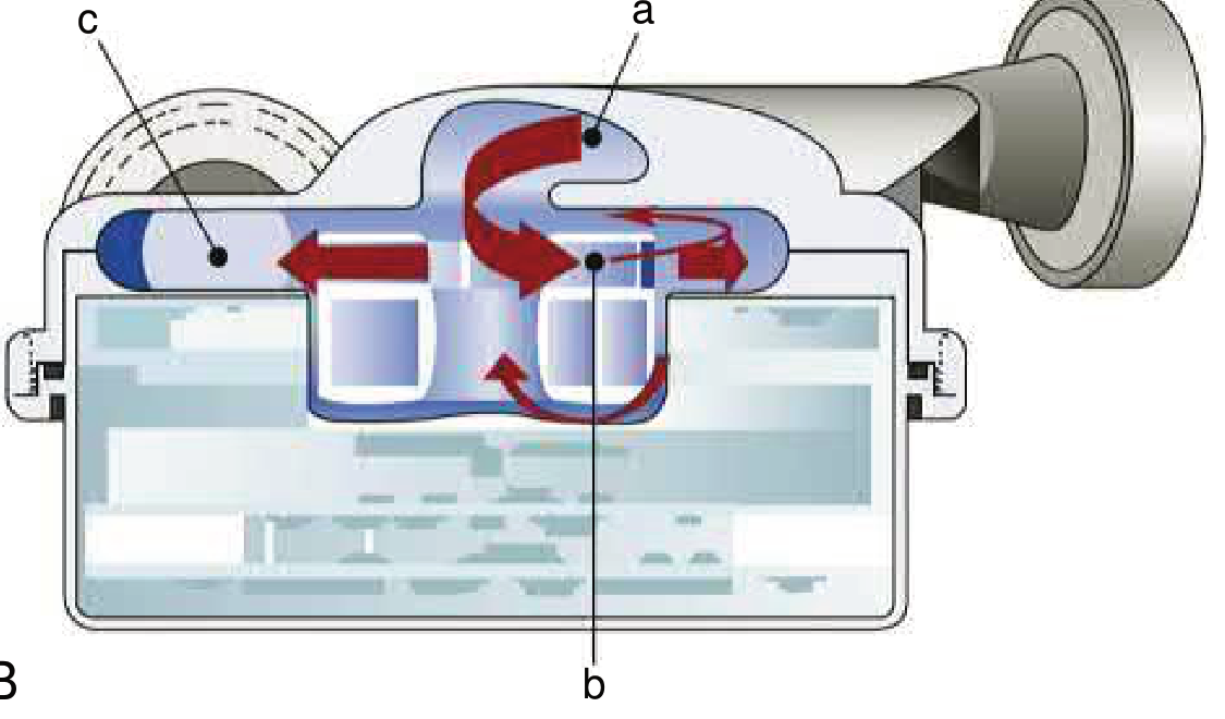
EFIGURE 59.1 - Axial (A) and centrifugal (B) rotary pump designs. From Braunwald's Heart Disease.
5. Axial vs. Centrifugal Flow Design
| Feature | Axial Flow | Centrifugal Flow |
|---|---|---|
| Blood path | Along the axis of rotation | Radially outward from center |
| Impeller support | Mechanical bearings or pivot | Magnetic or hydrodynamic levitation |
| Speed (RPM) | Higher (~8,000-12,000) | Lower (~2,000-3,500) |
| Examples | HeartMate II (HMII) | HeartMate 3 (HM3), TandemHeart |
| Shear stress | Higher | Lower |
Impeller suspension methods:
-
Mechanical bearings: Pivot contact design - potential frictional wear and heat generation over time, may lead to device failure
-
Hydrodynamic levitation: Fluid forces alone suspend the impeller; simpler electronics, smaller pump, but impeller may contact housing at very low speeds
-
Magnetic levitation (passive or active): Permanent magnets (passive, no power) or electromagnets (active, position sensing); fully contactless design eliminates bearing wear
-
Hybrid systems: Combination of hydrodynamic + magnetic levitation (e.g., HM3 uses passive magnetic + hydrodynamic)
-
Braunwald's Heart Disease, pp. 330-331
6. Hydrodynamic Properties
The fundamental operating principle of a continuous-flow rotary pump:
Flow = f(pump speed, pressure gradient)
- Flow is directly proportional to pump speed (RPM)
- Flow is inversely proportional to the pressure gradient across the pump (aortic pressure minus LV pressure)
This means:
- If aortic afterload rises, flow decreases at a fixed RPM
- If the native heart contracts weakly (high LV pressure), less gradient exists and pump can generate more flow
- Clinicians can titrate RPM to balance ventricular unloading and cardiac output
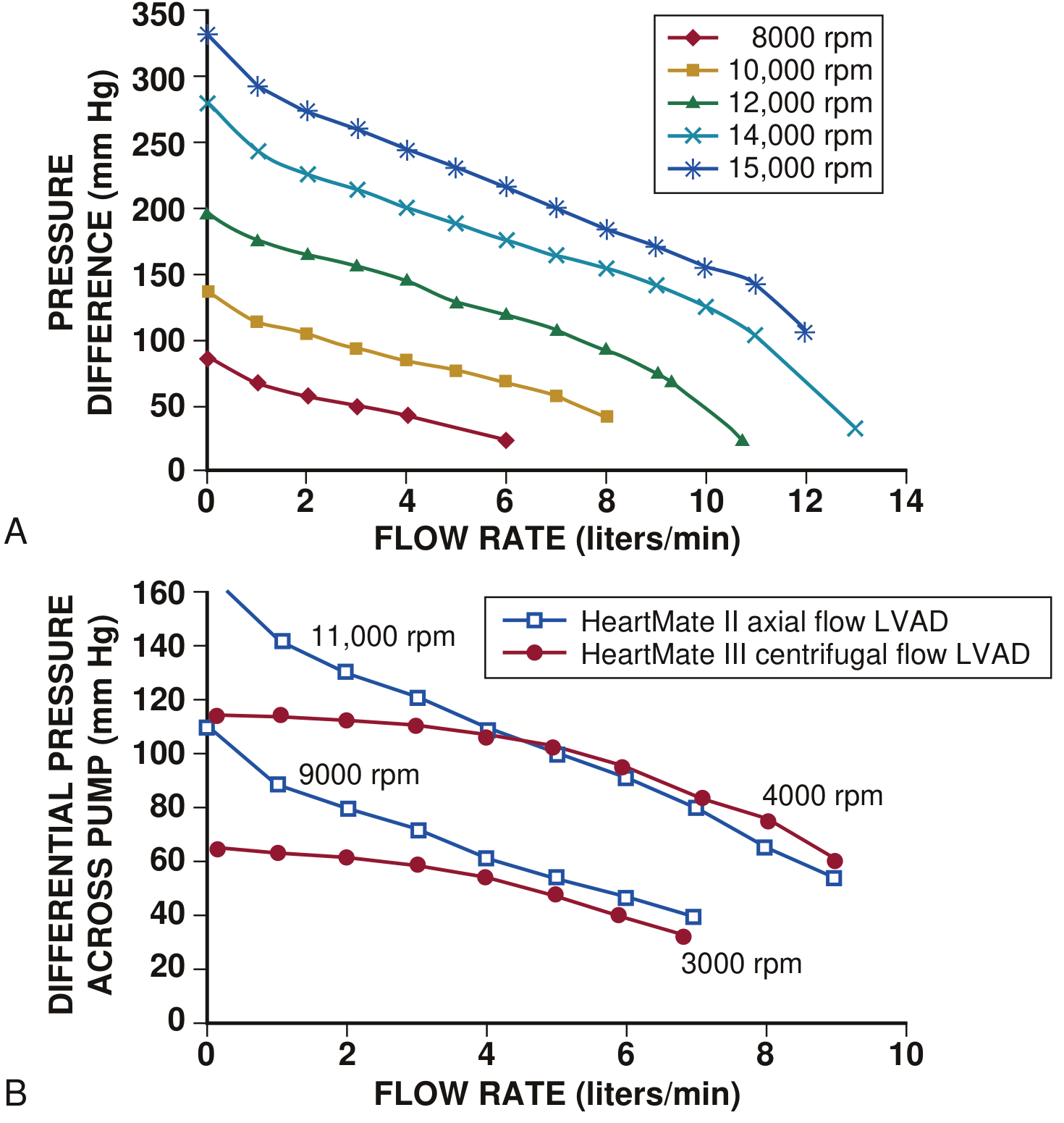
EFigure 59.3 - Hydrodynamic properties: flow is inversely related to pressure gradient and directly related to pump speed (RPM). From Braunwald's Heart Disease.
7. Specific Devices
Temporary/Short-term Devices
| Device | Type | Mechanism | Support |
|---|---|---|---|
| IABP (Intra-Aortic Balloon Pump) | Counterpulsation | Inflates in diastole, deflates in systole | LV (passive, ~0.5 L/min augmentation) |
| Impella | Microaxial rotary | Transvalvular catheter; aspirates from LV, ejects into aorta | LV (up to 5.5 L/min) |
| TandemHeart | Centrifugal | Transseptal LA drainage, returns to femoral artery | LV bypass (~4 L/min) |
| VA-ECMO | Centrifugal + membrane oxygenator | Drains venous blood, oxygenates, returns to artery | Biventricular + pulmonary |
The IABP-SHOCK II trial demonstrated that IABP does not reduce 30-day, 12-month, or 6-year mortality in cardiogenic shock with LV failure, and it is no longer recommended for this indication. Active percutaneous devices (Impella, TandemHeart) provide better hemodynamic support but have not yet demonstrated a clear mortality benefit in randomized trials. - Harrison's Principles of Internal Medicine 22E
Durable/Long-term Devices
- HeartMate II (HMII): First-generation axial-flow LVAD; most studied with >20,000 implantations worldwide
- HeartMate 3 (HM3): Centrifugal, fully magnetically levitated impeller; superior survival and lower stroke/GI bleeding vs. HMII in the MOMENTUM 3 trial
- HeartWare HVAD: Now discontinued (June 2021) after showing a 3.49x higher hazard ratio for mortality vs. HM3
8. Patient Selection Considerations
Before implantation, the following must be carefully assessed:
Renal function: Most common risk for morbidity/mortality. Cardiogenic shock elevates CVP, worsening renal congestion independent of low-flow injury.
Coagulation: Abnormal INR (without warfarin) reflects hepatic congestion/fibrosis. Combined coagulopathy + anticoagulation increases perioperative bleeding, RV failure, and multi-organ failure. Heparin-induced thrombocytopenia (HIT) must be screened.
Valvular disease:
- Mild-moderate AS: Not a contraindication
- Severe AS: Must be corrected (bioprosthesis) before LVAD to allow future weaning
- Moderate AI: Causes LV distension with LA-aorta cannulation; with LV apex-aorta devices, AI worsens as LV pressure drops (creates circular flow loop)
Right ventricular function: RV failure is a major post-implant complication; preoperative RV assessment is essential. A temporary right ventricular assist device (RVAD) may be needed post-LVAD.
Pulmonary hypertension, hepatic function, intracardiac shunts, and arrhythmias all require evaluation.
- Braunwald's Heart Disease, pp. 329-334
9. Adverse Events and Complications
The major complications common to all VADs:
| Complication | Mechanism |
|---|---|
| Driveline infection | Percutaneous driveline provides a conduit for bacteria; most common infection site |
| Gastrointestinal bleeding | Loss of high-molecular-weight von Willebrand factor multimers (shear-induced degradation), acquired vWD; arteriovenous malformations (AVMs) related to reduced pulse pressure |
| Stroke (ischemic/hemorrhagic) | Pump thrombosis causing emboli; inadequate anticoagulation; hypertension; hemorrhage from over-anticoagulation |
| Pump thrombosis | Thrombus in the pump itself - presents with power spikes, hemolysis, and worsening hemodynamics |
| Right heart failure | Increased preload to RV after LVAD decompresses LV; interventricular septal shift |
| Aortic insufficiency (acquired) | Reduced LV pressure from LVAD creates pressure gradient promoting AI development |
| Device failure | Bearing wear, lead fracture, driveline damage |
Anticoagulation (warfarin, target INR 2-3) plus antiplatelet therapy (aspirin) is standard for durable LVADs. The HM3's fully magnetically levitated design essentially eliminated pump thrombosis in MOMENTUM 3.
- Braunwald's Heart Disease; Miller's Anesthesia 10e; Mulholland and Greenfield's Surgery 7e
10. Monitoring and Management
- INTERMACS (Interagency Registry for Mechanically Assisted Circulatory Support): The primary registry tracking outcomes of all durable MCS devices in the United States. Provides risk stratification profiles (INTERMACS profiles 1-7, with 1 = critical cardiogenic shock, 7 = advanced NYHA III).
- Pump speed is titrated to optimize cardiac output while preventing ventricular suction (over-decompression of the LV)
- Echocardiography guides cannula position, RV function assessment, aortic valve opening frequency, and detection of complications
- Mean arterial pressure (not standard cuff systolic/diastolic) is the relevant blood pressure measurement in continuous-flow VAD patients
Summary
VADs work by mechanically unloading a failing ventricle and generating forward flow. The field has shifted from bulky pulsatile pumps to compact continuous-flow rotary pumps, with the centrifugal magnetically levitated design (HM3) now representing the gold standard for durable support. The three strategic goals - bridge to recovery, bridge to transplant, and destination therapy - frame all clinical decision-making. Complication management, particularly GI bleeding, stroke, driveline infection, and right heart failure, remains the central challenge of long-term VAD support.
Recent literature to note:
- Durable Mechanical Circulatory Support: JACC Scientific Statement (2023) - PMID 37758441: Comprehensive 2023 JACC statement covering current evidence and management
- Right Heart Failure with LVAD - ESC HFA Consensus Statement (2024) - PMID 38853659: Pre/peri/post-operative strategies for RV failure management
Matrial Valve stenosis
Reading File
Now let me search the library and PubMed in parallel for mitral stenosis.
Finding Sources
Searching PubMed
"mitral stenosis" AND management
Found excellent sources. Let me read the main sections from Braunwald's (comprehensive cardiology textbook) and the Echocardiography textbook in parallel.
Reading File
Reading File
Reading File
Excellent content. Now let me read the main comprehensive Braunwald's chapter section on MS (the block13 entry which has pathophysiology, clinical features, management) and continue reading the radiology textbook.
Reading File
Reading File
Good. Now let me search for the main comprehensive Braunwald's chapter on mitral stenosis pathophysiology and management (Chapter 75 referenced above).
Finding Sources
Finding Sources
Finding Sources
Reading File
Now let me read the dedicated Braunwald's chapter on MS - the block10 section with full pathophysiology detail.
Reading File
I need the main MS chapter - let me find it directly in Braunwald's. Let me search for the main chapter heading (Chapter 75).
Finding Sources
Finding Sources
Found the key sections. Let me now read the main Chapter 75 content - Rheumatic MS, treatment, and the non-rheumatic section.
Reading File
Reading File
Excellent - I have rich content on the etiology and definition sections. Now let me read the pathophysiology/natural history and clinical features sections which should be just below offset 553.
Reading File
Excellent! I have extremely rich and comprehensive content. Let me read just a bit more on the physical findings and management section, then compile the full answer.
Reading File
I now have comprehensive content from multiple textbooks. Let me compile the full, detailed answer.
Mitral Valve Stenosis
1. Definition and Overview
Mitral stenosis (MS) is a structural abnormality of the mitral valve that prevents proper opening during diastolic filling of the left ventricle. The resulting obstruction to left atrial (LA) outflow creates a pressure gradient across the mitral valve, chronically elevating LA pressure and causing progressive remodeling of the LA and pulmonary vascular bed.
Normal mitral valve area (MVA) is ≥ 4.0 cm². A gradient begins forming once MVA drops below 2.0 cm². Symptoms consistently appear at ≤ 1.5 cm².
- Braunwald's Heart Disease, p. 761; Grainger & Allison's Diagnostic Radiology, p. 334
2. Etiology and Epidemiology
Rheumatic Mitral Stenosis (RMS) - Most Common Cause
- Results from repeated group A Streptococcus (GAS) pharyngeal infections triggering an autoimmune response that mimics valve antigens (both humoral and cellular immune mechanisms)
- Acute rheumatic fever (ARF) typically occurs in the second decade; MS manifests 2-3 decades later
- The mitral valve is the most commonly involved valve in rheumatic heart disease (RHD)
- MS accounts for 40% of all rheumatic heart disease; ~60% of pure MS patients have a recalled history of rheumatic fever
- More common in women (2:1 for isolated MS) - even though ARF affects both sexes equally
- Global prevalence of RHD: ~33.4 million cases (concentrated in low-middle income countries - South/East Asia, sub-Saharan Africa, Oceania)
- In endemic regions: aggressive course, symptomatic in 2nd-4th decade
- In non-endemic regions: indolent course, present in 5th-7th decade
Degenerative (Non-Rheumatic) Mitral Stenosis (DMS)
- Primarily due to mitral annular calcification (MAC) - accounts for ~41% of severe MS on echo in western series
- Increasingly recognized in patients with aortic stenosis undergoing TAVR (11-18%)
- More common in older women, associated with atherosclerotic risk factors and severe CKD
Rare Causes
-
Congenital mitral stenosis
-
Prior chest radiation
-
Mucopolysaccharidosis
-
Ball valve thrombus
-
Left atrial myxoma (obstructive)
-
Cor triatriatum (membrane divides LA, mimicking MS)
-
Post-surgical/transcatheter mitral valve procedures
-
Braunwald's Heart Disease, pp. 759-761; Grainger & Allison's, p. 334
3. Pathology and Morphology
The rheumatic mitral valve shows characteristic changes:
- Leaflet thickening and nodularity
- Commissural fusion (primary mechanism in RMS)
- Leaflet calcification
- Chordal fusion and shortening (subvalvular apparatus)
- Valve narrows to a "fish-mouth" or "hockey stick" appearance (diastolic anterior leaflet bowing)
In MAC-related DMS, calcific deposits protrude into the valve orifice from the annulus rather than commissural fusion.
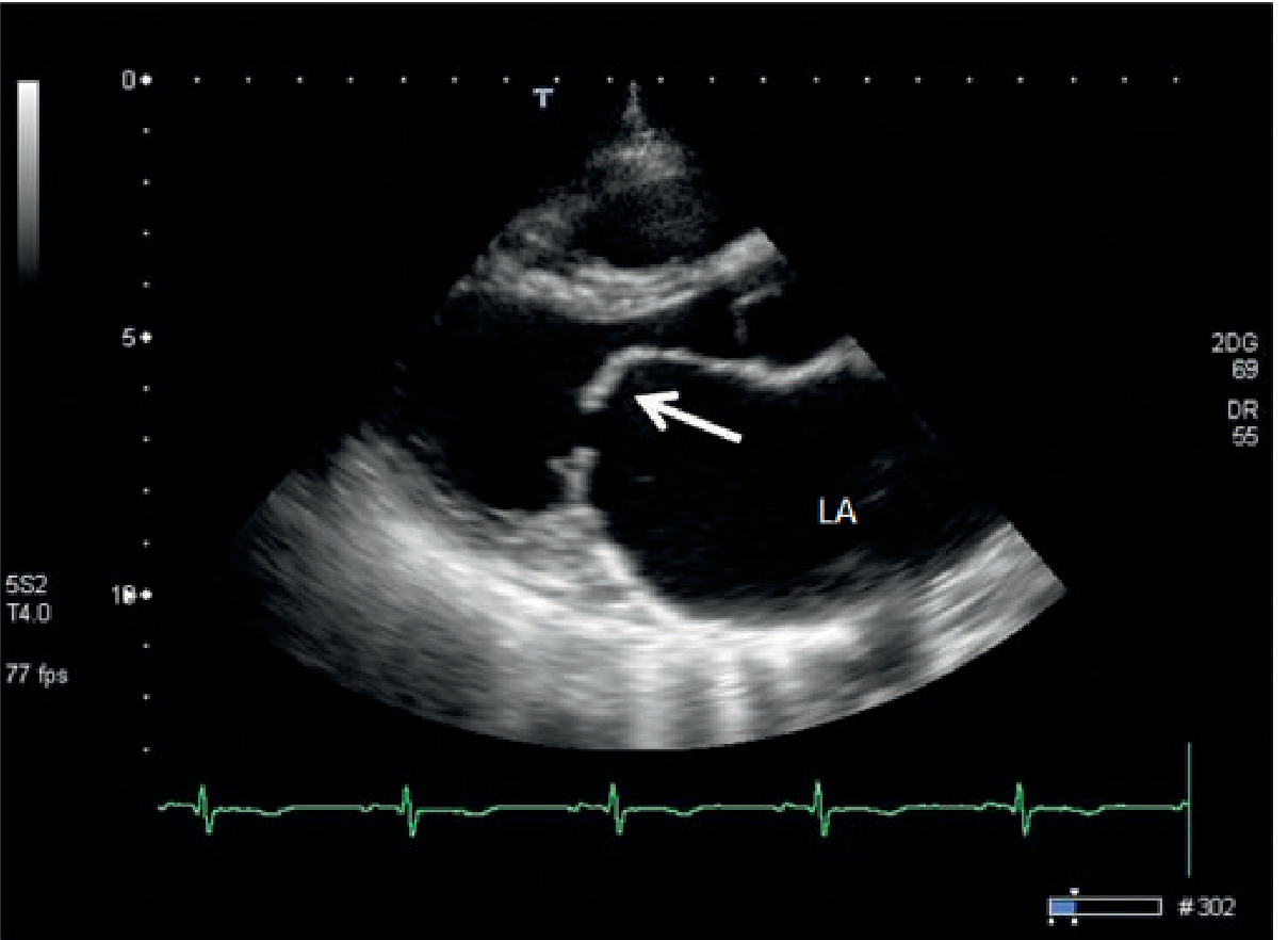
Fig. 14.38 - Mitral Stenosis. Echocardiography (parasternal long axis) shows marked thickening of mitral leaflets with restricted mitral valve orifice (doming anterior leaflet). Left atrial (LA) enlargement is evident. From Grainger & Allison's Diagnostic Radiology.
4. Pathophysiology
The core physiological consequence is obstruction to LV inflow:
MVA narrows → Pressure gradient across mitral valve →
↑ Left atrial pressure → LA dilation + pulmonary venous hypertension →
Dyspnea, pulmonary edema → Secondary pulmonary arterial hypertension →
RV pressure overload → RV dilation/failure + tricuspid regurgitation
Key physiological points:
- Cardiac output decreases as MVA decreases, especially on exertion
- Heart rate matters critically - tachycardia shortens diastole, reducing filling time across the stenotic valve, dramatically worsening the gradient. This is why AF with fast ventricular response is so hemodynamically dangerous in MS.
- Left ventricular function is generally preserved in pure MS (the LV is actually "protected" from volume overload), though reduced preload from the obstruction can reduce LV stroke volume
- Pulmonary hypertension (PAH): Chronic LA hypertension → pulmonary venous congestion → reactive pulmonary arterial hypertension. Severe PAH can become irreversible.
- Atrial fibrillation: LA dilation promotes AF. AF removes the atrial "kick" that contributes 20-30% of LV filling, further worsening hemodynamics
- Braunwald's Heart Disease; Grainger & Allison's, p. 334; Textbook of Clinical Echocardiography
5. Severity Classification
| Severity | MVA (cm²) | Mean Gradient (mm Hg) | PHT (msec) |
|---|---|---|---|
| Mild | > 1.5 | < 5 | < 150 |
| Moderate | 1.0-1.5 | 5-10 | 150-220 |
| Severe | < 1.0 | > 10 | > 220 |
AHA/ACC Disease Stages (2020 Guideline)
| Stage | Definition | Key Criteria |
|---|---|---|
| A | At risk | Mild doming; normal flow velocity; no hemodynamic consequences |
| B | Progressive MS | MVA > 1.5 cm²; diastolic PHT < 150 msec; mild-moderate LA enlargement |
| C | Asymptomatic severe MS | MVA ≤ 1.5 cm²; PHT ≥ 150 msec; severe LA enlargement; PASP > 50 mmHg |
| D | Symptomatic severe MS | Same as C + decreased exercise tolerance/exertional dyspnea |
- Braunwald's Heart Disease, Table 75.1, p. 762
6. Clinical Features
Symptoms
- Dyspnea - the cardinal symptom; initially on exertion, progressing to rest dyspnea, orthopnea, and paroxysmal nocturnal dyspnea
- Fatigue - especially with onset of PAH or over-diuresis
- Palpitations - often a consequence of AF, increase with age
- Hemoptysis - from pulmonary venous congestion or rupture of pulmonary-bronchial anastomoses
- Peripheral edema, ascites - when RV failure develops
- Systemic thromboembolism / stroke - due to LA stasis, especially with AF and LA appendage thrombus
- Pulmonary edema - precipitated by AF, pregnancy, infection, exercise
Precipitants that "unmask" MS: pregnancy, atrial fibrillation, fever/infection (increase heart rate and cardiac output demands)
Physical Examination Signs
| Finding | Description |
|---|---|
| Loud S1 | Due to increased force closing the wide-open (low-pressure) mitral valve at the onset of systole |
| Opening snap (OS) | High-pitched sound shortly after S2; produced by abrupt halt of mitral leaflet doming into LV; earlier OS (shorter S2-OS interval) = more severe MS |
| Rumbling mid-diastolic murmur | Low-pitched, best heard at apex with bell, in left lateral decubitus; accentuated pre-systolically (if sinus rhythm) due to atrial contraction |
| Presystolic accentuation | Lost in AF |
| Malar flush | Pinkish-purple discoloration of cheeks (low cardiac output + peripheral vasoconstriction) |
| Signs of PAH/RV failure | Loud P2, RV heave, JVD, hepatomegaly, peripheral edema |
In older adults, the loud S1 and opening snap may be absent due to calcified, fibrotic leaflets with reduced mobility.
- Braunwald's Heart Disease, pp. 762-764; Grainger & Allison's, p. 334
7. Investigations
Electrocardiogram
- P-mitrale: Broad, bifid P wave in lead II (> 0.12 sec) - LA enlargement
- Atrial fibrillation - common
- Right axis deviation, RV hypertrophy - with severe PAH
- Left atrial abnormality (negative terminal component of P wave in V1)
Chest X-Ray
- LA enlargement: double shadow at right cardiac border, elevation of left main bronchus, straightening of left heart border
- Pulmonary venous hypertension: upper lobe blood diversion, Kerley B lines, pulmonary edema
- Mitral valve calcification
- RV enlargement (late)
Echocardiography (Gold Standard)
2D Echo:
- Leaflet thickening, doming (hockey-stick deformity), calcification
- Commissural fusion
- LA enlargement; LA appendage thrombus (TEE superior)
- Subvalvular apparatus involvement
Doppler:
- Pressure half-time (PHT) method: MVA = 220/PHT (most widely used); PHT is the time for peak mitral gradient to fall by half
- Planimetry (direct measurement): most reliable; from parasternal short-axis at leaflet tips; 3D echo even more accurate
- Continuity equation: MVA = (LVOT TVI × LVOT area) / MV TVI
- Mean transmitral gradient: calculated from CW Doppler
Wilkins Score - assesses valve suitability for balloon mitral valvuloplasty (BMV): grades leaflet mobility, thickening, calcification, and subvalvular thickening (each 1-4 points; score ≤ 8 = favorable for BMV)
Cardiac MRI
- Useful when echo is suboptimal
- Cine-MRI shows restricted leaflets and anterograde flow void
- Direct MVA planimetry with good correlation to echo
Cardiac Catheterization
-
Indicated when non-invasive findings are inconclusive or discordant with symptoms
-
Simultaneous measurement of LA and LV diastolic pressures (or PCWP as LA surrogate)
-
Gorlin formula: MVA = CO / (DFP × HR × 44.3 × √mean gradient)
-
Braunwald's Heart Disease; Grainger & Allison's Diagnostic Radiology, pp. 334-337; Textbook of Clinical Echocardiography
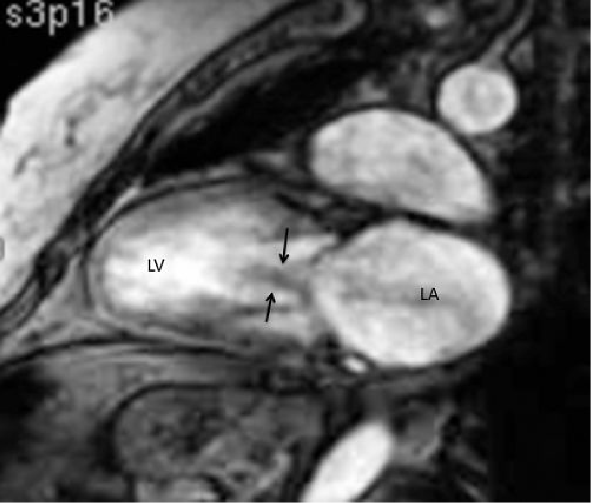
Fig. 14.39 - Cine-MRI of mitral stenosis. Flow void from LA to LV (arrows). LA is enlarged. From Grainger & Allison's Diagnostic Radiology.
8. Complications
| Complication | Mechanism |
|---|---|
| Atrial fibrillation | LA dilation disrupts electrical conduction; most common arrhythmia |
| Systemic thromboembolism/stroke | LA stasis (especially LA appendage) + AF |
| Pulmonary hypertension | Chronic LA pressure elevation → reactive PAH |
| Right heart failure | Pressure overload from PAH → RV dilation and failure |
| Tricuspid regurgitation | Secondary to RV dilation and annular dilatation |
| Infective endocarditis | Less common than in MR, but possible |
| Pulmonary edema | Acute decompensation (precipitation by AF, pregnancy, sepsis) |
| Hemoptysis | Pulmonary venous congestion, rupture of pulmonary venules |
9. Management
Medical Therapy
Medical therapy does not halt progression of MS but manages symptoms and prevents complications:
- Diuretics: Reduce pulmonary venous congestion and symptoms
- Beta-blockers / rate-limiting calcium channel blockers (diltiazem, verapamil): Control heart rate, prolong diastolic filling time - especially important in AF or with exertion
- Anticoagulation (warfarin, target INR 2-3): Indicated for MS with AF, prior embolism, or LA thrombus. Benefit in sinus rhythm with LA thrombus or severe MS is also recognized.
- Penicillin prophylaxis: Secondary prophylaxis against GAS infection to prevent recurrent ARF (essential in endemic regions in young patients)
- Digoxin: Rate control in AF
- Avoid aggressive reduction of preload with nitrates (can precipitate hypotension)
Indications for Intervention (AHA/ACC 2020 Guidelines)
Intervention is indicated to increase MVA when:
- Stage D (symptomatic severe MS, MVA ≤ 1.5 cm²) - Class I
- Asymptomatic severe MS (Stage C) with PASP > 50 mmHg - Class IIa
- Symptomatic moderate MS (MVA 1.5-2.0 cm²) with elevated gradient on exercise - selectively considered
Percutaneous Balloon Mitral Valvuloplasty / Commissurotomy (BMV/PTMC)
The preferred first-line intervention when anatomy is suitable.
Mechanism: A balloon catheter (Inoue balloon, most commonly) is advanced via transseptal puncture into the LA and inflated across the mitral valve, splitting the fused commissures.
Prerequisites (Wilkins Score ≤ 8):
- Pliable, non-heavily calcified leaflets
- No more than mild-moderate MR
- No LA thrombus (TEE essential prior to procedure)
- No significant subvalvular fusion
Outcomes:
- MVA typically doubles (from ~1.0 to ~2.0 cm²) with immediate symptomatic relief
- Success rates: >90% in LMICs/younger patients; <50% in older patients with calcified valves
- ~80% of patients remain free of death, mitral surgery, or repeat BMV over 15 years; ~66% at 20 years
Complications of BMV:
- Worsening MR: mild MR in ~20%; acute severe MR (anterior leaflet tear) < 1-2%
- Interatrial shunt (60-70% initially; persists <10%)
- Cardiac tamponade ~5%, stroke ~3%, mortality ~3% (higher in older patients)
Surgical Intervention
Indicated when BMV fails, is contraindicated, or when concurrent cardiac surgery is needed (tricuspid repair, CAD revascularization):
| Procedure | Details |
|---|---|
| Open Mitral Valvotomy (OMV) | Procedure of choice especially in the young; excellent results (96% survival at 10 years; 98% freedom from reoperation at 9 years); operative mortality < 2% |
| Closed Mitral Valvotomy (CMV) | Without cardiopulmonary bypass; used in LMICs; economical but inferior to OMV/BMV |
| Mitral Valve Replacement (MVR) | Mechanical or bioprosthetic; for unsuitable anatomy or failed repair; higher operative mortality (3-10%); lifelong anticoagulation with mechanical valve; high bioprosthesis failure rate in young patients |
| Maze procedure | Concomitant to reduce AF burden |
Degenerative/MAC-related MS
-
More complex; BMV generally less effective (calcific rather than commissural mechanism)
-
Transcatheter mitral valve replacement (TMVR) is an emerging option for MAC-related severe MS
-
High procedural risk; results still limited
-
Braunwald's Heart Disease, pp. 764-870; Grainger & Allison's, pp. 336-337
10. Special Situations
MS in Pregnancy
- Significant hemodynamic risk - increased plasma volume and heart rate of pregnancy worsens gradient
- May precipitate pulmonary edema, particularly in previously asymptomatic patients
- Management: beta-blockers for rate control, diuretics cautiously
- BMV can be performed safely during pregnancy (with lead shielding to minimize fetal radiation)
- Delivery should be planned in a high-risk obstetric center
MS in the Elderly
- Often MAC-related rather than rheumatic
- Loud S1 and OS may be absent (calcified leaflets)
- BMV success rate < 50%; higher complication rates
- MVR carries ≥ 10% perioperative mortality
Concurrent Aortic Valve Disease
- Present in ~1/3 of rheumatic MS patients
- AS severity can be overestimated in the presence of MS (reduced flow)
- BMV can be performed first if MS is predominant; caution - sudden LV preload increase post-BMV can precipitate pulmonary edema in undetected severe AS
Summary Table: Severity and Management
| Stage | MVA | Symptoms | PAH | Recommended Action |
|---|---|---|---|---|
| A | > 2.0 | None | None | Surveillance; GAS prophylaxis |
| B | > 1.5 | None | None | Medical therapy; exercise testing |
| C | ≤ 1.5 | None | PASP > 50 mmHg | Consider BMV (IIa) |
| D | ≤ 1.5 | Yes | Often present | BMV (if suitable) or Surgery (Class I) |
Recent literature:
- Valvular Heart Disease review 2024 (PMID 38278576) - Current primary care management overview
- Management of acute decompensated valvular heart disease 2025 (PMID 39663714) - European Heart Failure 2025 review covering acute MS management
Valvotomy
Finding Sources
Searching PubMed
"mitral valvotomy" OR "balloon mitral valvuloplasty" AND outcomes
Excellent sources found. Let me read the key sections from Fuster's, Harrison's, Braunwald's, and Sabiston's in parallel.
Reading File
Reading File
Reading File
Excellent content. Now let me read more from Fuster's (the step-by-step technique and outcomes), the Braunwald's PBMV section, and the Mulholland surgery section on open commissurotomy.
Reading File
Reading File
Reading File
Excellent, very rich content. Let me now fetch the key procedural diagrams and read the Sabiston section's Wilkins score table in full, plus the Washington Manual section.
Reading File
Reading File
I now have comprehensive, detailed content from multiple authoritative textbooks. Let me compile the full answer on valvotomy.
Valvotomy (Mitral Commissurotomy / Balloon Mitral Valvuloplasty)
Valvotomy refers to a surgical or percutaneous procedure to relieve valve stenosis by opening (incising or dilating) a stenotic valve. In clinical practice, the term is most commonly used for mitral valvotomy - the treatment of mitral stenosis (MS) by splitting fused commissures to increase the mitral valve area (MVA). The same principle extends to the pulmonary and aortic valves. This article focuses on mitral valvotomy in all its forms.
1. Rationale and Principle
In rheumatic MS, the primary mechanical problem is commissural fusion - the two commissures of the mitral valve become fused together by fibrous tissue, narrowing the orifice. Valvotomy directly addresses this by splitting those fused commissures, restoring leaflet mobility and increasing MVA.
This is why valvotomy is only effective in rheumatic MS (commissural fusion mechanism). It is not indicated in degenerative/MAC-related MS, where there is no commissural fusion and the calcification is annular.
- Washington Manual of Medical Therapeutics; Fuster and Hurst's The Heart, 15th Edition, p. 1012
2. Types of Valvotomy
| Type | Approach | CPB Required | Setting |
|---|---|---|---|
| Percutaneous Balloon Mitral Commissurotomy (PBMC/BMV) | Transseptal catheter | No | Cath lab |
| Open Mitral Commissurotomy (OMC) | Direct surgical, left atriotomy | Yes | Operating room |
| Closed Mitral Commissurotomy (CMC) | Surgical, transventricular dilator | No | OR (LMICs) |
| Mitral Valve Replacement (MVR) | Surgical excision + prosthesis | Yes | OR (when repair not possible) |
3. Percutaneous Balloon Mitral Commissurotomy (PBMC / BMV)
History
First performed by Inoue in 1984, followed by Lock in 1985. A meta-analysis of 7 randomized controlled trials showed no significant difference between BMV and surgical commissurotomy (OMV/CMV) in immediate results (MVA achieved, post-procedural MR, restenosis/reintervention at 30 months) - but BMV carries far less procedural morbidity.
Mechanism
Balloon inflation within the stenotic mitral orifice mechanically splits the fused commissures, permitting a dramatic increase in leaflet motion and valve area. MVA typically doubles immediately (e.g., from ~1.0 cm² to ~2.0 cm²).
Indications (AHA/ACC 2020 Guidelines)
| Class | Indication |
|---|---|
| Class I | Symptomatic severe MS (MVA ≤ 1.5 cm², Stage D) with favorable anatomy, no LA thrombus, no moderate-severe MR |
| Class IIa | Asymptomatic severe MS (MVA ≤ 1.5 cm²) with PASP > 50 mmHg and favorable anatomy |
| Class IIb | Asymptomatic severe MS + new-onset AF + suitable anatomy |
| Class IIb | Symptomatic MVA > 1.5 cm² with hemodynamically significant MS on exercise (gradient > 15 mmHg or PCWP > 25 mmHg) |
| Class IIb | Severely symptomatic (NYHA III-IV) with suboptimal anatomy at high surgical risk |
Additional considerations for BMV (even in asymptomatic patients):
-
Very high thromboembolic risk
-
Planning for pregnancy
-
Scheduled for major non-cardiac surgery
-
Fuster and Hurst's The Heart, 15th Ed; Braunwald's Heart Disease; Washington Manual
Absolute Contraindications
- LA/LAA thrombus (TEE must be performed before procedure)
- Moderate or severe MR (grade ≥ 3/4)
- Severe or bicommissural calcification
- Absence of commissural fusion (i.e., degenerative/MAC-related stenosis)
Patient Selection: The Wilkins Echo Score
The Wilkins Score grades four echocardiographic features, each on a scale of 1-4:
| Grade | Mobility | Leaflet Thickening | Calcification | Subvalvular Thickening |
|---|---|---|---|---|
| 1 | Highly mobile; only tips restricted | Near normal (4-5 mm) | Single area of brightness | Minimal thickening just below leaflets |
| 2 | Mid and base portions have normal mobility | Midleaflets normal; margins thickened (5-8 mm) | Scattered brightness at margins | Thickening to 1/3 of chordal length |
| 3 | Moves forward mainly from the base | Thickening through entire leaflet (5-8 mm) | Brightness into midportions of leaflets | Thickening to distal 1/3 of chords |
| 4 | No/minimal forward movement | Considerable thickening of all tissue (> 8-10 mm) | Extensive brightness throughout leaflet | Extensive thickening/shortening to papillary muscles |
Total score range: 4-16
-
≤ 8 = Favorable for PBMC - likely to be successful
-
> 8 = Surgery recommended (PBMC less likely to succeed)
-
Sabiston Textbook of Surgery, Table 112.7; Harrison's Principles of Internal Medicine 22E
Equipment / Balloon Systems
Three main techniques, all producing similar outcomes:
- Inoue balloon (hourglass/double lumen; triple lumen) - most widely used and preferred
- Accura balloon (double lumen; similar to Inoue)
- Single or double peripheral angioplasty balloon (Lock technique)
- Reusable metallic valvulotome (used in some LMICs)
The Inoue balloon's hourglass shape self-positions across the mitral valve and allows stepwise inflation.
Step-by-Step Procedure
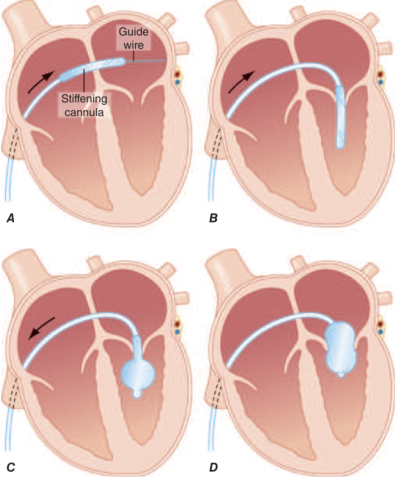
FIGURE 274-3 - Inoue balloon technique. A: Deflated balloon catheter advanced across interatrial septum, then across the mitral valve into the LV. B-D: Stepwise balloon inflation within the mitral orifice. From Harrison's Principles of Internal Medicine 22E.
- Venous access - typically via right femoral vein
- Transseptal puncture - needle punctures the interatrial septum under fluoroscopic/echocardiographic guidance, entering the LA from the RA (Fig. 274-3A)
- Septal dilation - the interatrial septum is dilated with a dilator to allow passage of the balloon catheter
- Balloon advancement - the balloon catheter (Inoue type) is advanced from the LA across the mitral valve and into the LV in its deflated state
- Stepwise balloon inflation (Fig. 274-3B-D):
- Distal portion inflated first (anchors in LV)
- Proximal portion inflated (anchors in LA)
- Middle portion then inflated - this splits the fused commissures
- Immediate assessment - Doppler echocardiography and pressure measurements confirm increased MVA and reduced gradient
Pre-procedure TEE is mandatory to exclude LA appendage thrombus and assess degree of MR. The procedure has a steep learning curve and should be performed only at high-volume centers.
- Fuster and Hurst's The Heart, p. 1013; Harrison's, p. 1548
Hemodynamic Results
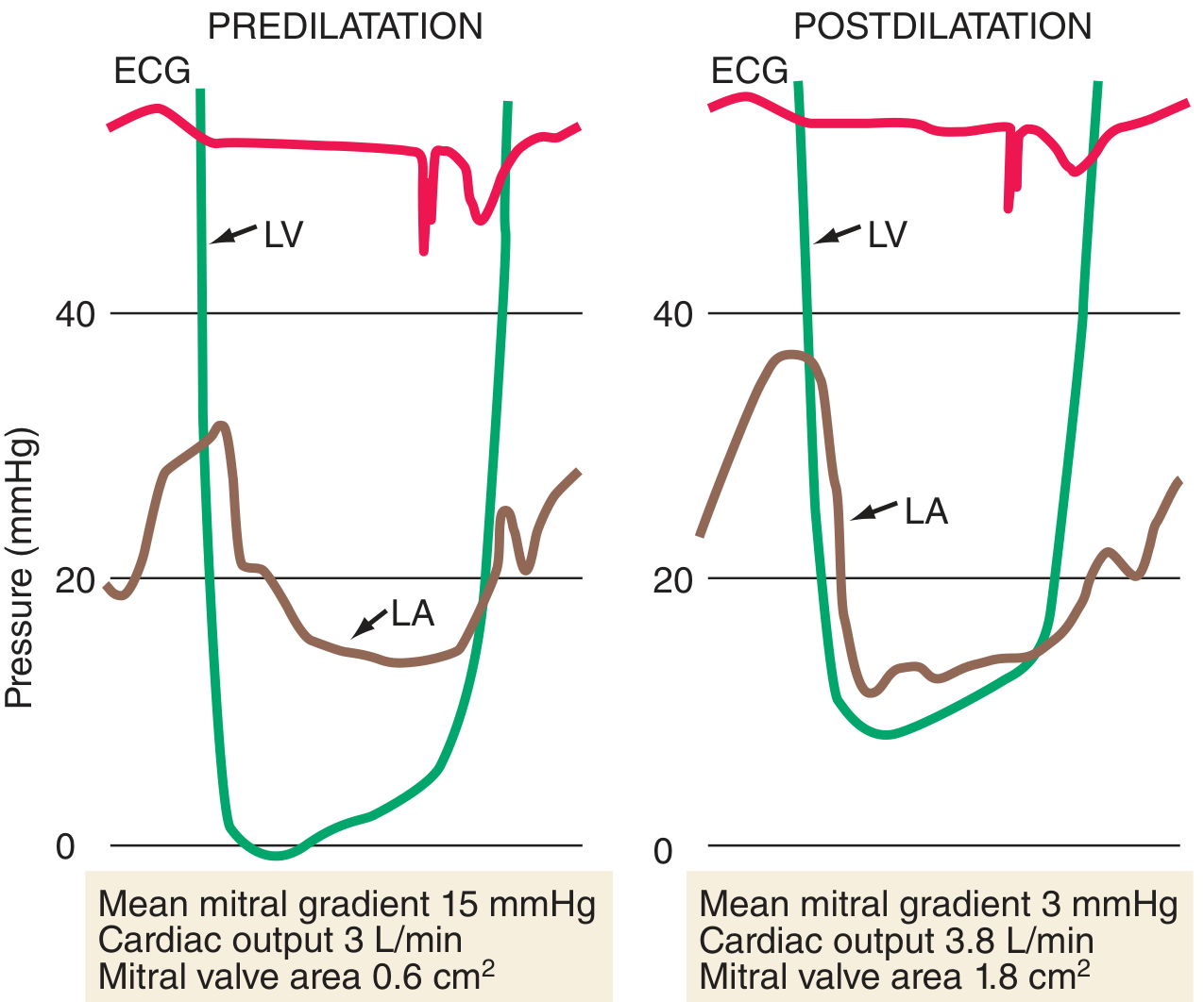
FIGURE 274-4 - Simultaneous LA and LV pressure before (left) and after (right) PBMC. The diastolic pressure gradient is abolished. From Harrison's Principles of Internal Medicine 22E.
Immediately after successful BMV:
- MVA doubles (e.g., 1.0 → 2.0 cm²)
- Mean transmitral gradient falls dramatically
- LA pressure decreases
- Symptoms resolve rapidly
Definition of Procedural Success
Post-dilation MVA > 1.5 cm² with MR grade < 2/4 - achieved in over 80% of appropriately selected patients
- Sabiston Textbook of Surgery
Complications
| Complication | Frequency |
|---|---|
| Worsening MR (mild, from commissural splitting) | ~20% - generally tolerated well; predicts good outcome |
| Acute severe MR (anterior leaflet tear / subvalvular damage) | < 1-2% - may require emergency surgery |
| Interatrial shunt (post-transseptal) | 60-70% acutely; persists < 10% long-term |
| Cardiac tamponade | ~5% |
| Systemic thromboembolism / stroke | ~3% |
| Procedural mortality | ~0.5-3% (higher in older, high-risk patients) |
- Braunwald's Heart Disease; Sabiston Textbook of Surgery; Fuster and Hurst's The Heart
Special Situation: BMV in Pregnancy
-
Treatment of choice for severe symptomatic MS in pregnancy
-
High success rates, minimal radiation (< 10 min fluoroscopy with lead shielding)
-
Fetal development unaffected when performed correctly
-
Best performed after 24 weeks (before 20 weeks: poorer fetal outcomes)
-
Far superior to surgical intervention in pregnancy (surgery carries ~9% maternal mortality and ~30% fetal loss)
-
Braunwald's Heart Disease, p. 765
4. Long-Term Outcomes After BMV
| Timeframe | Outcome |
|---|---|
| 3-7 years (younger, pliable valve) | 80-90% event-free survival |
| 15 years | ~80% free from death, mitral surgery, or repeat BMV |
| 20 years | ~66% remain free; ~30% have good functional results (France cohort, age 49 ± 14) |
Predictors of worse long-term outcome:
- Older age
- Higher NYHA class at baseline
- Suboptimal initial result (insufficient MVA increase)
- Higher Wilkins score
Post-BMV restenosis:
-
Occurs over years; if mechanism is re-fusion of commissures, repeat BMV can be performed with reasonable success
-
If restenosis is due to leaflet rigidity/degeneration, results are less favorable
-
Braunwald's Heart Disease; Fuster and Hurst's The Heart
5. Open Mitral Commissurotomy (OMC)
Indications
- PBMC not feasible or contraindicated
- Failed previous PBMC
- Restenosis after prior surgery
- Concurrent cardiac surgery needed (CAD, aortic valve disease, tricuspid repair)
Technique
- Performed under cardiopulmonary bypass (CPB)
- Mitral valve exposed through a left lateral atriotomy (anterior to pulmonary veins) or right atriotomy + incision through the atrial septum
- Under direct vision:
- Division of fused commissures with a knife or scissors
- Mobilization of scarred chordae and papillary muscles (subvalvular apparatus)
- Debridement of calcium deposits
- Removal of LA thrombus
- Ligation of LA appendage (reduces future embolic risk; a major advantage over BMV)
- Maze procedure can be added concurrently to reduce AF burden
Results
-
Operative mortality: < 2%
-
10-year survival: ~96%
-
10-year freedom from reoperation: ~90% (Sabiston) / ~98% at 9 years (Braunwald's)
-
Results comparable to MVR but avoids long-term prosthetic valve complications
-
More favorable than PBMC in older patients with subvalvular fusion, leaflet calcification, and reduced valve flexibility
-
Mulholland and Greenfield's Surgery 7e; Harrison's Principles; Sabiston Textbook of Surgery
6. Closed Mitral Commissurotomy (CMC)
-
Performed without cardiopulmonary bypass
-
Surgeon inserts a finger (transatrial) and a transventricular dilator (Tubbs dilator) to split the valve
-
Lower resource requirements - used predominantly in LMICs where CPB is unavailable
-
Results: inferior to OMC and BMV (less increase in MVA, higher restenosis)
-
When BMV is available, BMV should be preferred over CMC
-
Braunwald's Heart Disease; Fuster and Hurst's The Heart
7. Mitral Valve Replacement (MVR)
When valvotomy (percutaneous or open repair) is not possible or has failed:
Indications:
- Significant calcification or subvalvular fusion making repair unlikely to succeed
- Concomitant moderate-severe MR
- Failed PBMC or OMC
- Severe symptomatic MS in patients not candidates for repair
Key surgical principle: Preservation of the subvalvular apparatus (papillary muscle-chordal continuity with the annulus) is critical for maintaining LV geometry and function.
Valve choice:
- Mechanical: Durable; requires lifelong anticoagulation
- Bioprosthetic: No long-term anticoagulation; but high failure rate in young patients
Outcomes:
-
Operative mortality: 5-6% (higher than OMC)
-
Freedom from reoperation at 15 years: 50-75%
-
Sabiston Textbook of Surgery; Mulholland and Greenfield's Surgery
8. Comparison Summary
| Feature | BMV/PBMC | Open Commissurotomy (OMC) | Closed Commissurotomy (CMC) | MVR |
|---|---|---|---|---|
| CPB required | No | Yes | No | Yes |
| Invasiveness | Percutaneous | Open surgery | Open surgery | Open surgery |
| Mechanism addressed | Commissural splitting | Commissural splitting + subvalvular | Commissural splitting | Valve excision |
| LA appendage ligation | No | Yes | No | Yes |
| Immediate MVA increase | ~2x | ~2x | Less consistent | Prosthetic valve area |
| Operative mortality | 0.5-3% | < 2% | Low | 5-6% |
| 10-yr freedom from reoperation | ~80% | ~90% | Lower | 50-75% |
| Valve anatomy requirement | Favorable (Wilkins ≤ 8) | Broader indications | LMICs mainly | Unfavorable anatomy/failed repair |
| AF treatment possible | No | Yes (Maze) | No | Yes (Maze) |
9. Approach to Patient Selection (Algorithm)
Severe MS confirmed (MVA ≤ 1.5 cm², symptomatic OR PASP > 50 mmHg)
         ↓
Assess anatomy: Echo (Wilkins score), TEE (LA thrombus, MR degree)
         ↓
Wilkins score ≤ 8, no LA thrombus, MR < Grade 3?
    YES → BMV/PBMC (procedure of choice)
    NO  → Surgical options
              ↓
       Repair feasible? → Open Commissurotomy (preferred in young)
       Repair not feasible, significant MR/calcification → MVR
Key recent reference: Echocardiography Assessment of Rheumatic Heart Disease: Implications for Percutaneous Balloon Mitral Valvuloplasty (PMID 37544629) - 2023 review covering patient selection and echo scoring for BMV.
This is a shared conversation. Sign in to Orris to start your own chat.