
im having sever lower leg pain in my left leg...what is the possiable cause and write some immediate treatment
im having sever lower leg pain in my left leg...what is the possiable cause and write some immediate treatment
severe lower leg pain causes immediate treatment 2025
lower leg pain causes diagram DVT compartment syndrome
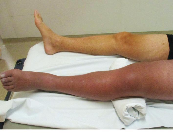
Clinical photograph providing a side-by-side comparison of a patient's lower extremities, illustrating the classic presentation of Phlegmasia Cerulea Dolens. The right leg displays a normal skin tone and anatomical contour. In contrast, the left leg exhibits significant pathology, characterized by diffuse, massive swelling (edema) extending from the thigh to the foot. The skin of the left leg shows deep erythematous and cyanotic discoloration, appearing smooth and taut, which is indicative of severe venous outflow obstruction and impending compartment syndrome. The left limb is slightly elevated on a bolster, while the patient is in a supine position. This image serves as a key diagnostic reference for identifying life-threatening deep vein thrombosis (DVT) complications, highlighting the hallmark triad of massive swelling, cyanosis, and pain (clinical context). It is intended for intermediate to advanced medical learners focusing on vascular surgery, emergency medicine, and hematology.
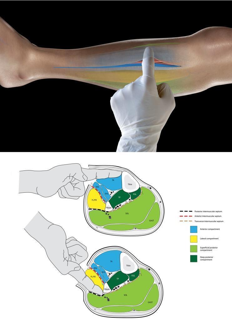
This educational graphic consists of a clinical photograph and two anatomical cross-sections illustrating the surgical technique for identifying leg compartments during a fasciotomy. The top photograph shows a gloved surgeon's finger inserted into an incision in the lower leg's fascia to palpate internal structures. Below, two schematic transverse cross-sections of the mid-leg categorize the four compartments: anterior (blue), lateral (yellow), superficial posterior (light green), and deep posterior (dark green). The top diagram demonstrates the 'tibia test': the finger enters the anterior compartment and palpates the medial tibia to confirm correct localization. The bottom diagram demonstrates the identification of the lateral (peroneal) compartment by palpating the fibula. The diagrams highlight the role of the intermuscular septa (anterior, posterior, and transverse) as physical barriers that prevent cross-compartment palpation, serving as a vital intraoperative check to ensure all compartments are adequately decompressed in cases of acute compartment syndrome.
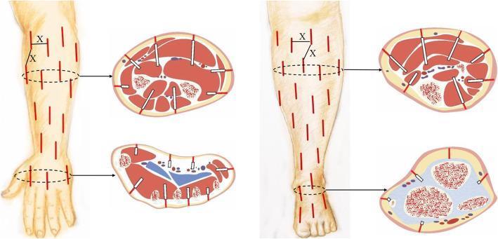
An anatomical and surgical diagram illustrating a reticular mini-incision decompression technique for osteofascial compartment syndrome (OCS). The figure displays two anatomical regions: the upper extremity (forearm and hand) and the lower extremity (leg and foot). A longitudinal view of each limb shows a pattern of multiple, short, parallel red lines representing skin incisions, approximately 0.5–1.0 cm apart (labeled as 'X'). Corresponding cross-sectional diagrams demonstrate the surgical depth of these incisions. The cross-sections show the incisions extending through the skin and subcutaneous tissue to reach the deep fascia. White instrument indicators (representing vascular clamps) are shown separating the subcutaneous and deep fascial layers to facilitate compartment release. Key anatomical structures visible in the cross-sections include muscle compartments, bones (tibia, fibula, radius, ulnar), and major vascular bundles. The diagram serves as a surgical protocol for minimally invasive fasciotomy, highlighting the spatial distribution of incisions required to achieve adequate tissue decompression while minimizing wound morbidity.
Severe Lower Left Leg Pain
⚠️ SEEK EMERGENCY CARE IMMEDIATELY IF YOU HAVE:
- Leg is pale, cold, or bluish/purple (loss of circulation)
- Sudden inability to move the foot/toes
- Extreme swelling with tight, shiny skin
- Chest pain or shortness of breath alongside leg pain
- High fever with redness spreading up the leg
Possible Causes of Severe Lower Leg Pain
1. Muscle Cramp / Charley Horse
- What it is: Sudden involuntary muscle contraction, most often in the calf
- Feels like: Intense, sharp cramping that may last seconds to minutes
- Common triggers: Dehydration, electrolyte imbalance (low potassium/calcium/magnesium), prolonged sitting or standing, overexertion
2. Deep Vein Thrombosis (DVT)
- What it is: A blood clot in a deep vein, usually in the calf or thigh
- Feels like: Dull aching, throbbing, or cramping pain - usually one leg only (your case: left leg)
- Warning signs: Swelling, redness/warmth, skin discoloration
- Risk factors: Long flights/bed rest, recent surgery, oral contraceptives, obesity, smoking
- This is a medical emergency - clot can travel to lungs (pulmonary embolism)
3. Acute Compartment Syndrome
- What it is: Dangerous pressure buildup inside a muscle compartment cutting off blood flow
- Feels like: Severe, burning/crushing pain that is out of proportion to any injury; worsened by stretching the muscles
- Causes: Trauma/fracture, crush injury, tight cast, reperfusion after blocked artery
- Time-sensitive: >12 hours without fasciotomy (surgical release) results in poor outcomes (only 8% good results vs 68% if treated within 12 hours) - Campbell's Operative Orthopaedics, 15th Ed
- Go to ER immediately if suspected
4. Shin Splints (Medial Tibial Stress Syndrome)
- What it is: Overuse injury causing pain along the inner shin bone (tibia)
- Feels like: Dull aching along the inner front of the lower leg, worsens with activity
- Common in: Runners, soldiers, dancers
5. Chronic Exertional Compartment Syndrome (CECS)
- What it is: Pressure that builds in leg compartments only during exercise
- Feels like: Cramping, burning, aching pain and/or numbness during activity that eases with rest
- Textbook of Family Medicine, 9th Ed
6. Peripheral Neuropathy / Nerve Entrapment
- What it is: Damage or compression of nerves (e.g., peroneal nerve at the fibular neck)
- Feels like: Burning, shooting, or electric pain; may have numbness/tingling
- Bradley and Daroff's Neurology in Clinical Practice
7. Peripheral Arterial Disease (PAD)
- What it is: Narrowed arteries reducing blood flow to the legs
- Feels like: Cramping pain in the calf with walking (claudication), relieved by rest
- In severe cases: Pain at rest, cold/pale foot
8. Cellulitis / Infection
- What it is: Bacterial skin/soft tissue infection
- Feels like: Pain with redness, warmth, swelling, and fever
- Needs antibiotics - can worsen rapidly
9. Stress Fracture
- What it is: Tiny crack in the tibia or fibula from overuse or trauma
- Feels like: Localized, pinpoint pain that worsens with weight-bearing
10. Tendonitis / Achilles Tendon Issues
- What it is: Inflammation of tendons around the ankle/lower leg
- Feels like: Pain and stiffness at the back of the leg near the heel, worse in the morning or with activity
- A sudden "pop" + severe pain = possible Achilles tendon rupture (needs urgent care)
Immediate Home Treatment (for non-emergency causes)
| Step | Action |
|---|---|
| R - Rest | Stop all activity; avoid putting weight on the leg |
| I - Ice | Apply an ice pack wrapped in a cloth for 15-20 min every 2 hours for the first 48 hours |
| C - Compression | Wrap with a compression bandage to reduce swelling (not too tight) |
| E - Elevation | Raise your leg above heart level while lying down to reduce swelling |
| Pain relief | Ibuprofen (400-600 mg with food) or paracetamol (500-1000 mg) for pain - avoid if you suspect a clot |
| Hydration | Drink water + electrolyte fluids if cramping is suspected |
| Gentle stretch | For calf cramps: stand, lean forward on the wall, stretch the calf muscle gently |
Note: The RICE/PEACE-LOVE protocol is the current standard for soft tissue sports injuries per Medical News Today (2025).
When to See a Doctor (Not Emergency, But Soon)
- Pain persists beyond 48-72 hours
- Pain worsens despite rest and ice
- Visible bruising or swelling that doesn't improve
- Pain started after a fall or impact
- You have diabetes, heart disease, or are on blood thinners
Bottom Line
Disclaimer: This information is educational. It does not replace a medical examination. Please consult a healthcare provider for diagnosis and treatment.