
Raas mechanism
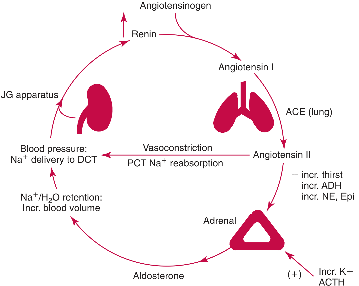
- Low renal perfusion/low Na+ → juxtaglomerular cells release renin
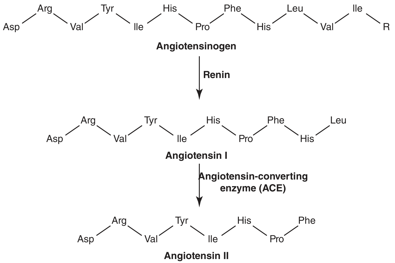
- Renin cleaves angiotensinogen (liver) → Angiotensin I
- ACE (lung endothelium) converts Ang I → Angiotensin II
- Ang II → vasoconstriction, aldosterone release, ADH release, thirst
- Aldosterone (adrenal cortex) → Na+/water retention, K+ excretion
RAAS renin angiotensin aldosterone system mechanism diagram
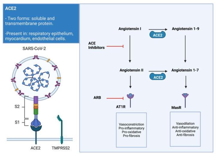
A medical illustration and pathophysiology diagram depicting the interaction between SARS-CoV-2 and the Renin-Angiotensin-Aldosterone System (RAAS). The left side shows the structural mechanism of viral entry, with the SARS-CoV-2 spike protein (S1 and S2 subunits) binding to the transmembrane ACE2 receptor, facilitated by the TMPRSS2 protease. The right side outlines the biochemical pathways of Angiotensin conversion. It illustrates the 'classic' pathway where Angiotensin I is converted to Angiotensin II, which activates the AT1R receptor to promote vasoconstriction, pro-inflammatory, pro-oxidative, and pro-fibrotic effects. This pathway is targeted by ACE inhibitors and Angiotensin Receptor Blockers (ARBs). The 'alternative' pathway shows ACE2 mediating the conversion of Angiotensin I to Angiotensin 1-9, and Angiotensin II to Angiotensin 1-7. Angiotensin 1-7 interacts with the Mas Receptor (MasR), triggering cardioprotective effects including vasodilation and anti-inflammatory, anti-oxidative, and anti-fibrotic responses. The diagram highlights the clinical significance of ACE2 downregulation during infection and its role in myocardial remodeling.
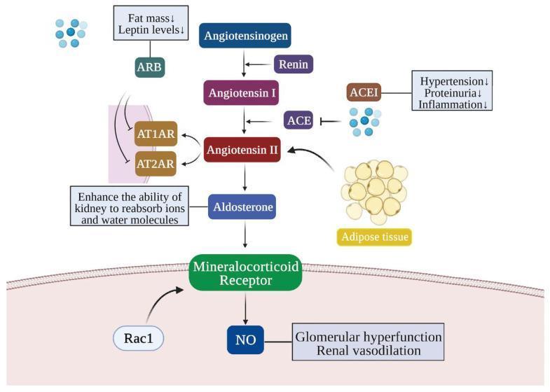
This pathophysiology diagram illustrates the Renin-Angiotensin-Aldosterone System (RAAS) pathway and its role in obesity-related kidney injury, highlighting pharmaceutical intervention points. The central pathway shows the enzymatic conversion of Angiotensinogen to Angiotensin I by Renin, and Angiotensin I to Angiotensin II via Angiotensin-Converting Enzyme (ACE). Angiotensin II acts on receptors AT1AR and AT2AR, facilitating kidney reabsorption of ions and water. The diagram specifically notes that adipose tissue contributes to increased Angiotensin II levels. Further down the cascade, Angiotensin II stimulates Aldosterone, which activates the Mineralocorticoid Receptor (supported by Rac1). This activation leads to the production of Nitric Oxide (NO), contributing to glomerular hyperfunction and renal vasodilation. Two major drug classes are highlighted: ACE Inhibitors (ACEI), which block the ACE enzyme to reduce hypertension, proteinuria, and inflammation; and Angiotensin Receptor Blockers (ARB), which inhibit AT1AR/AT2AR receptors and are associated with decreased fat mass and leptin levels.
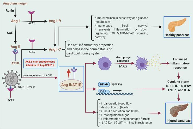
A medical pathophysiology diagram illustrating the Renin-Angiotensin-Aldosterone System (RAAS) signaling pathway and its disruption by SARS-CoV-2, leading to pancreatic injury. The cascade begins with the conversion of Angiotensinogen to Ang I by Renin, and Ang I to Ang II by ACE. The diagram highlights two divergent pathways: a protective arm involving ACE2, which converts Ang II to Ang 1-7 (promoting insulin sensitivity, beta-cell survival, and anti-inflammatory effects in a healthy pancreas), and a deleterious arm where Ang II binds to AT1R. The illustration shows that SARS-CoV-2 binds to and downregulates ACE2, leading to an overactive Ang II/AT1R axis. This overactivation triggers macrophage activation via MAS, NF-κB signaling, and a subsequent cytokine storm (IL-1β, IL-18, IFNγ, TNF-α, and IL-6). The cumulative effect results in an 'Injured pancreas' characterized by decreased blood flow, beta-cell destruction, reduced insulin secretion, hyperglycemia, fibrosis, and increased insulin resistance due to decreased GLUT4 and ACE2 expression.
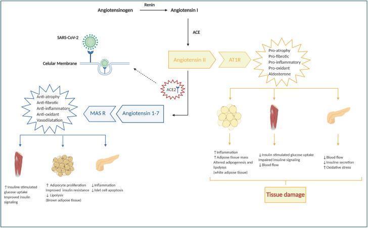
A pathophysiology diagram illustrating the Renin-Angiotensin-Aldosterone System (RAAS) and its deregulation in the context of COVID-19. The diagram outlines two competing metabolic pathways starting from Angiotensinogen. The primary pathway involves Renin converting Angiotensinogen to Angiotensin I, followed by ACE converting it to Angiotensin II. Binding of Angiotensin II to the AT1R receptor triggers pro-atrophy, pro-fibrotic, pro-inflammatory, and pro-oxidant effects, leading to tissue damage in white adipose tissue (increased inflammation and mass), skeletal muscle (impaired insulin signaling and blood flow), and the pancreas (reduced insulin secretion and blood flow). A counter-regulatory pathway shows ACE2 converting Angiotensin II into Angiotensin 1-7, which binds to the MAS receptor (MAS R) to produce protective anti-inflammatory, anti-fibrotic, and vasodilatory effects, improving insulin signaling in muscle and brown adipose tissue while reducing islet cell apoptosis in the pancreas. Critically, the diagram shows SARS-CoV-2 binding to the ACE2 receptor on the cellular membrane, inhibiting this protective pathway and shifting the balance toward tissue damage and systemic inflammation.
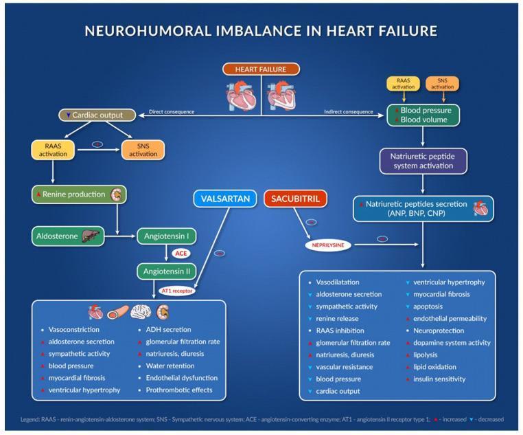
A complex pathophysiology diagram and pharmacological flowchart titled 'Neurohumoral Imbalance in Heart Failure'. It illustrates the interaction between the Renin-Angiotensin-Aldosterone System (RAAS), the Sympathetic Nervous System (SNS), and the Natriuretic Peptide (NP) system in the context of heart failure. The left side of the diagram outlines the RAAS cascade: Renin production leads to Angiotensin I, which is converted to Angiotensin II by ACE, ultimately binding to the AT1 receptor to cause deleterious effects like vasoconstriction, myocardial fibrosis, and water retention. The right side shows the NP system (ANP, BNP, CNP) which promotes diuresis and vasodilation. Centrally, the diagram depicts the pharmacological intervention of the ARNI class: Valsartan (an ARB) inhibiting the AT1 receptor, and Sacubitril (a neprilysin inhibitor) preventing the breakdown of natriuretic peptides. A comparison list at the bottom contrasts the pathological effects of uncontrolled RAAS/SNS activation against the beneficial physiological outcomes of ARNI therapy, including reduced ventricular hypertrophy, increased insulin sensitivity, and improved cardiac output. The diagram uses standard medical icons for the heart, kidneys, and liver to indicate anatomical sites of hormone production and action.
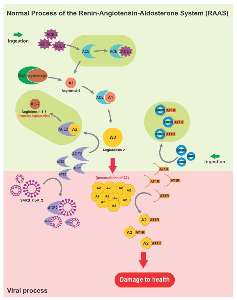
A pathophysiology diagram illustrating the interaction between SARS-CoV-2 and the Renin-Angiotensin-Aldosterone System (RAAS). The upper 'Normal Process' panel (green background) depicts the enzymatic cascade where Renin converts Angiotensinogen to Angiotensin I (A1), and Angiotensin-Converting Enzyme (ACE) converts A1 to Angiotensin II (A2). Under physiological conditions, Angiotensin-Converting Enzyme 2 (ACE2) converts A2 into Angiotensin 1-7 (A1-7), characterized as a 'harmless heptapeptide' with vasodilatory effects. The diagram also shows pharmaceutical interventions like ACE inhibitors (ACEi) and Angiotensin Receptor Blockers (ARBs) modulating these pathways. The lower 'Viral process' panel (red background) illustrates SARS-CoV-2 viruses binding to ACE2 receptors. This viral hijacking prevents the degradation of A2, leading to an 'Accumulation of A2'. The resulting excessive A2 binds to Angiotensin II Type 1 Receptors (AT1R), triggering a signaling cascade that results in 'Damage to health', representing the pro-inflammatory and hypertensive complications of COVID-19. This visual comparison highlights the loss of ACE2-mediated protection during viral infection.
Renin-Angiotensin-Aldosterone System (RAAS)
Step 1: Renin Release
- Decreased renal perfusion pressure - detected by baroreceptors in granular cells of the afferent arteriole
- Low Na+/Cl- at macula densa - decreased distal tubular NaCl delivery sensed by macula densa cells
- Beta-1 adrenergic stimulation - sympathetic nervous system activation (e.g., hemorrhage, stress)
In humans, angiotensinogen is normally in excess, so Angiotensin I and II production is rate-limited by the concentration of renin - Tietz Textbook of Laboratory Medicine, 7th Ed.
Step 2: Angiotensinogen → Angiotensin I
Step 3: Angiotensin I → Angiotensin II (via ACE)
Step 4: Actions of Angiotensin II
| Effect | Site | Mechanism |
|---|---|---|
| Vasoconstriction | Systemic vasculature | Direct smooth muscle contraction via AT1R |
| Na+ reabsorption | Proximal convoluted tubule | Direct stimulation |
| Aldosterone release | Adrenal zona glomerulosa | Stimulates cholesterol desmolase + aldosterone synthase |
| ADH (AVP) release | Posterior pituitary (via SFO) | AT1R in subfornical organ (lacks blood-brain barrier) |
| Thirst | Hypothalamus | Via SFO-SON/PVN neural pathways |
| Sympathetic activation | Adrenal medulla | Increases NE and epinephrine release |
| Cardiac/renal fibrosis | Heart, kidney (local RAAS) | Via TGF-β, AT1R in paracrine/autocrine manner |
Step 5: Aldosterone
- Na+ reabsorption (via ENaC and Na+/K+-ATPase upregulation)
- K+ secretion
- H+ secretion
- Net effect: increased blood volume and blood pressure
Feedback / Negative Regulation
- Restored blood pressure and Na+ delivery reduce further renin secretion
- Angiotensin II itself causes efferent arteriolar constriction, raising filtration fraction
- ANP/BNP and nitric oxide inhibit renin secretion via cGKII activation in JG cells
- ACE2 (an alternative enzyme) cleaves Ang II to Ang 1-7, which acts on Mas receptors to produce vasodilation, anti-inflammatory, and anti-fibrotic effects - the counter-regulatory arm of RAAS
Local (Tissue) RAAS
Pharmacological Targets
| Drug Class | Target | Example |
|---|---|---|
| ACE inhibitors | Block ACE | Enalapril, Ramipril |
| ARBs | Block AT1R | Losartan, Valsartan |
| Direct renin inhibitors | Block renin | Aliskiren |
| Aldosterone antagonists | Block mineralocorticoid receptor | Spironolactone, Eplerenone |
| ARNI | ARB + Neprilysin inhibitor | Sacubitril/Valsartan |