
Inflammation
Reading File
The topic is broad. Let me clarify scope before diving in.
Asking for Preferences
What aspect of inflammation would you like to explore?
Searching sources...
Finding Sources
Finding Sources
Robbins & Kumar Pathologic Basis of Disease is the gold standard for this. Let me pull the core inflammation content from it, plus search for mediators and cells.
Reading File
Finding Sources
Finding Sources
Good. Now let me pull the mediators section, chronic inflammation cells, and outcomes from Robbins Pathologic Basis of Disease in parallel.
Reading File
Reading File
Reading File
Now let me get the chronic inflammation and outcomes sections.
Reading File
Reading File
Reading File
I now have comprehensive material to write a thorough overview. Let me fetch a key image too.
Loading Image
Reading File
I now have all the material needed for a comprehensive overview. Let me compose the full answer.
Inflammation - Overview
Inflammation is a protective host response to foreign invaders (microbes, toxins) and necrotic tissue. While fundamentally beneficial, it can also cause tissue damage when poorly controlled or misdirected. The entire response can be summarized in the Five R's:
- Recognition of the injurious agent
- Recruitment of leukocytes
- Removal of the agent
- Regulation (control) of the response
- Repair (resolution)
- Robbins, Cotran & Kumar Pathologic Basis of Disease, p. 2564-2566
Types of Inflammation
| Feature | Acute | Chronic |
|---|---|---|
| Onset | Minutes to hours | Days to years |
| Duration | Short (days) | Long (months-years) |
| Principal cells | Neutrophils | Macrophages, lymphocytes, plasma cells |
| Tissue injury | Usually mild/reversible | Often progressive with fibrosis |
| Vascular changes | Prominent (vasodilation, exudation) | Less prominent |
1. Acute Inflammation
Cardinal Signs (Latin)
- Rubor (redness) - vasodilation
- Calor (heat) - increased blood flow
- Tumor (swelling) - fluid exudation
- Dolor (pain) - mediators acting on nerves
- Functio laesa (loss of function) - combined effect
Vascular Events
Acute inflammation has three major components:
- Vasodilation of small vessels (especially postcapillary venules), increasing blood flow. Histamine is the principal early mediator. This causes heat and redness.
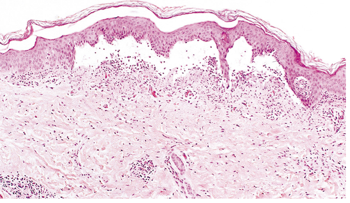
- Increased vascular permeability, allowing protein-rich fluid (exudate) to escape into extravascular tissue. Unlike a transudate (a low-protein ultrafiltrate due to hydrostatic/osmotic imbalance), an exudate signals active vascular permeability change.
- Leukocyte emigration - neutrophils accumulate along the endothelium, adhere, then transmigrate into tissues.
Exudate vs. Transudate:
- Exudate = high protein, high cells, inflammatory cause
- Transudate = low protein, osmotic/hydrostatic imbalance (e.g., heart failure, cirrhosis)
- Pus = purulent exudate rich in neutrophils + cell debris
Cellular Events (Leukocyte Recruitment)
The sequence of events in leukocyte recruitment:
- Margination - leukocytes move to the vessel periphery as blood flow slows
- Rolling - mediated by selectins (L-, E-, P-selectin)
- Firm adhesion - mediated by integrins (e.g., LFA-1, Mac-1) binding to endothelial ICAMs (upregulated by TNF, IL-1)
- Transmigration (diapedesis) - through interendothelial junctions via CD31 (PECAM-1)
- Chemotaxis - migration along a chemical gradient toward the injury site
Key chemoattractants:
- Bacterial products (N-formylmethionine peptides)
- C5a (complement)
- LTB4 (leukotriene B4)
- IL-8/CXCL8 (chemokine)
Genetic deficiency of leukocyte adhesion molecules (Leukocyte Adhesion Deficiency, LAD) causes recurrent severe bacterial infections - proof of how critical this process is.
Morphologic Patterns of Acute Inflammation
| Pattern | Features | Example |
|---|---|---|
| Serous | Watery, protein-poor fluid; no microbes | Skin blister (viral), pleuritis |
| Fibrinous | Fibrin-rich exudate; "bread-and-butter" pericarditis | Rheumatic pericarditis, lobar pneumonia |
| Suppurative/Purulent | Pus (neutrophils + debris); abscess formation | Bacterial infection (Staph abscess) |
| Ulcerative | Surface epithelial erosion exposing underlying tissue | Peptic ulcer, aphthous ulcer |
2. Chemical Mediators of Inflammation
Mediators are either cell-derived (rapidly secreted from granules or synthesized de novo) or plasma-derived (inactive precursors activated by proteolytic cleavage, mainly produced in the liver).
| Mediator | Source | Key Actions |
|---|---|---|
| Histamine | Mast cells, basophils, platelets | Vasodilation, increased vascular permeability, endothelial activation |
| Prostaglandins (PGE2, PGI2) | Mast cells, leukocytes | Vasodilation, pain, fever |
| Leukotrienes (LTB4, LTC4/D4/E4) | Mast cells, leukocytes | LTB4: chemotaxis; LTC4/D4/E4: increased permeability, bronchoconstriction |
| TNF, IL-1 | Macrophages, endothelial cells, mast cells | Endothelial activation (adhesion molecules), fever, acute phase response, shock |
| Chemokines (e.g., IL-8) | Leukocytes, macrophages | Chemotaxis, leukocyte activation |
| Platelet-Activating Factor (PAF) | Leukocytes, mast cells | Vasodilation, permeability, leukocyte adhesion, chemotaxis, degranulation |
| Complement (C3a, C5a, MAC) | Plasma (liver) | C3a/C5a: mast cell degranulation, chemotaxis (C5a), opsonization (C3b); MAC: cell lysis |
| Kinins (bradykinin) | Plasma (liver) | Increased permeability, smooth muscle contraction, pain, vasodilation |
Mediator-to-reaction mapping:
-
Vasodilation: Histamine, prostaglandins
-
Increased permeability: Histamine, C3a/C5a, leukotrienes LTC4/D4/E4
-
Chemotaxis: TNF, IL-1, chemokines, C5a, LTB4
-
Fever: IL-1, TNF, prostaglandins
-
Pain: Prostaglandins, bradykinin, substance P
-
Tissue damage: Lysosomal enzymes, reactive oxygen species (ROS)
-
Robbins, Cotran & Kumar Pathologic Basis of Disease, p. 2906-2909
3. Outcomes of Acute Inflammation
Acute inflammation has three possible outcomes:
-
Complete resolution - the ideal outcome. The injurious agent is eliminated, edema is resorbed via lymphatics, macrophages clear debris, and the tissue regenerates. Seen when injury is limited and parenchymal cells can regenerate.
-
Scarring (fibrosis) - occurs when tissue destruction is substantial, tissue cannot regenerate, or abundant fibrin cannot be cleared. Connective tissue replaces the damaged area ("organization").
-
Progression to chronic inflammation - when the acute response cannot resolve the injurious agent or normal healing is disrupted.
4. Chronic Inflammation
Chronic inflammation is characterized by the combination of:
- Ongoing leukocyte infiltration (predominantly mononuclear - macrophages, lymphocytes, plasma cells)
- Tissue destruction (by leukocyte products)
- Healing attempts (fibrosis, angiogenesis)
Causes
- Persistent infections (e.g., Mycobacterium tuberculosis, H. pylori, certain fungi, viruses) that resist clearance
- Immune-mediated diseases (autoimmune diseases, prolonged immune reactions to environmental antigens)
- Prolonged toxic exposure (e.g., silica - silicosis; atherosclerosis)
Principal Cells
Macrophages are the dominant cells. They:
- Phagocytose microbes and dead tissue
- Secrete pro-inflammatory cytokines (TNF, IL-1, chemokines, eicosanoids)
- Initiate tissue repair (growth factors)
- Activate T lymphocytes by antigen presentation + costimulation
Tissue macrophage names: Kupffer cells (liver), microglia (CNS), alveolar macrophages (lung), sinus histiocytes (lymph nodes/spleen).
Lymphocytes (T and B):
- CD4+ T cells:
- Th1 subset: produces IFN-γ (activates macrophages) - involved in defense against bacteria/viruses and autoimmunity
- Th17 subset: produces IL-17 - recruits neutrophils; also involved in autoimmunity
- Th2 subset: produces IL-4, IL-5, IL-13 - important in parasitic infections and allergic inflammation
- T cells and macrophages interact bidirectionally, creating a self-amplifying cycle that sustains chronic inflammation
- Plasma cells produce antibodies against persistent antigens or self-antigens
Other cells:
- Eosinophils - prominent in Th2-mediated reactions (parasites, allergies); contain major basic protein (toxic to helminths but damages host tissue)
- Mast cells - bridge acute and chronic inflammation; express FcεRI for IgE binding; degranulate in immediate hypersensitivity
Granulomatous Inflammation
A special form of chronic inflammation defined by aggregates of activated macrophages (epithelioid cells) surrounded by lymphocytes, often with multinucleate giant cells (formed by fusion of macrophages). Forms when the causative agent cannot be eliminated.
Causes: TB, sarcoidosis, Crohn disease, syphilis, cat-scratch disease, fungal infections, foreign body reactions, berylliosis.
Types of giant cells:
- Langhans giant cell - nuclei at the periphery in a horseshoe pattern (classic for TB)
- Foreign body giant cell - nuclei randomly distributed
Tertiary Lymphoid Structures
In some long-standing chronic inflammatory states, lymphocytes organize into lymph node-like follicles at the site of injury. Examples: synovium in rheumatoid arthritis, thyroid in Hashimoto thyroiditis.
5. Systemic Effects - Acute Phase Response
When the inflammatory mediators (especially IL-1, TNF, IL-6) enter the bloodstream, they produce systemic effects:
| Effect | Mediator(s) | Mechanism |
|---|---|---|
| Fever | IL-1, TNF, IL-6 | Act on hypothalamus; induce PGE2 via COX |
| Acute phase proteins | IL-6 | Liver produces CRP, fibrinogen, serum amyloid A, complement |
| Leukocytosis | IL-1, TNF, CSFs | Accelerated leukocyte release from bone marrow |
| Malaise/anorexia | IL-1, TNF | CNS effects |
| Septic shock (extreme) | TNF | Hypotension, DIC, multiorgan failure |
Summary Diagram (Conceptual)
Injury/Infection
       |
       v
Recognition by sentinel cells (macrophages, mast cells, dendritic cells)
       |
       v
Release of mediators (histamine, cytokines, eicosanoids, complement)
       |
   ┌───┴────────────────────────────────┐
   v                                    v
Vascular events                   Cellular events
(vasodilation,                     (leukocyte rolling,
 increased permeability,            adhesion, diapedesis,
 exudate formation)                 chemotaxis, phagocytosis)
   │
   └──────────────── Outcomes ──────────────────┐
           |                   |                |
     Resolution             Fibrosis      Chronic inflammation
Source: Robbins, Cotran & Kumar Pathologic Basis of Disease (10th ed.), Chapter 3 - General Pathology of Inflammation.
This is a shared conversation. Sign in to Orris to start your own chat.