


Ans of all q
temporal bone parts squamous petrous mastoid
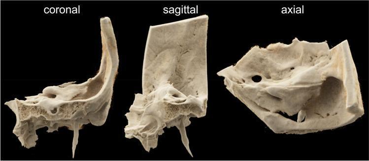
This diagnostic image displays a high-resolution Volume Rendering Technique (VRT) reconstruction of a human temporal bone specimen, presented in three standard anatomical planes: coronal, sagittal, and axial. The reconstruction highlights the complex morphology of the petrous and squamous portions of the temporal bone. The coronal view reveals the vertical orientation of the squamous part and the styloid process extending inferiorly, while showing the porous trabecular nature of the mastoid region. The sagittal view illustrates the anteroposterior depth and the density variations between the cortical surface and the internal cancellous bone. The axial view provides a superior perspective, showcasing the internal acoustic canal, various foramina, and the irregular contours of the petrous ridge. This visual is intended for educational purposes in radiology and otolaryngology, demonstrating the capabilities of advanced CT scanning protocols (such as MS-CT and FD-CT) in visualizing minute anatomical structures, bony landmarks, and surface textures of the ear's structural foundation.
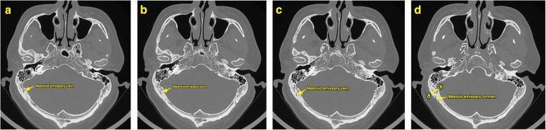
This diagnostic image consists of a series of four high-resolution axial computerized tomography (CT) scans (labeled a–d) of the human skull base, displayed in a bone window. The sequence demonstrates the surgical anatomy of the temporal bone, specifically highlighting the mastoid emissary vein (MEV) and its associated bony canal. In images a, b, and c, yellow arrows trace the course of the mastoid emissary vein as it traverses the mastoid part of the temporal bone. Image d, designated as 'Slice B', identifies the mastoid emissary foramen on the outer cortex. Key landmarks visible across all slices include the petrous and squamous parts of the temporal bone, the mastoid air cells, the sphenoid bone with the sella turcica, and the occipital bone. Annotations in frame d use Greek letters gamma (γ) and delta (δ) to indicate specific measurement points on the inner and outer cortical surfaces, respectively. This visual serves as an anatomical guide for preoperative planning in retrosigmoid approaches to the posterior cranial fossa, helping to avoid intraoperative hemorrhage from emissary veins.
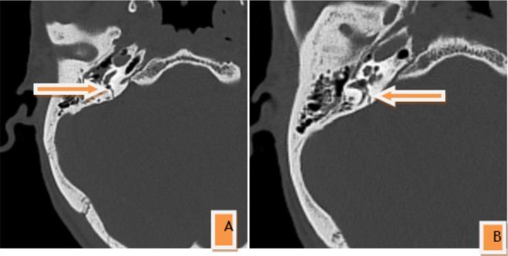
Two-panel axial non-contrast CT scan of the head in bone window settings demonstrating a right petrous temporal bone fracture. Panel A illustrates the fracture line traversing the petrous pyramid with involvement of the internal carotid artery (ICA) canal. Panel B shows the extension of the fracture into the jugular fossa walls. White arrows indicate the fracture site. The fracture is characterized as a transverse pattern, which is clinically significant due to its violation of the otic capsule, the dense bony housing of the inner ear. The images display disruption of the bony architecture surrounding the cochlea and vestibular system, posing a high risk for sensorineural hearing loss and facial nerve injury. The surrounding mastoid air cells appear largely aerated, and the squamous part of the temporal bone remains intact. This diagnostic image is representative of high-impact craniofacial trauma in adult patients.
Q. NO. 4
A) Name the Condition
- Proptosis (forward protrusion of the eyeball)
- Periorbital edema and necrosis
- Black/necrotic discoloration of surrounding tissue
- This is the classic appearance in a diabetic patient with DKA (diabetic ketoacidosis)
B) Management of Rhinocerebral Mucormycosis
1. Treat Underlying Disease (Critical)
- Control hyperglycemia and reverse diabetic ketoacidosis
- Taper/stop glucocorticoids if applicable
- Stop deferoxamine (iron chelation) if used
- Correct neutropenia if present
2. Antifungal Therapy
| Drug | Dose | Role |
|---|---|---|
| Liposomal Amphotericin B | 5-10 mg/kg/day IV | Drug of choice (first-line) |
| Isavuconazole | 372 mg q8h x 6 doses, then 372 mg daily | Equally efficacious alternative |
| Posaconazole | 300 mg oral daily | Salvage/step-down therapy |
- Mucorales are resistant to: fluconazole, voriconazole, flucytosine, echinocandins, ketoconazole
- Combining liposomal amphotericin B + an echinocandin may be better for diabetic patients with rhino-orbital disease
3. Aggressive Surgical Debridement
- Prompt surgical removal of all necrotic tissue
- CT or MRI guides the extent of surgery
- Intraoperative frozen sections help ensure complete excision
- Enucleation of the eye may be required if orbital involvement is extensive
- Repeated debridement is often necessary
4. Adjunctive Therapies
- Hyperbaric oxygen (especially in diabetic rhinocerebral disease - beneficial but not routinely recommended)
- Granulocyte transfusion in refractory neutropenic patients
- G-CSF / GM-CSF as immune stimulation
Source: Goldman-Cecil Medicine, Chapter 312 - Mucormycosis
Q. NO. 10
A) Identify the Bone and Its Different Parts
| Part | Key Features |
|---|---|
| Squamous part | Flat, thin, fan-shaped; forms the lateral wall of the skull; bears the zygomatic process and supramastoid crest (temporal line) |
| Petrous part (Petrous pyramid) | Dense, pyramid-shaped; contains the inner ear (cochlea, semicircular canals), internal carotid artery canal, internal acoustic meatus |
| Mastoid part | Posterior projection; contains mastoid air cells; mastoid process is its inferior prominence |
| Tympanic part | C-shaped; forms the floor and anterior/inferior walls of the external auditory canal (EAC) |
| Styloid process | Slender projection below the tympanic part; gives attachment to stylohyoid and stylomandibular ligaments |
- Superior border = Supramastoid crest
- Anterior border = Suprameatal spine (spine of Henle)
- Posterior border = Line tangential to the posterior border of the external auditory canal
B) Identify the Triangle Shown in the Picture
MacEwen's Triangle:
- Superior border - Supramastoid crest (temporal line)
- Anterior border - Suprameatal spine (spine of Henle) / posterior wall of the EAC
- Posterior border - Line drawn tangential to the posterior border of the external auditory canal
Clinical Importance:
- It is a surface landmark for cortical mastoidectomy - the surgeon enters the mastoid antrum through this triangle
- The mastoid antrum lies approximately 1.5 cm deep to MacEwen's triangle
- It overlies the antrum, which connects the middle ear (epitympanum) with the mastoid air cells
- Used to locate the antrum during surgery for mastoiditis or cholesteatoma
- In adults: the antrum is ~1.5 cm deep; in children, it is shallower and more medially placed
Source: Scott-Brown's Otorhinolaryngology Head & Neck Surgery, Vol. 2 - Temporal Bone