
Type 1 respiratory impairment
Reading File
Quick answer: Type 1 respiratory failure (impairment) is defined by hypoxaemia (PaO2 < 8 kPa / 60 mmHg) with a normal or low PaCO2, indicating a failure of oxygenation without ventilatory failure. It is caused by ventilation-perfusion (V/Q) mismatch, diffusion impairment, or intracardiac/intrapulmonary shunts.
Searching sources...
Finding Sources
Finding Sources
Reading File
Reading File
Reading File
Finding Sources
Reading File
Reading File
Finding Sources
Reading File
Finding Sources
Now I have comprehensive information from multiple authoritative sources. Let me compile the full answer.
Type 1 Respiratory Failure (Hypoxemic Respiratory Failure)
Definition
Type 1 respiratory failure (also called hypoxemic respiratory failure or Type I: Acute Hypoxemic) is defined as:
- PaO2 < 60 mmHg (or SaO2 < 90%)
- PaCO2 normal or low (in contrast to Type 2, where PaCO2 is elevated)
- Usually associated with tachypnea and hypocapnia; however, progression can lead to hypercapnia as well
The key distinction from Type 2 (hypercapnic) failure: there is a problem with oxygenation but not necessarily with ventilation.
- The Washington Manual of Medical Therapeutics
- Tintinalli's Emergency Medicine
Pathophysiology: The 5 Mechanisms
Type 1 failure results from the lung's reduced ability to deliver oxygen across the alveolocapillary membrane. Hypoxemia arises via one of five mechanisms, assessed using the alveolar-arterial (A-a) gradient:
1. V/Q Mismatch (most common)
Perfusion does not match ventilation or vice versa. A-a gradient is elevated. Supplemental oxygen raises PaO2. Note: in emphysema, supplemental O2 paradoxically worsens V/Q mismatch by reversing hypoxic vasoconstriction.
- Examples: emphysema, pneumonia, pulmonary edema, pulmonary embolism
2. Shunt
Mixed venous blood bypasses lung units entirely, entering arterial circulation un-oxygenated. A-a gradient is elevated. Supplemental oxygen does NOT correct hypoxaemia in pure shunt.
- Pulmonary shunts: pneumonia (pus), cardiogenic/non-cardiogenic pulmonary edema (water), diffuse alveolar haemorrhage (blood), atelectasis, pleural effusion
- Cardiac shunts: patent foramen ovale, ASD, VSD
- Vascular shunts: arteriovenous malformation
3. Diffusion Abnormality
The interstitium is thickened, making gas equilibration take longer than red blood cell transit time through pulmonary capillaries. A-a gradient is elevated.
- Examples: pulmonary fibrosis, pulmonary hypertension
4. Hypoventilation
Decreased minute ventilation raises PaCO2, displacing oxygen. A-a gradient is normal. Responds to supplemental oxygen. (This mechanism more commonly produces Type 2 failure when severe.)
5. Low Inspired Oxygen (FiO2)
Low partial pressure of inspired O2 (e.g., high altitude). A-a gradient is normal. Responds to supplemental oxygen.
- Washington Manual of Medical Therapeutics, p. 277
Common Clinical Causes
| Category | Examples |
|---|---|
| Infection | Pneumonia, COVID-19, sepsis |
| Pulmonary oedema | Cardiogenic (LVF, mitral disease), non-cardiogenic (ARDS) |
| Lung injury | Gastric aspiration, inhalational injury, near-drowning |
| Vascular | Pulmonary embolism |
| Interstitial | Pulmonary fibrosis, sarcoidosis |
| Structural | Pleural effusion, lobar collapse, mucous plugging |
Harrison's Principles of Internal Medicine 22E lists sepsis, gastric aspiration, pneumonia, and COVID-19 as the archetypes of Type I acute hypoxemic respiratory failure.
ARDS: The Paradigm of Type 1 Failure
Acute Respiratory Distress Syndrome (ARDS) is the most important form of hypoxemic respiratory failure. It results from acute lung injury causing disruption of the alveolocapillary membrane, increased vascular permeability, and protein-rich inflammatory fluid in the alveolar space.
Berlin Definition criteria:
- Onset within 1 week of a known clinical insult
- Bilateral opacities on imaging not explained by effusions or collapse
- Respiratory failure not explained by cardiac failure or fluid overload
- PaO2/FiO2 ratio ≤ 300 mmHg (with PEEP ≥ 5 cmH2O)
ARDS severity by P/F ratio:
| Severity | PaO2/FiO2 |
|---|---|
| Mild | 200 - 300 mmHg |
| Moderate | 100 - 200 mmHg |
| Severe | ≤ 100 mmHg |
- Washington Manual of Medical Therapeutics, p. 276-277
Management of Type 1 Respiratory Failure
The priority is optimising oxygenation (unlike Type 2, which also requires supporting ventilation).
1. Supplemental Oxygen (Non-invasive)
| Device | FiO2 Delivered | Notes |
|---|---|---|
| Nasal cannula | ~24-44% (4% per L/min) | Max 6 L/min |
| Simple face mask | 35-55% | 5-12 L/min |
| Venturi mask | 24-50% (precise) | Preferred when accurate FiO2 needed |
| Non-rebreather mask | Up to ~80-90% | Reservoir bag + one-way valve |
2. High-Flow Nasal Cannula (HFNC)
- Up to 60 L/min, 100% FiO2
- Washes out dead space from upper airway, provides small degree of PEEP
- Meta-analysis: HFNC reduced need for intubation and escalation of oxygen therapy vs. low-flow O2
- Preferred in pneumonia and post-surgical hypoxaemic failure
- NOT preferred in cardiogenic pulmonary oedema - NIV is superior there
3. Non-Invasive Positive Pressure Ventilation (NPPV)
- CPAP: Prevents alveolar collapse, recruits atelectatic units - particularly useful in cardiogenic pulmonary oedema and OSA. Start at 5 cmH2O, titrate to 10-15 cmH2O.
- BiPAP: Reduces work of breathing; IPAP 5-10 cmH2O, EPAP 5 cmH2O starting point
- Strong evidence in: neuromuscular disease, COPD, pulmonary oedema, post-operative failure
- Requires: conscious, cooperative patient who can protect airway and is haemodynamically stable
- Helmet-delivered NIV showed intubation reduction + improved survival in ARDS (2016 trial)
4. Invasive Mechanical Ventilation (Intubation)
Indicated when HFNC/NIV fail, patient cannot protect airway, or is haemodynamically unstable. In ARDS, lung-protective ventilation is the standard:
-
Tidal volume: 6 mL/kg ideal body weight
-
PEEP titrated to maintain alveolar recruitment
-
Plateau pressure < 30 cmH2O
-
Washington Manual of Medical Therapeutics, pp. 278-280
Type 1 vs. Type 2: Quick Comparison
| Feature | Type 1 | Type 2 |
|---|---|---|
| PaO2 | Low (< 60 mmHg) | Low |
| PaCO2 | Normal or low | High (> 45 mmHg) |
| Primary problem | Oxygenation failure | Ventilatory failure |
| A-a gradient | Usually elevated | Normal (if pure hypoventilation) |
| Examples | Pneumonia, ARDS, PE | COPD exacerbation, neuromuscular disease, opioid overdose |
| O2 response | Often improves with O2 | Improves with O2 (caution in COPD) |
| Treatment goal | Optimise oxygenation | Optimise oxygenation + support ventilation |
Alveo
I want to make sure I cover exactly what you need before diving in.
Asking for Preferences
What aspect of 'alveoli' would you like covered?
Searching sources...
Finding Sources
Finding Sources
Finding Sources
Reading File
Reading File
Reading File
Reading File
Reading File
Reading File
Reading File
Reading File
Loading Image
Loading Image
Loading Image
I now have rich, comprehensive content from multiple histology textbooks with diagrams. Here is the full answer:
Alveolar Anatomy & Structure
Overview
Alveoli are the terminal air spaces of the respiratory system - small, sac-like evaginations (~200 µm in diameter) arising from respiratory bronchioles, alveolar ducts, and alveolar sacs. They are responsible for the spongy appearance of lung tissue and are the actual site of gas exchange between inhaled air and blood.
Key numbers:
-
~150-250 million alveoli per adult lung
-
Total internal surface area: ~75 m² (roughly the size of a tennis court)
-
Blood-air barrier thickness: 0.1 to 1.5 µm
-
Junqueira's Basic Histology, 17e; Histology: A Text and Atlas
Structural Organisation
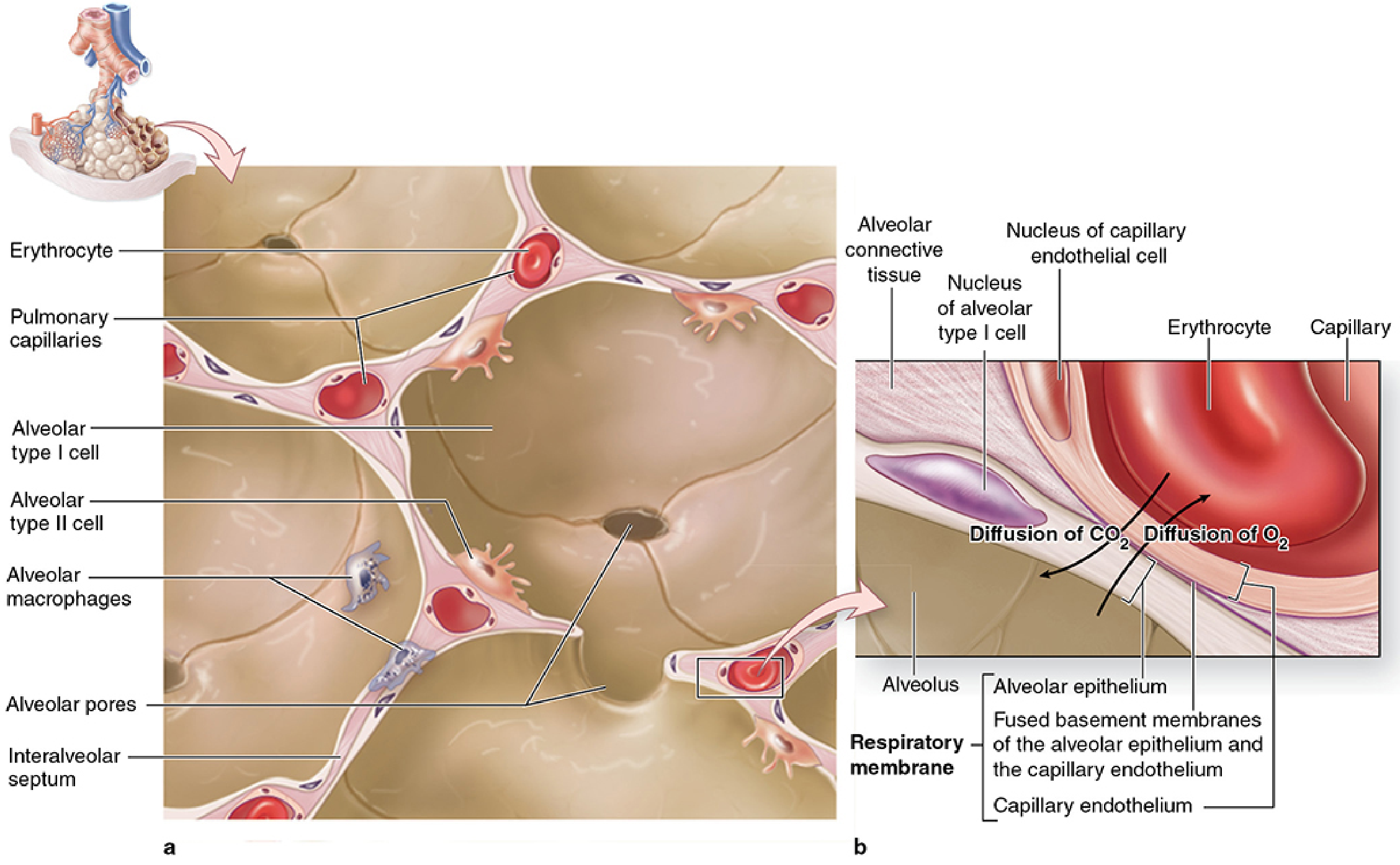
Alveolar Ducts and Sacs
- Alveolar ducts: Elongated airways whose peripheral boundary is almost entirely alveoli; rings of smooth muscle are present at interalveolar septal knobs
- Alveolar sacs: Spaces surrounded by clusters of alveoli; occur at the termination of an alveolar duct (or anywhere along its length)
- Each alveolus opens into an alveolar duct or alveolar sac on one side
Interalveolar Septa
Between neighboring alveoli lie thin interalveolar septa (septal walls) containing:
- Scattered fibroblasts and sparse extracellular matrix (ECM)
- Elastic fibers - allow alveoli to expand with inspiration and contract passively with expiration
- Reticular fibers - prevent collapse and excessive distention
- The richest capillary networks in the body, densely anastomosing within the septa
The Blood-Air Barrier (Respiratory Membrane)
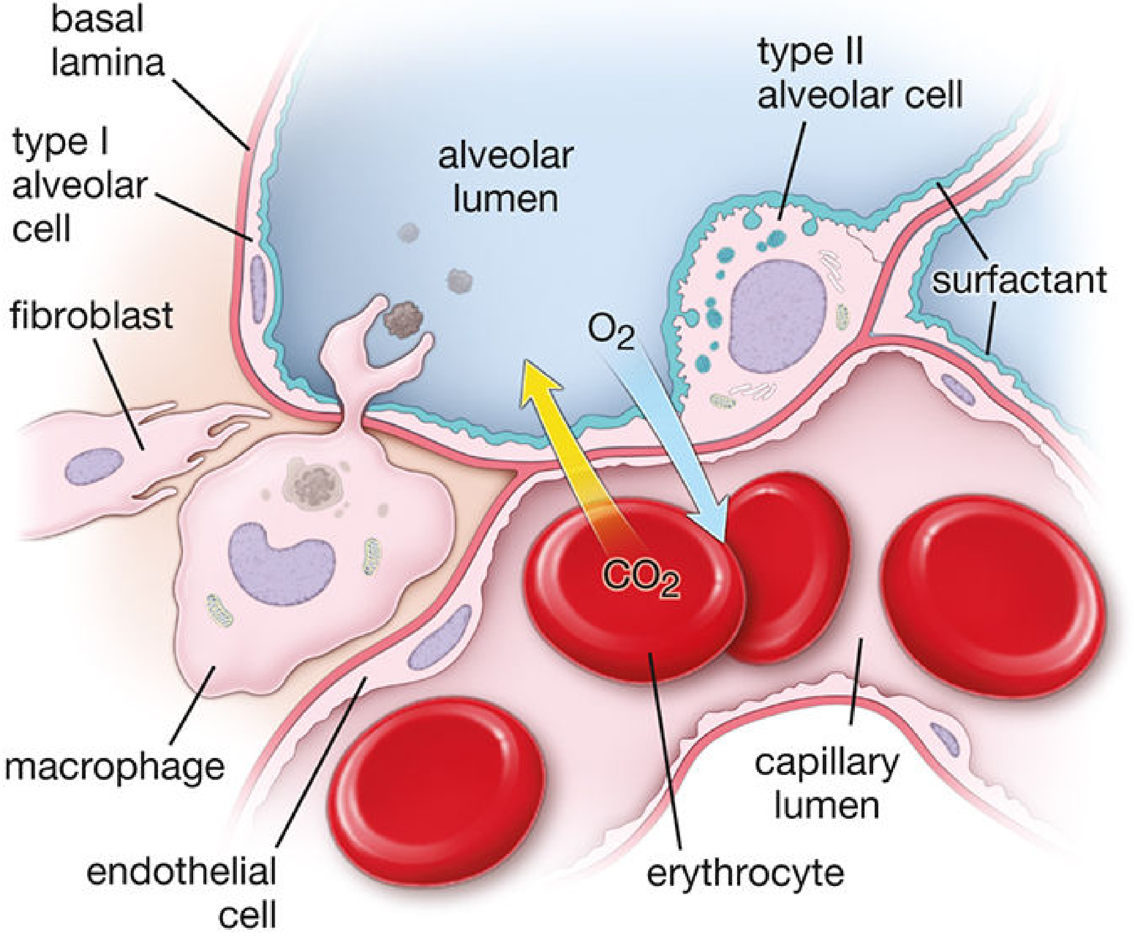
Alveolar air is separated from capillary blood by three layers (total thickness: 0.1-1.5 µm):
- Alveolar epithelium (type I pneumocyte + surfactant layer)
- Fused basal laminae of the alveolar epithelial cell and capillary endothelial cell
- Capillary endothelium (continuous, non-fenestrated)
The septum has a thin portion (main site of gas exchange) and a thick portion (where connective tissue cells and fibres widen the barrier - site of fluid accumulation in pathological states). Lymphatic vessels in the terminal bronchiole connective tissue drain fluid from the thick portion.
Cell Types of the Alveolar Wall
Type I Alveolar Cells (Type I Pneumocytes)
| Feature | Detail |
|---|---|
| Proportion of lining cells | 40% of cells |
| Surface area covered | ~95% of alveolar surface |
| Shape | Extremely thin, squamous (attenuated) |
| Function | Gas exchange surface; water transport; fluid homeostasis |
| Cell division | Not capable - cannot divide |
| Junctions | Occluding junctions (tight junctions) with each other and type II cells - form an effective air-blood barrier |
| Marker | RAGE receptor (receptor for advanced glycation end products) |
Type I cells are so thin that organelles cluster around the nucleus, leaving the rest of the cell as a near-transparent sheet maximally adapted for diffusion.
Type II Alveolar Cells (Type II Pneumocytes / Septal Cells)
| Feature | Detail |
|---|---|
| Proportion of lining cells | 60% of cells |
| Surface area covered | ~5% of alveolar surface |
| Shape | Cuboidal, dome-shaped, bulging into airspace |
| Location | Interspersed among type I cells; congregate at septal junctions, often in groups of 2-3 |
| Function | Surfactant production and secretion; ion transport; immune surveillance; progenitor cells |
| Cell division | Yes - can divide and regenerate both type I and II cells after injury |
| Key organelles | Lamellar bodies (100-400 nm granules containing surfactant) |
| Marker | Pro-surfactant protein C (SP-C) |
Type II cell hyperplasia is an indicator of alveolar injury and repair.
Alveolar Macrophages ("Dust Cells")
- Large phagocytic cells found in alveolar spaces and within the interalveolar septa
- Part of the innate immune defence - engulf inhaled particles, pathogens, and cellular debris
- Can be seen in alveolar spaces or within the septa
Brush Cells
- Present in small numbers in the alveolar wall
- Function as chemoreceptors that monitor air quality in the lung
Surfactant
Produced and secreted by type II alveolar cells via lamellar bodies. It forms a thin film of phospholipids and lipoproteins over the alveolar epithelium.
Primary lipid: Dipalmitoylphosphatidylcholine (DPPC) - responsible for almost all surface tension-reducing properties.
Function:
- Lowers surface tension at the air-epithelium interface
- Prevents alveolar collapse on expiration
- Allows inflation with less inspiratory force (reduces work of breathing)
- Participates in alveolar innate immunity
Surfactant Proteins:
| Protein | Function |
|---|---|
| SP-A (most abundant) | Regulates surfactant homeostasis; modulates immune responses to viruses, bacteria, fungi |
| SP-B | Critical for transforming lamellar body into surface film; adsorption and spreading |
| SP-C | Aids DPPC orientation within surfactant layer |
| SP-D | Host defence; binds Gram-negative bacteria and lymphocytes; modulates response to inhaled antigens |
Surfactant synthesis begins after the 35th week of gestation, modulated by cortisol, insulin, prolactin, and thyroxine. Deficiency leads to neonatal respiratory distress syndrome (RDS) in preterm infants.
- Histology: A Text and Atlas, p. 1806-1807
Alveolar Pores of Kohn
- 10-15 µm diameter openings that penetrate the interalveolar septa
- Connect neighboring alveoli on different bronchioles
- Two functions:
- Equalise air pressure between alveoli
- Permit collateral ventilation - allow air circulation if a bronchiole becomes obstructed
Summary Table: Key Cell Types
| Cell | % of cells | % surface | Divides? | Key role |
|---|---|---|---|---|
| Type I pneumocyte | 40% | 95% | No | Gas exchange, fluid homeostasis |
| Type II pneumocyte | 60% | 5% | Yes | Surfactant, progenitor cell |
| Alveolar macrophage | Few | - | Yes | Innate immunity, phagocytosis |
| Brush cell | Very few | - | - | Chemoreception |
- Junqueira's Basic Histology, 17e, pp. 880-887; Histology: A Text and Atlas, pp. 1800-1809; Murray & Nadel's Textbook of Respiratory Medicine
In Type 1 respiratory impairment alveolar ventilation is normal?
Quick answer: Yes - in Type 1 respiratory failure, overall alveolar ventilation is typically normal or even increased (hence normal or low PaCO2). The problem is not how much air is moved, but how efficiently oxygen crosses into the blood. The hypoxaemia comes from V/Q mismatch, shunt, or diffusion failure - not from hypoventilation.
Reasoning...
Finding Sources
Finding Sources
Finding Sources
Finding Sources
Reading File
Reading File
This is now very clear. Here is the precise, nuanced answer:
Is Alveolar Ventilation Normal in Type 1 Respiratory Failure?
The answer is: yes - normal or above normal - and this is actually the defining feature.
The Core Concept
Type 1 respiratory failure is defined by failure of oxygenation, not failure of ventilation. The total minute ventilation and alveolar ventilation are intact - the problem lies in how efficiently the alveoli transfer oxygen to the blood.
Because the patient is hypoxaemic, chemoreceptors are stimulated, typically driving a reflex increase in alveolar ventilation. This is why the typical ABG pattern shows:
| Parameter | Finding | Explanation |
|---|---|---|
| PaO2 | Low (< 60 mmHg) | Oxygenation failure |
| PaCO2 | Normal or low | Ventilation preserved / increased |
| pH | Normal or slightly raised (respiratory alkalosis) | Hyperventilation blows off CO2 |
"Usually associated with tachypnea and hypocapnia."
- Washington Manual of Medical Therapeutics
Why Normal Ventilation Cannot Fix the Hypoxaemia
This is the key physiological point. Increasing alveolar ventilation (VA) helps eliminate CO2 efficiently, but cannot adequately compensate for impaired oxygenation in the two main mechanisms:
1. V/Q Mismatch
Blood from low V/Q units has low O2 and high CO2. The high CO2 stimulates an increase in overall alveolar ventilation, which:
- Does correct the hypercapnia - CO2 is blown off from the well-ventilated units
- Cannot correct the hypoxaemia - because pulmonary end-capillary blood from well-ventilated units is already maximally saturated with O2; there is no reserve capacity to carry extra oxygen
"Arterial CO2 tension often decreases from a hypoxemia-induced reflex increase in alveolar ventilation. An appreciable compensatory increase in O2 uptake cannot take place in remaining areas where V/Q is normal because pulmonary end-capillary blood is usually already maximally saturated with O2."
- Morgan & Mikhail's Clinical Anesthesiology, 7e
2. Shunt
In absolute shunt, blood bypasses ventilated alveoli entirely. No amount of increased ventilation can oxygenate blood that never contacts alveolar air. This is why pure shunt does not respond to supplemental O2 (a key clinical test).
3. V/Q mismatch (type 1) vs. hypoventilation (type 2)
In Type 2 failure, the problem IS alveolar ventilation - it is globally reduced, so PaCO2 rises, displacing O2 from the alveolus. In Type 1, global ventilation is normal or increased; it's the distribution (V/Q mismatch) or completeness (shunt) of gas exchange that fails.
Summary
| Type 1 | Type 2 | |
|---|---|---|
| Global alveolar ventilation | Normal or increased | Reduced |
| PaCO2 | Normal or low | High (> 45 mmHg) |
| Problem | Oxygenation (V/Q, shunt, diffusion) | Ventilation failure |
| Can increasing VA correct hypoxaemia? | No (for shunt); Partial (for V/Q mismatch) | Yes |
In Type 1 failure, systemic oxygen content depends mainly on haemoglobin concentration and V/Q matching - and tends to plateau even as alveolar ventilation is increased (e.g., with NIV). This is why treatment priorities focus on recruiting alveoli (PEEP, CPAP) and improving V/Q matching rather than simply increasing minute ventilation.
- Fishman's Pulmonary Diseases and Disorders; Morgan & Mikhail's Clinical Anesthesiology, 7e; Washington Manual of Medical Therapeutics
This is a shared conversation. Sign in to Orris to start your own chat.