
Hi
Hi! I'm Orris. How can I help you today?

1000430766.jpg
60yr male with kco dm2 and htn with s/p PTCA to RCA , patient on anti htn, oha and Ecosprin gold 20 with normal lipid profile and cbc ,hba1c 8.5 patient develop this maculo papular lesion which is painless and without any sensors loss lesion present all over body suggest me different diagnosis
analyse_media
Reading File
Now let me search the medical library and look for images in parallel.
Finding Sources
Finding Sources
Reading File
Reading File
Finding Sources
Loading Image
Finding Sources
I now have enough information to give a thorough, well-referenced differential diagnosis. Let me compile the answer.
Clinical Summary
Patient: 60-year-old male | KCO: DM2 (HbA1c 8.5 - poorly controlled) + HTN + s/p PTCA to RCA
Current medications: Anti-HTN agents, OHA, Ecosprin Gold 20 (Aspirin 75mg + Atorvastatin 20mg)
Lesion characteristics: Multiple flesh-colored to pinkish-brown, dome-shaped, smooth, firm papules and small nodules (2-8mm); distributed on extensor surface of arm/elbow; discrete + clustered; painless, no sensory loss, no scale/crust/ulceration
Differential Diagnosis (in order of likelihood)
1. Eruptive Xanthoma (Most Likely)
This is the top diagnosis for this patient given his poorly controlled DM2 (HbA1c 8.5) even with a currently normal lipid profile.
- Flesh-colored to yellowish-orange/reddish-brown dome-shaped papules appearing in crops over the entire body, with preference for extensor surfaces of arms, thighs, knees, buttocks
- Painless; pruritus is variable; may koebnerize
- Strongly suggest elevated triglyceride levels - importantly, eruptive xanthomas occur most commonly in uncontrolled diabetes mellitus even with a "normal" routine lipid panel (a fasting triglyceride-specific panel is needed)
- Uncontrolled DM causes decreased lipoprotein lipase activity → hypertriglyceridemia → eruptive xanthoma formation
- Key point: A "normal" lipid profile does not rule this out - triglycerides must be specifically checked when fasting
Andrews' Diseases of the Skin, Clinical Dermatology - "Eruptive xanthomas are seen most often in poorly controlled type 2 diabetes mellitus." Harrison's Principles of Internal Medicine 22E - "The most common setting for eruptive xanthomas is uncontrolled diabetes mellitus."
Reference image (eruptive xanthoma):
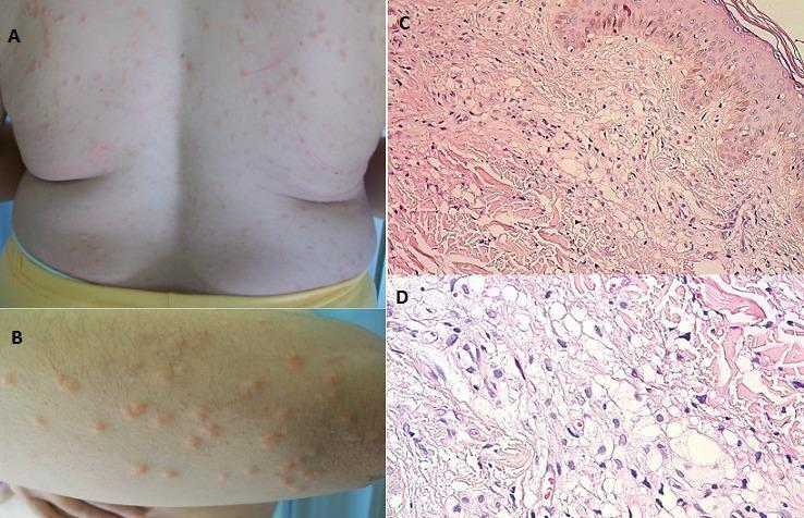
2. Prurigo Nodularis
- Firm, dome-shaped, hyperkeratotic nodules (5-12mm), usually on extensor limbs
- Typically intensely pruritic - but in diabetic neuropathy, the itch sensation may be blunted
- Associated with diabetes (peripheral neuropathy can alter itch sensation), chronic liver disease, renal disease
- No sensory loss per se, but altered sensation in DM is a confounding factor
- Surface is usually more excoriated/crusted in classic cases; smooth lesions early
3. Drug-Induced Eruption
- Aspirin (Ecosprin) and statins (Atorvastatin) can both cause cutaneous reactions
- Maculopapular drug eruptions are the most common type of drug reaction (95% of all cutaneous drug reactions)
- Usually appears weeks after starting the drug; can be widespread
- The lesion morphology here (smooth, firm, dome-shaped nodules rather than flat macules/papules) makes a classic morbilliform drug eruption less likely, but cannot be excluded
4. Secondary Syphilis
- Classic maculopapular rash that is widespread, involving the whole body including palms and soles
- Painless, non-pruritic - fits the clinical description
- In an older male, sexual history must be taken
- Usually accompanied by systemic symptoms (lymphadenopathy, malaise, mucous patches, condyloma lata)
- VDRL/RPR screening is inexpensive and should be done in any widespread maculopapular rash without a clear cause
5. Molluscum Contagiosum (Disseminated)
- Flesh-colored, dome-shaped, umbilicated papules (2-5mm)
- Usually in immunocompromised patients - DM2 is a risk factor for atypical/disseminated MC
- The lesions in the image do not show central umbilication, making classic MC less likely - but giant/confluent lesions may lose the umbilication
- Widespread distribution fits
6. Generalized Eruptive Histiocytoma
- Rare, non-Langerhans cell histiocytosis
- Multiple, firm, reddish-brown to flesh-colored papules appearing in crops across the trunk and extremities
- Painless, no systemic involvement
- Confirmed by biopsy (histiocytic infiltrate without foam cells)
- Associated with DM in some case reports
7. Disseminated Granuloma Annulare
- Ring-shaped or papular lesions on extensor surfaces
- Strongly associated with DM2
- Usually flesh-colored to erythematous, smooth papules
- Generalized form (>10 lesions or widespread distribution) is more common in middle-aged/older adults with DM
- Confirmed by biopsy (palisading granuloma with mucin)
Recommended Workup
| Investigation | Rationale |
|---|---|
| Fasting triglycerides + lipid panel | Rule out hypertriglyceridemia (eruptive xanthoma) |
| VDRL / RPR | Rule out secondary syphilis |
| Skin biopsy (H&E + PAS) | Definitive diagnosis - foam cells = xanthoma; histiocytes = GEH; mucin = GA |
| Fasting blood glucose + HbA1c | Already done; optimize DM control regardless |
| TSH | Hypothyroidism causes secondary hypertriglyceridemia |
| Renal function (BUN/Cr) | Chronic renal failure is another cause of eruptive xanthoma |
| HIV screening | If disseminated MC/prurigo nodularis is suspected |
Most Likely Diagnosis
Given the clinical context - poorly controlled DM2 (HbA1c 8.5), painless dome-shaped papules in crops on extensor surfaces of both arms, widespread distribution - Eruptive Xanthoma is the most likely diagnosis. The triglyceride level may be elevated even with an apparently "normal" standard lipid report. Optimizing glycemic control alone can cause these lesions to involute spontaneously.
Immediate action: Order a fasting triglyceride level and refer for skin biopsy.
This is a shared conversation. Sign in to Orris to start your own chat.