
Inverted papilloma Complete from basic to clinical aspects involving management and its procedures
"inverted papilloma" sinonasal management
inverted papilloma sinonasal histology endoscopic
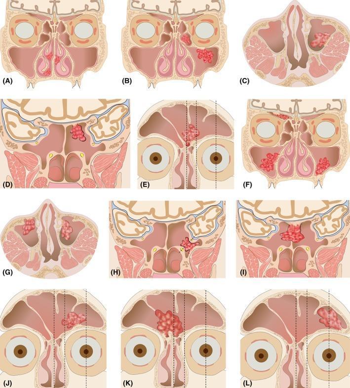
A multi-panel schematic anatomical diagram illustrating an endoscopic staging system for sinonasal inverted papilloma (SNIP). The visual set (A–L) categorizes stages 1 through 4 based on primary tumor origin sites within the paranasal sinuses and nasal cavity. Stage 1 (A) shows isolated nasal cavity involvement. Stage 2 (B–E) demonstrates tumors originating in the ethmoid sinus, superior/lateral/posterior maxillary walls, medial sphenoid sinus, or frontal sinus between the midline and lamina papyracea. Stage 3 (F–K) illustrates involvement of the anterior/medial maxillary walls, lateral sphenoid recess, bilateral sphenoid sinus, or frontal sinus regions extending from the lamina papyracea to the central pupil line. Stage 4 (L) depicts tumor origin lateral to the central pupil in the frontal sinus. Anatomical landmarks highlighted include the facial midline, lamina papyracea, and central pupil for surgical accessibility mapping. This diagnostic illustration serves as a clinical guide for otolaryngologists to determine endoscopic versus external surgical approaches based on the degree of anatomical difficulty and recurrence risk associated with specific tumor origin sites.
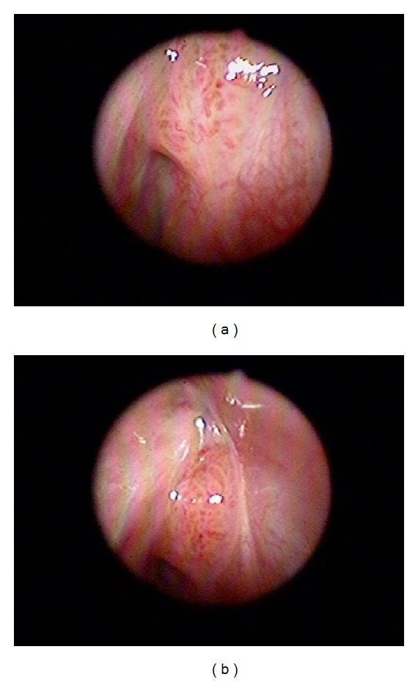
This clinical image presents two sinoscopic (endoscopic) views, labeled (a) and (b), of the nasal cavity. The imaging demonstrates a sinonasal inverted papilloma that has undergone malignant transformation. The mucosal lining is predominantly pinkish-red, indicating significant vascularity and underlying inflammation. Central to both views is an abnormal, localized tissue lesion characterized by an irregular, exophytic texture with subtle folds and crevices. Notable visual features include prominent surface vascularization and small red punctate spots, which are typical of the increased angiogenesis seen in neoplastic changes. The overall visual impression is one of disordered mucosal architecture and tumor growth within the sinonasal region. These endoscopic findings are clinically significant for identifying premalignant or malignant progression in patients with recurrent inverted papillomas, necessitating further histological evaluation for squamous cell carcinoma transformation.
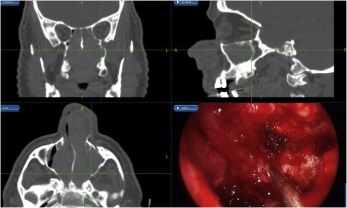
This composite medical image displays a case of sinonasal inverted papilloma managed through image-guided surgery. The layout includes three non-contrast Computed Tomography (CT) panels (coronal, sagittal, and axial views) and one intra-operative endoscopic photograph. The CT scans reveal extensive, homogenous soft-tissue opacities filling the bilateral nasal cavities, ethmoid sinuses, and maxillary sinuses, causing significant expansion and structural distortion. Green intersecting crosshairs across the CT panels denote the use of an intra-operative navigation system to localize a specific point in 3D space. The bottom-right panel shows the real-time endoscopic surgical field during a navigation-assisted bilateral medial maxillectomy. It demonstrates a view of the nasal mucosa, visible hemorrhage, and surgical instrumentation during tumor resection. This visual content illustrates the diagnostic imaging of sinonasal masses and the clinical application of surgical navigation in otorhinolaryngology for precise anatomical orientation during complex endoscopic sinus surgeries.
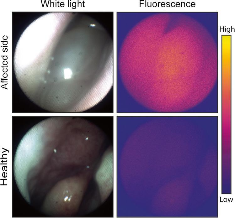
This composite diagnostic image demonstrates in vivo fluorescence molecular imaging (FMI) during endoscopic sinus surgery for sinonasal inverted papilloma (SNIP). The figure is arranged in a 2x2 grid comparing the 'Affected side' (top row) with the 'Healthy side' (bottom row) under 'White light' (left column) and 'Fluorescence' (right column) imaging modalities. In the white light images, the affected tissue appears pale, smooth, and less vascularized compared to the reddish, highly vascularized normal mucosa on the healthy side. The corresponding fluorescence images utilize a pseudocolor heatmap (indicated by a vertical scale from blue/low to yellow/high) to represent bevacizumab-800CW tracer uptake, which targets VEGF-A. The affected side shows a high-intensity, concentrated yellowish-orange signal, indicating significant tracer accumulation and VEGF-A expression. Conversely, the healthy side exhibits a low-intensity, dark purple/blue signal, representing minimal background fluorescence. This comparison illustrates the clinical utility of targeted FMI in differentiating pathological sinonasal tissue from healthy mucosa based on molecular markers.
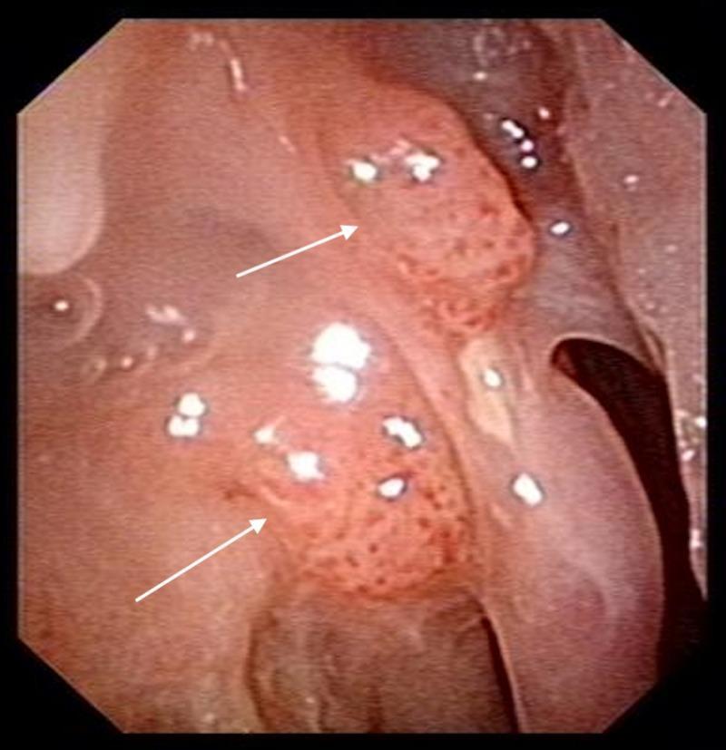
This nasoendoscopy image depicts the right lateral nasal wall, demonstrating a recurrence of an inverted papilloma one year post-operatively. The clinical photograph identifies two primary sites of recurrence, indicated by white arrows. The papillomatous masses exhibit a characteristic exophytic, irregular, and 'cobblestone' surface texture with prominent vascular punctae. These localized lesions are situated along the lateral nasal mucosa, showing a clear morphological transition from the smoother, surrounding healthy mucosal tissue. The endoscopic view provides critical visual evidence for postoperative surveillance in otolaryngology, highlighting the typical growth pattern and morphology of Schneiderian papillomas. The educational focus is on identifying local recurrence and the visual characteristics of sinonasal tumors during diagnostic endoscopy.
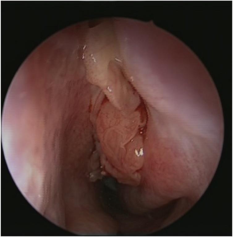
This clinical diagnostic image is an endoscopic view of the nasal cavity showing a large, fleshy, intranasal mass consistent with a sinonasal inverted papilloma. The mass is primarily pinkish-red and exhibits a distinctively lobulated, cerebriform surface texture with irregular folds and crevices. Occasional translucent or whitish spots are visible on its surface. The lesion originates from the lateral nasal wall or middle meatus and occupies a significant portion of the nasal passage, causing mechanical obstruction. The surrounding nasal mucosa appears erythematous. This visual is characteristic of inverted papilloma, a benign but locally aggressive neoplasm known for its high recurrence rate and risk of malignant transformation. The image serves as an educational example of otolaryngological endoscopy for diagnosing obstructive sinonasal tumors and differentiating them from simple inflammatory polyps.
Inverted Papilloma - Complete Review: Basic to Clinical
1. Definition and Overview
2. Epidemiology
| Parameter | Data |
|---|---|
| Incidence | 0.74 - 2.3 new cases per 100,000/year |
| % of nasal tumors | 0.4% - 4.7% of surgically removed nasal tumors |
| Sex ratio | Male:Female = 2-3:1 |
| Peak age | 5th and 6th decades |
| Bilaterality | Exceptional (rare) |
3. Etiology and Risk Factors
3a. Human Papillomavirus (HPV)
- Overall HPV incidence in IP: 22-26% (Lawson et al. 2008 meta-analysis)
- High-risk HPV subtypes detected in: dysplasia (56%) and carcinoma ex-IP (55%)
- However, transcriptionally active HPV was not detected in a series of 52 IPs including those with malignant transformation (Rooper et al.)
- Low-risk subtypes 6 and 11 found in ~8%; high-risk 16, 18, 45, 56 in ~5%
- Conclusion: most malignant transformations from IP appear to be driven by non-HPV mechanisms; p16 staining is a poor surrogate marker
3b. Occupational / Chemical Exposure
- Organic solvent exposure is significantly associated with IP development (dose-response relationship confirmed)
- No proven association with alcohol
3c. Smoking
- Smoking is NOT a causative factor for IP itself
- BUT is a recognized risk factor for recurrence and malignant transformation
- 12-fold higher risk of malignant transformation in smokers (26.4% in smokers vs. 2.8% in nonsmokers, p<0.001; Hong et al., cohort of 162 patients)
4. Pathology
4a. Gross Appearance
- Typically unilateral, polypoid, fleshy, lobulated, cerebriform (brain-like surface)
- Pink-red color with irregular folds and crevices
- Visually distinct from simple inflammatory polyps (which are smooth and pale)
4b. Histology (KEY FEATURES)
- Hyperplastic ribbons of basement membrane-enclosed epithelium growing downward (inward) into the underlying stroma - the defining "inversion"
- Multilayered epithelium: squamous cells + ciliated columnar cells mixed with mucocytes
- Transmigrating neutrophils within the epithelial ribbons (characteristic finding)
- Intact basement membrane (distinguishes it from carcinoma)
- May occasionally be associated with sinonasal hamartomas
4c. Malignant Transformation
- Occurs in 5-15% of cases
- Synchronous (coexisting at time of IP diagnosis) > metachronous
- Most common malignancy: Squamous cell carcinoma (SCC)
- Rare: Sinonasal undifferentiated carcinoma, mucoepidermoid carcinoma, verrucous carcinoma
5. Sites of Origin
- Most common: Lateral nasal wall and medial wall of maxillary sinus (particularly the region of the fontanelles)
- Ethmoid region: 48% (per Scott-Brown's)
- Lateral nasal wall/maxillary sinus: 28%
- Frontal sinus: 1.6-15% (rare primary involvement; only 2.5% per Scott-Brown's)
- Sphenoid sinus: rarely involved primarily
- Multifocal in ~30% of cases - makes precise origin localization difficult
- Middle ear IP: extremely rare; similar behavior and HPV association as sinonasal IP
6. Clinical Features
Symptoms
- Unilateral nasal obstruction (most common presenting symptom)
- Unilateral epistaxis
- Rhinorrhea
- Facial pain / pressure
- Anosmia
- Symptoms of sinusitis (chronic)
- Proptosis / diplopia (if orbital extension occurs)
- Epiphora (if lacrimal pathway involved)
Signs
- Unilateral polypoid mass on nasal endoscopy - cerebriform/lobulated surface (unlike smooth inflammatory polyp)
- Arising from lateral nasal wall (middle meatus most common endoscopic location)
7. Imaging
CT Scan (Primary Imaging)
- Soft tissue opacity, typically unilateral and involving multiple sinuses
- Focal hyperostosis at the point of tumor origin - seen in majority; can predict the attachment site (95% accuracy for intraoperative prediction)
- CT is used for:
- Defining extent of disease
- Predicting site of origin (via hyperostosis)
- Surgical planning
MRI (Complementary - Strongly Recommended)
- T2-weighted MRI: shows the characteristic cerebriform (convoluted) striated pattern - alternating bands of high and low signal (highly specific for IP)
- Key advantage: distinguishes IP tissue from retained secretions (which appear bright on T2 but lack the striated pattern) - critical for frontal sinus cases
- Useful for:
- Assessing skull base / orbital / intracranial involvement
- Differentiating IP from inflammatory mucosa
- Detecting malignant transformation
8. Staging Systems
Krouse Staging (2000)
| Stage | Description |
|---|---|
| T1 | Confined to nasal cavity; no extension to sinuses |
| T2 | Involving the ostiomeatal complex, ethmoid sinuses, or medial portion of maxillary sinus |
| T3 | Involving walls of maxillary sinus, frontal sinus, or sphenoid sinus |
| T4 | All tumors with extranasal/extrasinus extension; any tumor with malignancy |
Han et al. / Endoscopic-Based Systems
9. Management
9a. General Principle
Subperiosteal dissection of involved mucosa + drilling of the underlying bone at the site of tumor attachment/origin.
9b. Biopsy
- Confirm diagnosis
- Rule out synchronous malignancy (presence of squamous cell carcinoma changes the management entirely)
9c. Approach Selection
Endoscopic Approaches (Standard of Care)
- Lower morbidity vs. open approaches
- Equivalent recurrence rates when complete removal is achieved
- Requires clear identification of the pedicle
| Type | Description |
|---|---|
| Type A | Middle antrostomy |
| Type B | Inferior antrostomy (partial inferior meatal window) |
| Type C | Total inferior meatal window |
| Type D | Sturmann-Canfield procedure - includes removal of medial portion of anterior maxillary wall |
Pedicle-Oriented / Conservative Resection
- Introduced by Landsberg (2008), popularized by Kamel and Pagella
- Technique: debulk tumor first → identify site of origin → subperiosteal dissection + bone drilling limited to the insertion point only
- Comparable recurrence rates to extensive demucosualization, with shorter operative times and fewer complications
Open / Combined Approaches (Selected Cases)
- Extensive frontal sinus involvement (lateral attachment, anterior/superior walls)
- Failed previous endoscopic surgery
- Malignant transformation (SCC)
- Lateral rhinotomy with medial maxillectomy: traditional open approach; allows excellent exposure; scar along nasal sidewall
- Midfacial degloving: no external scar; bilateral sublabial + piriform rim incision; good for bilateral or extensive disease
- Osteoplastic flap (OPF): for frontal sinus IP - coronal or brow incision; lifts frontal bone flap; excellent exposure of frontal sinus
- Cranioendoscopic / combined approaches: for extensive skull base involvement
9d. Frontal Sinus IP - Special Considerations
| Site of attachment | Approach |
|---|---|
| Medial wall (MW) or lower anterior wall (AW)/posterior wall (PW), ± multifocal | Draf IIa/IIb or MELP (Modified Endoscopic Lothrop Procedure) |
| Superior AW, superior PW, or lateral wall | OPF (Osteoplastic flap) or MELP ± external trephine |
| Lateral to midpoint of orbit | OPF or MELP with external trephine |
- Draf IIa: Frontal sinusotomy opening the frontal recess unilaterally
- Draf IIb: Removes floor of frontal sinus from septum to lamina papyracea
- Draf III / Modified Lothrop / MELP: Creates a large common frontal neo-ostium by removing interfrontal septum and nasal beak; gold standard for bilateral or recurrent frontal IP
9e. Adjuncts
- Image-guided surgery (IGS/navigation): Especially valuable for revision cases, extensive disease, and frontal/sphenoid sinus involvement
- Angled endoscopes (30°, 45°, 70°): Critical for visualizing and resecting disease in sinuses
- Intraoperative frozen section: Guides extent of resection
10. Malignant Transformation - Management
- Treatment follows principles of sinonasal SCC management
- Surgery (typically open) + adjuvant radiotherapy
- Prognosis is significantly worse than IP alone
11. Recurrence
- The main determinant of recurrence is incomplete removal of the origin/pedicle - not tumor size or stage
- Overall recurrence rates:
- Endoscopic surgery: ~3-15% (varies by site and technique)
- Frontal sinus: up to 22%
- Open surgery: historically similar to endoscopic when complete
- Recurrence typically occurs within 2-3 years post-surgery
- Recurrent IP has higher risk of malignant transformation
12. Postoperative Follow-Up
- Minimum 3 years of follow-up recommended (Scott-Brown's)
- Endoscopic surveillance: ideally direct visualization via the surgical cavity
- When direct surveillance is not possible (e.g., after OPF without MELP): MRI is recommended
- Watch for:
- Recurrence (most within first 3 years)
- Malignant transformation on biopsy of any new lesion
13. Summary Table
| Feature | Key Point |
|---|---|
| Origin | Schneiderian membrane; lateral nasal wall/medial maxillary sinus most common |
| Histology | Endophytic (inverting) epithelial ribbons into stroma; intact BM; transmigrating neutrophils |
| HPV role | Present in ~22-26%; role in malignant transformation debated; mainly non-HPV mechanism |
| Malignancy risk | 5-15%; mainly SCC; smoking increases risk 12-fold |
| Key symptom | Unilateral nasal obstruction |
| Imaging | CT (hyperostosis = origin predictor) + MRI (striated T2 pattern) |
| Surgery | Subperiosteal dissection + bone drilling at pedicle - mainstay of treatment |
| Preferred approach | Endoscopic (pedicle-oriented or complete) for most; open for extensive/frontal/recurrent |
| Recurrence determinant | Completeness of pedicle/origin removal |
| Follow-up | Minimum 3 years; endoscopy ± MRI |
- Cummings Otolaryngology Head and Neck Surgery, Chapter 50 (Benign Sinonasal Tumors) and Chapter 46 (Frontal Sinus Surgery)
- Scott-Brown's Otorhinolaryngology Head & Neck Surgery, Vol 1, Chapter 99