
a. A 52-year-old woman presents with a palpable lump in her right breast. On examination, the lump is hard, irregular, and fixed to the underlying tissue. There are palpable axillary lymph nodes. A mammogram shows a mass with micro calcifications. A biopsy is performed. Based on the clinical presentation and imaging findings, what is the most likely histological diagnosis?(05 Mark) II. What are the key prognostic factors for breast carcinoma?(05 Mark) Describe the significance of immune histochemical staining in the diagnosis and prognosis of breast carcinoma.(05 Mark) IV. What are the histological features of Paget's disease of the breast?(05 Mark)
invasive ductal carcinoma breast histology microcalcifications
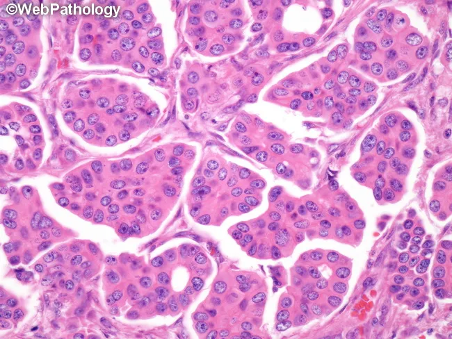
Comprehensive Description: Histopathology image of breast tissue illustrating invasive micropapillary carcinoma with accompanying ductal carcinoma in situ (DCIS). This bright-field micrograph was produced from Hematoxylin and Eosin stained sections of formalin-fixed, paraffin-embedded tissue and viewed under high-power magnification (approximate 400x total). The primary breast lesion is localized to the mammary gland with nests and clusters of malignant epithelial cells forming micropapillary fronds, tubuloalveolar, or morular configurations. Clusters are surrounded by conspicuous lacunar spaces that mimic lymphovascular channels, a pattern that frequently overcalls vascular invasion; true lymphovascular invasion is commonly present. Tumor cells exhibit moderate eosinophilic cytoplasm, high-grade nuclear pleomorphism, and frequent mitotic figures. Notably, the cells display reverse polarity, with apical surfaces facing the surrounding stroma; desmoplastic stroma supports invasive growth. Microcalcifications are commonly observed in association with in situ components. DCIS of micropapillary and cribriform types is often present adjacent to the invasive component. Clinically, this histology portends a higher risk of nodal metastasis and recurrence, guiding axillary staging and adjuvant therapy decisions. Radiologic-pathologic correlation is aided by mammographic calcifications and ultrasound features; management implications include surgical planning, sentinel lymph node assessment, and systemic therapy consideration. This image serves educational and research purposes for tumor morphology and diagnostic pattern recognition.
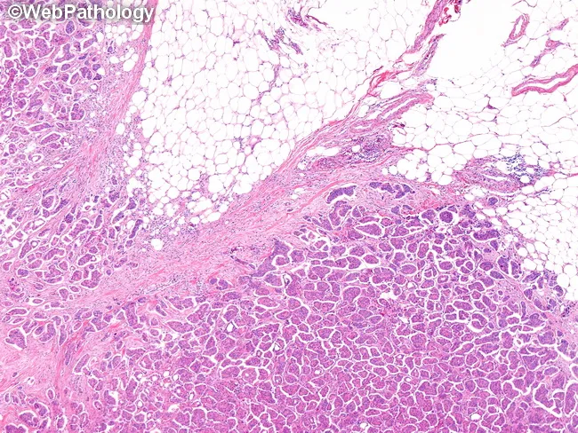
This histopathology image is a hematoxylin and eosin stained paraffin-embedded breast tissue section examined under light microscopy. The primary subject is micropapillary carcinoma (MPC) of the breast, an aggressive variant of invasive ductal carcinoma, frequently presenting as a palpable mass in the upper outer quadrant and often associated with nodal metastases. The visual architecture shows cohesive tumor cell clusters arranged in micropapillary, tubular-alveolar, and morular patterns embedded within lacunar spaces that resemble lymphovascular channels. The tumor nests are small-to-medium in size, with high nuclear grade, enlarged, pleomorphic nuclei, conspicuous nucleoli, and brisk mitotic activity; cytoarchitecture demonstrates reverse polarity with apical-like cell surfaces facing the lacunar spaces. Adjacent benign or fibroadipose breast tissue is present, with conspicuous desmoplastic stroma at the invasive front. Microcalcifications may be detected radiographically; however, in this histology image, calcifications are not visualized. The image highlights the diagnostic hallmarks of MPC: micropapillary clusters, lacunar spaces, and a propensity for nodal/distant spread. Clinically, this variant portends a worse prognosis than conventional invasive ductal carcinoma, ductal NOS, underscoring the need for precise histologic subclassification, thorough sentinel lymph node assessment, and consideration of systemic therapy in management planning for pathology.
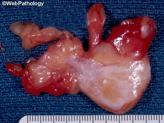
Gross pathology photograph of a breast tumor mass demonstrating invasive ductal carcinoma features. The specimen shows an irregular, often stellate or nodular mass with variable consistency due to desmoplastic stroma; firm to hard in desmoplastic-dominated areas and soft or cystic when extensive necrosis or hemorrhage is present. Gray-white fibrous strands radiate into adjacent fat, with focal yellow-white necrotic streaks and occasional hemorrhagic foci. The tumor margins may be indistinct or well-circumscribed in discrete sections, reflecting a mix of infiltrative and circumscribed growth patterns. Such stellate, infiltrative gross morphology is associated with larger size at presentation, higher likelihood of necrosis, and increased risk of axillary lymph node metastasis, imparting a poorer prognosis relative to circumscribed lesions. Microcalcifications—not always visible grossly—can influence gross texture by imparting a gritty consistency. The gross appearance underscores tumor heterogeneity, sampling considerations for histology, and clinical staging implications. This image supports education on gross features of breast invasive ductal carcinoma, grossing technique, and correlation with radiologic-pathologic findings in diagnostic practice. The description facilitates searchability by including synonyms (invasive ductal carcinoma, breast cancer), descriptive descriptors (stellate margins, desmoplasia, necrosis, hemorrhage, cystic change), and prognostic cues (size, invasion, nodal tendency) valuable for clinicians, educators, and researchers in pathology practice.
Paget disease breast nipple histology Paget cells epidermis
Bright-field light microscopy of a hematoxylin-eosin stained histologic section from the nipple-areolar complex of a human breast. The specimen demonstrates mammary Paget disease with extension of malignant glandular epithelial cells into the epidermis of the nipple and within lactiferous duct lumina. Paget cells are characterized by large, round to oval cells with abundant pale cytoplasm, enlarged hyperchromatic nuclei, and prominent nucleoli. These cells often line or disrupt the keratinizing epidermis and may form small clusters or single-cell infiltrates set against a desmoplastic stroma in adjacent dermis. In many cases, intraepithelial spread is accompanied by focal mucin production and occasional signet-ring-like cytoplasmic clearing, supporting an adenocarcinoma origin. The observed pattern suggests an underlying ductal carcinoma in situ or invasive carcinoma in continuity with the epidermal involvement, although the surface epithelium can sometimes appear non-specific in isolation. Clinically, this histology confirms mammary Paget disease and flags the likelihood of underlying breast carcinoma, mandating comprehensive imaging (mammography, ultrasound, MRI) and staged surgical management. This image is invaluable for dermatologic-pathologic correlation, breast-conserving therapy planning, educational demonstrations of Paget pathology, and differential considerations against nipple dermatitis or melanoma. Overall, the case exemplifies the classic Pagetoid spread of malignant cells from ducts to epidermis, with clinical implications for prognosis and treatment.
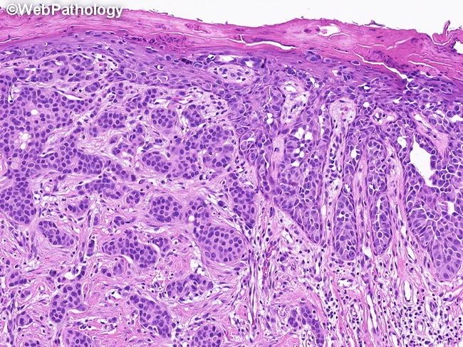
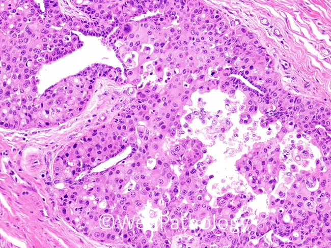
High-fidelity light microscopy of a breast tissue section stained with hematoxylin and eosin demonstrates the classic biphasic appearance of Paget disease of the nipple with an associated invasive ductal carcinoma. The right half of the field shows Paget cells—large, round to oval cells within the epidermis and epidermal appendages, with ample pale cytoplasm, large hyperchromatic nuclei, conspicuous nucleoli, and occasional mitotic figures arranged in a pagetoid pattern along the basal and suprabasal layers. Interspersed keratinocytes and a mild inflammatory infiltrate may be present; the epidermal surface lies atop underlying dermal stroma. The left half reveals an invasive ductal carcinoma component characterized by irregular glandular formations, nests of malignant epithelial cells, cytologic atypia, and a desmoplastic stromal reaction. Invasive tumor cells infiltrate the breast parenchyma beyond ducts, concordant with ductal pathology; mucinous or papillary variants are rare possibilities but not clearly proven in this specimen. The image highlights the strong association of Paget disease with underlying in-situ or invasive carcinoma, most commonly ductal type, with reported incidence around 82–94%. Clinically, this histology supports a diagnosis of PD with concurrent IDC, guiding surgical planning, lymph node assessment, and adjuvant therapy considerations. Diagnostic significance lies in confirming nipple-centric Paget spread with concurrent malignancy.
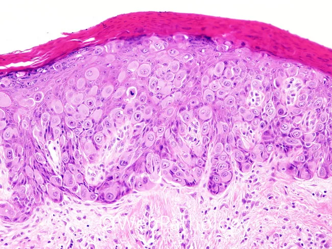
This histopathology image depicts classic mammary Paget disease involving nipple-areola skin. Prepared as a hematoxylin and eosin (H&E) stained paraffin section, the epidermis shows intraepidermal Paget cells with voluminous pale eosinophilic cytoplasm, large vesicular nuclei, and prominent nucleoli. These malignant glandular cells extend through the full thickness of the epidermis, exhibiting marked cytologic atypia and brisk mitotic activity. The Paget cells often form cohesive clusters within the epidermis and may infiltrate the entire epithelial layer, with occasional discohesive cells at the dermal–epidermal junction. Background dermis may display mild chronic inflammatory changes; adnexal structures can be variably preserved. This pattern is highly suggestive of mammary Paget disease and is typically associated with an underlying ductal carcinoma in situ or invasive breast carcinoma, although the underlying neoplasm may not be represented in a single slide. Clinically, nipple-areola erythema, scaling, and crusting prompt biopsy; histology confirming Paget cells establishes diagnosis and guides management. Differential considerations include extramammary Paget disease, melanoma in situ with pagetoid spread, and Bowen disease. The finding has critical diagnostic significance, informing surgical planning, staging workup, and adjuvant therapy decisions in breast cancer care, including sentinel lymph node assessment and radiotherapy decisions.
Breast Carcinoma - Comprehensive Pathology Answer
Part I: Most Likely Histological Diagnosis (5 Marks)
| Clinical Feature | Significance |
|---|---|
| Hard, irregular, fixed lump | Desmoplastic stroma causing fibrosis; fixation = invasion into deep tissue |
| Palpable axillary lymph nodes | Regional lymph node metastasis (N+ disease) |
| Mammographic microcalcifications | Dystrophic calcification within necrotic areas of DCIS or invasive cancer |
| Age 52 (postmenopausal) | Peak incidence period; falling estrogen - higher risk |
- Irregular nests, cords, trabeculae, or sheets of malignant epithelial cells infiltrating fibrous stroma
- Marked nuclear pleomorphism - enlarged, hyperchromatic nuclei with prominent nucleoli
- Increased mitotic figures (contributes to high Nottingham grade)
- Desmoplastic stromal reaction (scirrhous pattern) - accounts for the hard, gritty texture palpated and the stellate appearance on mammogram
- Microcalcifications within areas of comedonecrosis or in the stroma
- No specific differentiation pattern (distinguishes NST from special subtypes like tubular, mucinous, etc.)
Part II: Key Prognostic Factors for Breast Carcinoma (5 Marks)
A. Anatomic / Stage-Related Factors
-
Distant Metastases (M) - The single most important prognostic factor. Once present, cure is unlikely. 10-year survival drops to ~10%.
-
Lymph Node Status (N) - The most important factor in the absence of distant metastases:
- No nodal involvement: 10-year disease-free survival = 70-80%
- 1-3 positive nodes: 35-40%
-
10 positive nodes: 10-15%
-
Tumor Size (T):
- Node-negative, <1 cm: >90% 10-year survival
-
2 cm: falls to ~77%
-
Locally Advanced Disease: Skin invasion, ulceration, or chest wall invasion = poor local control
B. Histopathological Factors
-
Histologic Grade (Nottingham Grade): Assesses tubule formation, nuclear pleomorphism, and mitotic count. Higher grade = worse prognosis. Incorporated into AJCC prognostic staging.
-
Histologic Subtype: Tubular and cribriform carcinomas = excellent prognosis. Metaplastic carcinoma = variable but often poor. Invasive lobular carcinoma = intermediate.
-
Lymphovascular Invasion (LVI): Seen in ~50% of cases; poor prognosis in node-negative patients; associated with higher local recurrence risk.
-
Inflammatory Carcinoma: Due to extensive dermal lymphovascular invasion; 3-year survival only 3-10%. Accounts for 1-5% of cases but higher incidence in African-descent and younger women.
C. Biomarker-Related Prognostic Factors
-
ER (Estrogen Receptor): ER-positive (≥1% nuclear staining) = better prognosis; responds to endocrine therapy (tamoxifen, aromatase inhibitors). "Low positive" (1-10%) = worse than high ER.
-
PR (Progesterone Receptor): Typically co-expressed with ER. Low PR with positive ER = worse outcome.
-
HER2 (Human Epidermal Growth Factor Receptor 2): Overexpression = aggressive behavior; but responds well to HER2-targeted agents (trastuzumab, pertuzumab).
-
Gene Expression Profiling: Oncotype DX, MammaPrint - identify ER-positive, slow-growing cancers that can be spared chemotherapy.
-
Tumor-Infiltrating Lymphocytes (TILs): High TIL count = better prognosis and better response to neoadjuvant chemotherapy, especially in HER2-positive and triple-negative breast cancer (TNBC).
-
Response to Neoadjuvant Chemotherapy: Pathologic complete response (pCR) = better prognosis; common in TNBC and HER2-positive subtypes.
-
ESR1 Mutations: Drive endocrine resistance in ER+/HER2- metastatic disease; tested at recurrence.
Part III: Significance of Immunohistochemical (IHC) Staining in Diagnosis and Prognosis (5 Marks)
A. Diagnostic Role
| IHC Marker | Use |
|---|---|
| CK7, CK8, CK18 (low-mol-wt cytokeratins) | Confirm epithelial origin of the tumor |
| E-cadherin | Positive in ductal carcinoma; absent in lobular carcinoma (distinguishes the two) |
| p63, smooth muscle actin (SMA) | Identify myoepithelial cells; absence helps confirm invasion vs. in situ |
| Ki-67 | Proliferation index; high Ki-67 = high-grade, aggressive tumor |
B. Prognostic and Predictive Role
| Molecular Subtype | ER | PR | HER2 | Features & Prognosis |
|---|---|---|---|---|
| Luminal A | + | + | - | Low grade, low Ki-67; best prognosis; responds to endocrine therapy |
| Luminal B | + | +/- | -/+ | Higher grade, higher Ki-67; moderate prognosis |
| HER2-enriched | - | - | + | Aggressive; responds to trastuzumab/pertuzumab |
| Triple Negative (TNBC) | - | - | - | Worst prognosis; no targeted therapy; responds to chemo +/- immunotherapy |
- ER positivity (≥1% nuclear staining): qualifies for endocrine therapy; both prognostic and predictive
- PR expression: co-predictor; low PR in ER+ = worse outcome
- Survival is highest for ER+/PR+/HER2- and lowest for ER-/PR-/HER2- (TNBC)
- IHC score 0/1+ = negative; 2+ = equivocal (needs FISH confirmation); 3+ = positive
- HER2+ tumors respond to HER2-targeted therapies; without treatment = poor prognosis
- HER2 "low" (IHC 1+ or 2+/FISH non-amplified) is now a new therapeutic category eligible for antibody-drug conjugates (trastuzumab deruxtecan)
- In TNBC: PD-L1 expression predicts response to immune checkpoint inhibitors (atezolizumab, pembrolizumab)
- Proliferation marker; high Ki-67 (>20%) = aggressive tumor, high grade, poorer prognosis
- Guides the distinction between Luminal A and Luminal B subtypes
Part IV: Histological Features of Paget's Disease of the Breast (5 Marks)
Gross/Clinical Appearance
- Scaly, raw, vesicular, or ulcerated lesion starting at the nipple, spreading to the areola
- Unilateral; associated with bloody nipple discharge, pain, burning, and pruritus
- Clinically mimics eczema or dermatitis
Key Histological Features
-
Paget Cells (pathognomonic):
- Large, round to oval malignant glandular epithelial cells within the epidermis of the nipple
- Abundant pale (clear) cytoplasm - due to mucin content
- Large, hyperchromatic nuclei with prominent nucleoli
- Cells occur singly or in small clusters within the epidermis (pagetoid spread)
- Found in the basal and suprabasal layers of the epidermis
-
Intraepidermal spread:
- Paget cells infiltrate the full thickness of the epidermis without forming a separate layer
- May extend into adnexal structures (hair follicles, sweat ducts)
-
Mucin production:
- PAS-positive, diastase-resistant intracytoplasmic mucin granules in Paget cells
- Mucicarmine stain highlights mucin - helps distinguish from melanoma in situ (Melan-A/S100 positive vs. Paget cells)
-
Associated underlying carcinoma:
- In 85-88% of cases, an underlying DCIS or invasive ductal carcinoma is present in the breast parenchyma
- If no palpable mass and normal mammogram: more likely associated with DCIS
- If palpable mass present: more likely invasive carcinoma
-
IHC profile of Paget cells:
- CK7 positive (low-molecular-weight cytokeratin) - confirms glandular epithelial origin
- HER2 overexpressed in 70-90% of cases
- ER/PR positive in ~50% of cases
- CEA positive in many cases
- Negative for S100, HMB-45, Melan-A (differentiates from melanoma)
- Negative for high-mol-wt cytokeratins (differentiates from Bowen's disease/squamous carcinoma in situ)
Differential Diagnosis (Histologically)
| Feature | Paget Disease | Melanoma in situ | Bowen's Disease |
|---|---|---|---|
| Cell type | Glandular cells | Melanocytes | Squamous cells |
| CK7 | + | - | - |
| S100/Melan-A | - | + | - |
| Mucin (PAS) | + | - | - |
| HER2 | +++ | - | - |

- Robbins, Cotran & Kumar, Pathologic Basis of Disease (10th ed.) - Invasive Carcinoma of the Breast, Prognostic and Predictive Factors, AJCC 8th Edition Staging
- Mulholland & Greenfield's Surgery: Scientific Principles and Practice (7e) - Paget Disease of the Breast
Answer according to Robbins and exams format