

Uhhh, can you please, uhhh, briefly explain the extrinsic pathway from the stable only so that I can understand and also write in exam and give in a such a way that, uhhh, it should be more understanding and exam prospective in a simple language as much as you can? Uh...
extrinsic pathway blood coagulation cascade diagram
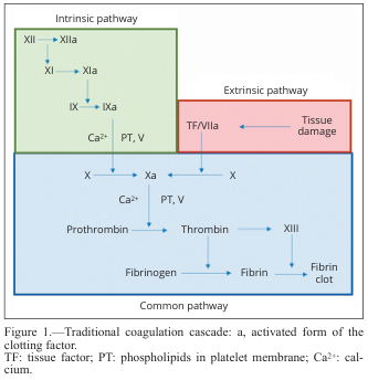
Summary : This diagram illustrates the traditional coagulation cascade, detailing the sequence of events and factors involved in blood clotting via the intrinsic, extrinsic, and common pathways. flowchart: # Pathways : • Intrinsic pathway (green box): Initiated by factor XII, proceeds through XI, IX, and VIII, with activation steps indicated by "a" (e.g., XIIa, XIa, IXa). • Extrinsic pathway (red box): Initiated by tissue damage, which activates tissue factor (TF) and factor VIIa. • Common pathway (blue box): Both intrinsic and extrinsic pathways converge at factor X, leading to the activation of prothrombin to thrombin, which then converts fibrinogen to fibrin, forming a fibrin clot. # Nodes : • XII → XIIa (Intrinsic) • XI → XIa (Intrinsic) • IX → IXa (Intrinsic) • Ca²⁺, PT, V (Intrinsic, Common) • TF/VIIa (Extrinsic) • Tissue damage (Extrinsic) • X → Xa (Common) • Prothrombin → Thrombin (Common) • Fibrinogen → Fibrin (Common) • XIII (Common) • Fibrin clot (Common) # Connectors : • Arrows indicate the direction of activation or conversion between factors. • Intrinsic and extrinsic pathways both feed into the activation of factor X in the common pathway. • Ca²⁺ and PT, V are required cofactors at specific steps. # Layout : • Three main colored sections: green (intrinsic, top left), red (extrinsic, top right), blue (common, bottom). • Flow proceeds from top (initiation) to bottom (clot formation). • Arrows show sequential activation and convergence of pathways. # Analysis : • The diagram clearly separates the intrinsic and extrinsic initiation mechanisms, both converging on the common pathway at factor X. • The cascade is highly sequential, with each activated factor catalyzing the next step. • Calcium ions (Ca²⁺) and phospholipids (PT) are essential cofactors at multiple steps. • The final outcome is the formation of a stable fibrin clot, highlighting the coordinated nature of the coagulation process.
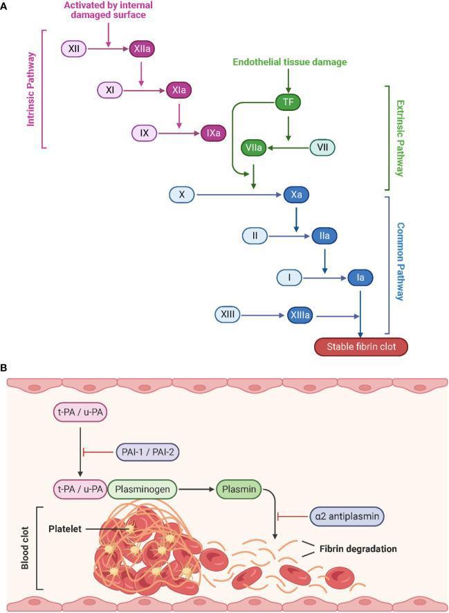
This medical schematic illustrates the physiological processes of hemostasis and fibrinolysis. Part A depicts the Coagulation Cascade, divided into the Intrinsic, Extrinsic, and Common pathways. The Intrinsic pathway (purple) is triggered by internal damaged surfaces, activating Factor XII to XIIa, followed by XI and IX. The Extrinsic pathway (green) begins with endothelial tissue damage and Tissue Factor (TF), activating Factor VII to VIIa. Both pathways converge at the Common pathway (blue) starting with the activation of Factor X to Xa. This leads to the conversion of prothrombin (II) to thrombin (IIa), and fibrinogen (I) to fibrin (Ia), culminating in a stable fibrin clot stabilized by Factor XIIIa. Activated factors are denoted by an 'a' suffix. Part B illustrates Fibrinolysis, where tissue-type (t-PA) and urokinase-type (u-PA) plasminogen activators convert plasminogen to plasmin, which then degrades the fibrin network of the blood clot. Inhibitory regulators including PAI-1, PAI-2, and ̡2-antiplasmin are also shown. The diagram represents the structural breakdown of a clot containing platelets and red blood cells into fibrin degradation products.
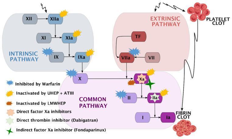
A comprehensive pathophysiology diagram illustrating the human coagulation cascade and the specific pharmacodynamic sites of action for various anticoagulants. The diagram is divided into three functional zones: the Intrinsic Pathway (blue), starting with Factor XII; the Extrinsic Pathway (red), initiated by Tissue Factor (TF) and Factor VII; and the Common Pathway (purple), where the pathways converge at Factor X. The cascade terminates in the conversion of Fibrinogen (I) to Fibrin (Ia), forming a fibrin clot. A secondary visual element depicts a platelet clot associated with the extrinsic pathway. Key clinical pharmacotherapeutic targets are marked with color-coded symbols: Warfarin inhibits Factors IX, VII, X, and II; Unfractionated Heparin (UHEP) + Antithrombin III (ATIII) inactivates Factors XIIa, XIa, IXa, Xa, and IIa; Low-Molecular-Weight Heparin (LMWHEP) and Fondaparinux target Factor Xa; Direct Factor Xa inhibitors act on Factor Xa; and Dabigatran acts as a direct thrombin inhibitor (Factor IIa). This schematic is designed for medical education regarding hemostasis and thromboprophylaxis.
Extrinsic Pathway of Blood Coagulation
What is it? (One-liner for exam)
Step-by-Step Explanation
STEP 1 - Trigger: Tissue Damage
- When a blood vessel is damaged, the tissue underneath (called subendothelial tissue) gets exposed to blood.
- This tissue releases a substance called Tissue Thromboplastin (also called Tissue Factor / Factor III).
- Tissue thromboplastin is made up of Glycoprotein + Phospholipid (exactly as in your notes).
Exam tip: Tissue factor is normally hidden inside the vessel wall. Injury exposes it to blood plasma.
STEP 2 - Activation of Factor VII
- Small amounts of Factor VII are already circulating in the blood in an active form (VIIa).
- Tissue Factor (III) + Factor VIIa + Calcium form a complex on the phospholipid surface.
- This complex activates Factor X → Factor Xa (the "a" means activated).
Exam tip: The tissue factor/Factor VII complex is the key trigger. Factor VII is the ONLY factor unique to the extrinsic pathway.
STEP 3 - Positive Feedback Loop
- The same TF/VIIa complex also feeds back to further activate Factor X → Xa, amplifying the reaction (shown by the curved arrow in your notes = positive feedback).
STEP 4 - Prothrombin Activator Formation (Common Pathway begins)
- Factor V (cofactor)
- Calcium ions (Ca²+)
- On a phospholipid membrane surface
Exam tip: This complex is the bridge between the extrinsic pathway and the common pathway.
STEP 5 - Prothrombin → Thrombin
- The prothrombin activator converts Prothrombin (Factor II) → Thrombin (Factor IIa).
- Thrombin then loops back to activate more Factor V and more Factor X = another positive feedback.
This is why clotting amplifies rapidly after it starts!
STEP 6 - Fibrinogen → Fibrin
- Thrombin cleaves Fibrinogen (Factor I) → Fibrin monomers.
- These fibrin monomers undergo polymerization = they link together to form loose strands of fibrin.
STEP 7 - Stabilization by Factor XIII + Calcium
- Thrombin also activates Factor XIII → Factor XIIIa.
- Factor XIIIa, along with Calcium, cross-links the loose fibrin strands.
- Result = Tight, stable fibrin clot (insoluble, resistant to breakdown).
Summary Table (Quick Revision)
| Step | What happens | Key Factor |
|---|---|---|
| 1 | Tissue damage → releases Tissue Factor | Factor III (Tissue Thromboplastin) |
| 2 | TF + VIIa activate Factor X | Factor VII (unique to extrinsic) |
| 3 | Positive feedback amplifies reaction | TF/VIIa complex |
| 4 | Xa + Va + Ca²+ = Prothrombin Activator | Factor Xa, V, Ca²+ |
| 5 | Prothrombin → Thrombin | Factor II → IIa |
| 6 | Fibrinogen → Fibrin (polymerization) | Factor I |
| 7 | Loose fibrin → Tight clot | Factor XIII + Ca²+ |
Flow Chart (Memorize This!)
TISSUE DAMAGE
↓
Tissue Thromboplastin (Glycoprotein + Phospholipid)
↓ + Factor VII + Ca²+
Factor X → Factor Xa ←←← (Positive Feedback)
↓ + Factor V + Ca²+
PROTHROMBIN ACTIVATOR
↓
Prothrombin → THROMBIN ←←← (Positive Feedback)
↓
Fibrinogen → Fibrin (polymerization)
↓ + Ca²+ + Factor XIII
TIGHT FIBRIN CLOT ✓
Key Points for Exam
- Factor VII = the only factor unique to the extrinsic pathway (most asked MCQ)
- Calcium is needed at multiple steps - without it, clotting fails
- The word "extrinsic" = trigger comes from outside the vessel (tissue)
- Positive feedback by thrombin = makes clotting fast and amplified
- Factor XIII = stabilizes the final clot (cross-linking agent)
- Tissue factor is normally hidden - exposed only after vascular injury
Can you please give me point wise answer? Like make a five, six point in a more simple language like, अ, this is converted into this, first this happened, after this, this happened and this is converted to this, with this, like that give. Okay.
Extrinsic Pathway - Point Wise (Simple Language)
- When a blood vessel gets injured, the tissue outside the vessel gets damaged.
- This damaged tissue releases Tissue Thromboplastin (also called Tissue Factor / Factor III), which is made of Glycoprotein + Phospholipid.
- Tissue Thromboplastin combines with Factor VII + Calcium (Ca²+) in the blood.
- This combination converts Factor X → Factor Xa (activated form).
- (Remember: "a" after any factor = it is now activated)
- Factor Xa alone cannot do the next step.
- So Factor Xa combines with Factor V + Calcium (Ca²+) on a phospholipid surface.
- Together they form the Prothrombin Activator (also called Prothrombinase complex).
- The Prothrombin Activator now acts on Prothrombin (Factor II).
- Prothrombin → Thrombin (Factor IIa)
- Thrombin is a very important enzyme - it drives the next steps.
- Also, Thrombin gives positive feedback - it loops back and activates more Factor V and Factor X, making clotting faster and stronger.
- Thrombin now acts on Fibrinogen (Factor I), which is a soluble protein dissolved in blood.
- Fibrinogen → Fibrin monomers
- These fibrin monomers join together (polymerization) to form loose, weak strands of fibrin.
- The loose fibrin strands are not strong enough yet.
- Thrombin activates Factor XIII → Factor XIIIa.
- Factor XIIIa + Calcium (Ca²+) cross-links the loose fibrin strands together.
- Result = Tight, stable fibrin clot = the final blood clot is formed! ✅
One-Line Flow (Read this like a story):
Tissue damage → Tissue Thromboplastin released → activates Factor VII → Factor X converted to Xa → Xa + Factor V + Ca²+ = Prothrombin Activator → Prothrombin converted to Thrombin → Thrombin converts Fibrinogen to Fibrin → Fibrin polymerizes into loose strands → Factor XIII + Ca²+ converts loose fibrin into tight stable clot ✓
- Helps activate Factor X (Step 2)
- Helps form Prothrombin Activator (Step 3)
- Helps stabilize the final fibrin clot (Step 6)
Not in subheading and description. Only simply five, six point in a simple language.
-
Tissue gets damaged → it releases Tissue Thromboplastin (Glycoprotein + Phospholipid).
-
Tissue Thromboplastin + Factor VII + Calcium → converts Factor X into Factor Xa.
-
Factor Xa + Factor V + Calcium → forms Prothrombin Activator.
-
Prothrombin Activator converts Prothrombin → Thrombin (Thrombin also gives positive feedback to speed up clotting).
-
Thrombin converts Fibrinogen → Fibrin monomers → they join together (polymerization) → form loose fibrin strands.
-
Thrombin activates Factor XIII → Factor XIIIa + Calcium → cross-links the loose fibrin strands → forms a tight, stable blood clot. ✅
I think tissue damage plus tissue thromboplastin. अ, it release glycoprotein and phospholipid. After that, अ, factor seven convert. Factor 10 into factor 10 activator. And then, hmm, factor, uh, 10 plus calcium plus factor V plus thrombin gives prothrombin activator. Prothrombin activator is, uh, converted to thrombin with prothrombin, and thrombin also give positive feedback to set up, uh, speed up clotting. After that, thrombin convert fibrinogen A to fibrin. Fibrinogen A polymerize and, uh, lose, uh, strand of fibrin in which calcium it, uh, convert fibrin tight blood clot. I think according to my desk, this is answer. Now can you please correct and give answer in a point-wise?
-
Tissue damage occurs → Tissue Thromboplastin is released → it is made of Glycoprotein + Phospholipid.
-
Tissue Thromboplastin + Factor VII + Calcium → converts Factor X into Factor Xa (activated).
-
Factor Xa + Factor V + Calcium → forms Prothrombin Activator.
-
Prothrombin Activator converts Prothrombin → Thrombin → Thrombin gives positive feedback to speed up clotting.
-
Thrombin converts Fibrinogen → Fibrin → Fibrin monomers undergo polymerization → form loose strands of fibrin.
-
Loose fibrin strands + Calcium + Factor XIII → cross-linked → form tight, stable blood clot. ✅