
Peritonitis
Reading File
Now let me search the library for comprehensive peritonitis coverage, running parallel queries.
Finding Sources
Searching PubMed
peritonitis management
Good results. Now let me read the most comprehensive sources in parallel - Bailey & Love (surgery), Harrison's, Robbins (pathology), and Tintinalli's (emergency).
Reading File
Reading File
Reading File
Reading File
Excellent content from multiple sources. Let me now read more from Harrison's (which was truncated with rich content on primary and secondary peritonitis) and Bailey & Love for surgical management:
Reading File
Reading File
I now have comprehensive content from multiple authoritative sources. Let me also fetch the peritoneal anatomy diagram from Harrison's:
Loading Image
Reading File
I now have extensive, comprehensive content from multiple authoritative textbooks. Let me compile the full answer.
Peritonitis
Peritonitis is inflammation of the peritoneum - the serous membrane lining the abdominal cavity and viscera. It is a potentially life-threatening condition that may be localised or diffuse, acute or chronic, bacterial or chemical.
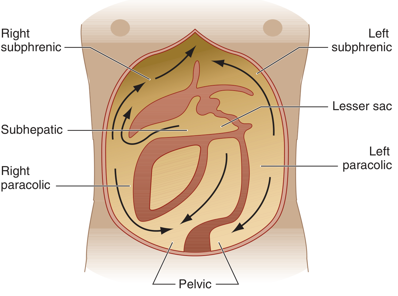
Classification
By Source
| Type | Definition | Key Features |
|---|---|---|
| Primary (Spontaneous) | No apparent source of GI contamination | Usually monomicrobial; gram-negatives dominant |
| Secondary | Arises from an identifiable intraabdominal source | Polymicrobial; gram-negatives + anaerobes |
| Tertiary | Persistent/recurrent peritonitis after treatment | Often drug-resistant organisms, fungi |
By Extent
- Localised - involves a focal area; parietal peritoneal involvement causes somatic pain with guarding and rebound
- Diffuse (generalised) - involves large regions of the parietes; signifies life-threatening pathology
Causes / Aetiology
Bacterial (most common):
- Perforation of abdominal viscus (peptic ulcer, appendix, diverticulum, colon in obstruction)
- Transmural bacterial translocation without frank perforation (pancreatitis, ischaemic bowel, primary bacterial peritonitis)
- Exogenous contamination (peritoneal dialysis catheters, trauma, open surgery)
- Female genital tract (pelvic inflammatory disease, salpingitis)
- Haematogenous spread (rare)
Chemical (sterile initially):
- Bile leakage - highly irritating, usually complicated by secondary bacterial infection
- Pancreatic enzymes (acute haemorrhagic pancreatitis) - fat necrosis and enzymatic peritonitis
- Barium (from contrast studies)
Other:
- Starch peritonitis (allergic/foreign body reaction to surgical glove powder)
- Endometriosis (haemorrhage acts as irritant)
- Ruptured dermoid cysts (keratin-induced granulomatous reaction)
- Familial Mediterranean fever
- Bailey and Love's Short Practice of Surgery, 28th Ed., p. 1109
- Robbins, Cotran & Kumar Pathologic Basis of Disease
Pathophysiology
Normal peritoneal fluid contains <30 g/L protein and <300 WBC/µL (mainly mononuclear). When bacteria or chemical irritants enter the peritoneal cavity:
- Early phase - rapid influx of polymorphonuclear leukocytes (PMNs) driven by resident-cell chemokine synthesis
- Exudative phase - serous fluid accumulates, becomes turbid, then frankly purulent
- Fibrinous phase - fibrin plaques coat viscera and abdominal wall, loops of bowel adhere to each other and to the parietes
- Late phase - frank pus, abscess formation in dependent spaces (pelvis, subphrenic, Morrison's pouch), or systemic sepsis/bacteraemia
At surgery the inflamed peritoneum appears reddened, thickened and has a velvety texture, with yellow/white fibrin plaques and turbid exudate.
Harrison's Principles of Internal Medicine, 22e, p. 1113-1114
Primary (Spontaneous) Bacterial Peritonitis (SBP)
Epidemiology: Occurs in ≤10% of cirrhotic patients with ascites. Also seen in nephrotic syndrome (children), congestive heart failure, SLE, and lymphoedema.
Pathogenesis:
- Haematogenous seeding of ascitic fluid (a good growth medium)
- Diseased liver + altered portal circulation → defective bacterial filtration
- Reduced opsonic/phagocytic capacity of PMNs in advanced liver disease
- Gut dysbiosis with increased Enterobacteriaceae + small intestinal bacterial overgrowth → pathologic bacterial translocation
- Reduced Paneth cell defensins, portal hypertensive enteropathy
Microbiology: Monomicrobial - E. coli most common; also streptococci, enterococci, Klebsiella. Anaerobes are rare (their presence should prompt reconsideration of the diagnosis toward secondary peritonitis). Important current trend: increasing gram-positive bacteria and ESBL-producing Enterobacteriaceae due to widespread quinolone prophylaxis.
Clinical features:
- Fever (up to 80% of patients) - most common sign
- Abdominal pain and peritoneal signs may be present but are often subtle
- Non-localising symptoms: malaise, fatigue, hepatic encephalopathy, acute kidney injury, jaundice
Diagnosis: Sample peritoneal fluid in any cirrhotic patient with ascites and fever. >250 PMN/µL in ascitic fluid is diagnostic (this threshold does not apply to secondary peritonitis).
Treatment:
- IV third-generation cephalosporin (e.g., cefotaxime, ceftriaxone)
- IV albumin (1.5 g/kg at diagnosis + 1 g/kg on day 3) to reduce acute kidney injury and mortality
- Prophylaxis with norfloxacin in high-risk patients (low-protein ascites, prior SBP episode)
Secondary Peritonitis
The most common form, arising from a perforated or inflamed intraabdominal viscus. It is polymicrobial - aerobic gram-negative bacilli (E. coli, Klebsiella) plus anaerobes (Bacteroides fragilis, C. perfringens) are the rule.
Clinical Features
Localised peritonitis:
- Pain in the affected region (somatic - parietal peritoneum is extensively innervated)
- Involuntary guarding (reflex abdominal wall muscle contraction)
- Rebound tenderness (pain on releasing examiner's hand)
- Pyrexia and tachycardia
Diffuse peritonitis (surgical emergency):
- Severe, generalised abdominal pain, worse on movement, coughing, and respiration
- Patient lies motionless, often with knees drawn up
- "Board-like rigidity" - entire abdominal musculature in reflex contraction
- Hippocratic facies (gravely ill appearance)
- Generalised ileus → abdominal distension
- Haemodynamic instability: hypotension, tachycardia, fever
- Progressive confusion → loss of consciousness if untreated
Additional features:
- Shoulder tip ("phrenic") pain if inflammation reaches the diaphragm - referred via C5 dermatome
- Pelvic tenderness on digital rectal/vaginal examination if pelvic source
- Signs may be blunted in obese patients or those on immunosuppressants
Bailey and Love's Short Practice of Surgery, 28th Ed., pp. 1109-1111 Harrison's Principles of Internal Medicine, 22e, p. 1114
Summary of clinical features (Bailey & Love):
- Abdominal pain, worse on movement, coughing and deep respiration
- Constitutional upset: anorexia, malaise, fever, lassitude
- GI upset: nausea +/- vomiting
- Pyrexia (may be absent)
- Raised pulse rate
- Tenderness +/- guarding / rigidity / rebound
Investigations
| Investigation | Finding / Purpose |
|---|---|
| CT abdomen (with contrast) | Investigation of choice; identifies source, free gas, collections |
| Erect CXR | Subdiaphragmatic free gas (pneumoperitoneum) in perforation |
| Lateral decubitus XR | Alternative to erect CXR in unstable patients |
| Abdominal ultrasound | Limited specificity except for tubo-ovarian pathology |
| FBC | Leukocytosis with left shift (band forms) |
| CRP, procalcitonin | Support diagnosis; rarely diagnostically specific |
| Ascitic fluid tap (SBP) | Cell count >250 PMN/µL is diagnostic; culture in blood culture bottles |
| Laparoscopy | If CT/USS inconclusive |
| Peritoneal fluid Gram stain | Insensitive for bacteria; useful mainly for early yeast detection |
Microbiology Summary
| Setting | Organisms |
|---|---|
| SBP / cirrhosis | E. coli, streptococci, Klebsiella (monomicrobial) |
| Secondary (GI perforation) | E. coli, streptococci, S. aureus, enterococci, C. perfringens, Bacteroides (polymicrobial) |
| Peritoneal dialysis (PD) | S. epidermidis (mild), S. aureus, gram-negatives, fungi |
| Fungal peritonitis | Candida spp. (PD patients; immunocompromised) |
| Mycobacterial | M. tuberculosis (chronic presentation) |
Treatment
Surgical / Interventional (Secondary Peritonitis)
- Expedient surgical intervention - remove the underlying cause (seal perforation, resect gangrenous bowel, drain abscess)
- Peritoneal lavage - dilute and evacuate contamination
- At laparotomy: drain all pus, irrigate, consider open abdomen / "damage control" surgery in severe cases
Antibiotic Therapy
Community-acquired, mild-to-moderate:
- Piperacillin/tazobactam 3.375 g IV q6h, OR
- Ticarcillin/clavulanate 3.1 g IV q4-6h, OR
- Fluoroquinolone (e.g., levofloxacin 750 mg IV q24h) OR ceftriaxone 2 g IV q24h + metronidazole 500 mg IV q8h
Health-care-associated / ICU / severe:
- Imipenem 500 mg IV q6h, OR
- Meropenem 1 g IV q8h, OR
- Piperacillin/tazobactam 4.5 g IV q6h (higher dose), OR
- Cefepime/ceftazidime 2 g IV q8h + metronidazole
Enterococcal coverage (add if very ill, cephalosporin-based regimen): ampicillin or vancomycin; use linezolid/daptomycin for VRE.
Antifungal coverage: warranted if Candida grows from a sterile site.
Newer agent: Eravacycline (tetracycline class) 1 mg/kg IV q12h - FDA-approved for complicated intraabdominal infections.
Harrison's Principles of Internal Medicine, 22e, p. 1115
Peritonitis in Peritoneal Dialysis (PD-Related)
A distinct and important entity in nephrology:
Diagnosis (requires 2 of 3):
- Abdominal pain
- Cloudy peritoneal effluent / WBC >100/mm³ (>50% PMN)
- Positive culture of dialysis effluent
Routes of infection:
- Transluminal (through catheter lumen) - most common
- Pericatheter route
- Transmural bacterial migration (associated with constipation, diarrhoea, recent colonoscopy, diverticulitis)
- Haematogenous spread
- Ascending via female genital tract
Key points:
- S. epidermidis causes milder symptoms than S. aureus or gram-negatives
- Fungal peritonitis (Candida) is an indication for catheter removal
- PD-related peritonitis is rarely associated with bacteraemia (unlike surgical peritonitis)
- Gram stain of effluent is insensitive; culture by inoculating 5-10 mL into blood culture bottles
Brenner and Rector's The Kidney, 2-Volume Set; Comprehensive Clinical Nephrology, 7th Ed.
Complications
- Intraabdominal abscess formation - subphrenic (R > L due to falciform ligament barrier), subhepatic (Morrison's pouch), pelvic
- Sepsis syndrome / bacteraemia - especially in secondary peritonitis
- Paralytic ileus
- Adhesions - risk of future bowel obstruction
- Multi-organ failure - in advanced or untreated cases
- Death - untreated diffuse peritonitis is rapidly fatal
Special Forms
| Type | Key Features |
|---|---|
| Tuberculous peritonitis | Chronic; granulomatous; reaction typically remains superficial |
| Fungal (Candida) peritonitis | PD patients; immunocompromised; mandates catheter removal |
| Chemical (bile/pancreatic enzyme) | Sterile initially; intense peritoneal reaction; secondary infection common |
| Eosinophilic peritonitis | >10% eosinophils in effluent; early after PD catheter placement or drug reactions |
| Sclerosing peritonitis | Dense fibrosis; may represent IgG4-related disease |
| Familial Mediterranean fever | Episodic, self-limiting sterile peritonitis |
Sources:
- Harrison's Principles of Internal Medicine, 22e (2025), Chapter 137
- Bailey and Love's Short Practice of Surgery, 28th Ed., Chapter 65
- Robbins, Cotran & Kumar Pathologic Basis of Disease
- Brenner and Rector's The Kidney, 2-Volume Set
- Comprehensive Clinical Nephrology, 7th Ed.
This is a shared conversation. Sign in to Orris to start your own chat.