
Avn best medicine grade 1 age 70 male Post traumaric
avascular necrosis femoral head treatment early stage
PMID: 38172990
PMID: 41283259
avascular necrosis femoral head osteonecrosis MRI classification stages
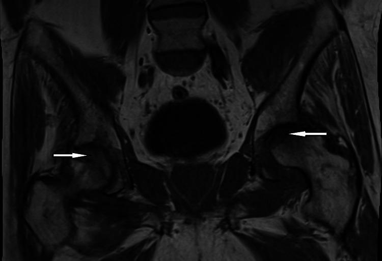
Coronal T1-weighted MRI of the pelvis demonstrating bilateral osteonecrosis (avascular necrosis) of the femoral heads. The image reveals heterogeneous signal intensity and irregular morphology within both femoral heads. White arrows highlight areas of subchondral collapse, which is most pronounced on the left side, where there is a clear loss of the normal spherical contour of the femoral head. These findings are consistent with advanced-stage osteonecrosis leading to articular surface fragmentation and secondary degenerative joint disease (osteoarthritis) of the hip. The pelvic girdle, including the acetabula and surrounding soft tissues, shows signs of chronic remodeling associated with altered joint mechanics. This diagnostic image serves as a clinical example of the progression from bone marrow ischemia to structural joint failure in a patient with systemic risk factors such as hemochromatosis.
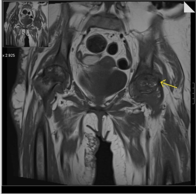
This diagnostic image is a coronal view Magnetic Resonance Imaging (MRI) of the pelvis, primarily demonstrating bilateral avascular necrosis (AVN) of the femoral heads. Both femoral heads exhibit characteristic serpiginous, low-signal intensity lines circumscribing the infarcted areas, consistent with late-stage osteonecrosis. There is evidence of significant structural progression, including an advanced loss of joint space and articular cartilage bilaterally. A yellow arrow points specifically to a subchondral collapse at the superolateral aspect of the left femoral head, indicating secondary osteoarthritis and joint surface irregularity. Surrounding soft tissues show evidence of complex effusions and synovial thickening, potentially indicating secondary synovial osteochondromatosis. Additionally, the image displays diffuse muscular oedema and atrophy of the surrounding pelvic and proximal thigh musculature, most notably in the right rectus muscle. This visual material is clinically relevant for orthopedic surgical planning, such as total hip arthroplasty, and for teaching the radiological hallmarks of Stage IV/V avascular necrosis using the Ficat or Steinberg classification systems.
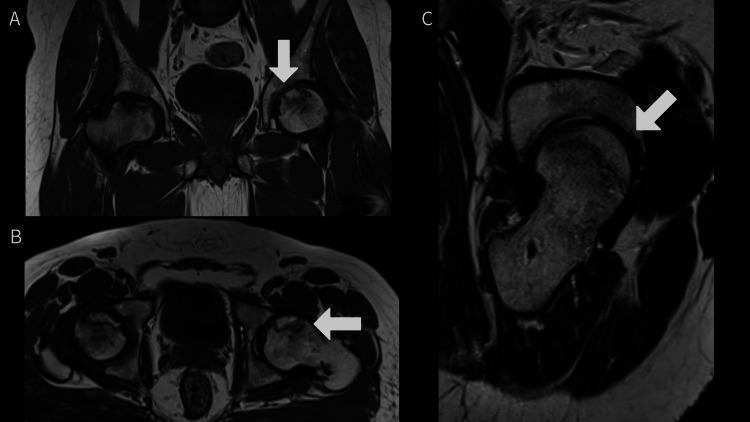
This composite of three Magnetic Resonance Imaging (MRI) scans—comprising coronal (A), axial (B), and sagittal (C) views—demonstrates bilateral avascular necrosis (AVN) of the femoral heads at varying stages. The right femoral head (indicated by white arrows) exhibits features consistent with Grade 3 AVN, characterized by significant low-signal intensity (sclerosis) in the weight-bearing region, subchondral collapse, and subtle flattening of the articular contour. In contrast, the left femoral head demonstrates Grade 2 AVN, showing focal areas of marrow signal alteration and a well-defined 'double-line sign' or sclerotic rim, but with a relatively preserved spherical shape and no evidence of articular surface depression. These images illustrate the radiological progression of osteonecrosis of the hip, highlighting the transition from early marrow edema and focal lesions to mechanical subchondral failure. The series serves as an educational tool for differentiating Ficat and Arlet or Steinberg stages of femoral head necrosis using multi-planar cross-sectional imaging.
AVN (Osteonecrosis) of the Femoral Head - Grade/Ficat Stage 1
70-year-old Male, Post-Traumatic
Understanding Stage 1 (Ficat Classification)
| Feature | Stage 1 Ficat |
|---|---|
| Symptoms | None to mild |
| Radiograph (X-ray) | Normal |
| MRI | Abnormal (diagnostic modality of choice) |
| Bone scan | Cold spot on femoral head |
| Pathology | Infarction of weight-bearing portion; dead marrow cells |
Stage 1 = pre-collapse, femoral head still intact - this is the ideal window for intervention.
Management - Graded Approach for a 70-year-old
1. Non-Operative (First Line at Stage 1)
| Treatment | Evidence | Notes |
|---|---|---|
| Protected weight bearing (crutches/walker) | Standard | Prevents collapse while awaiting definitive treatment |
| Bisphosphonates (alendronate) | Weak - multicenter RCT failed to show benefit on THA rate or progression at 2 years | Can still be considered for osteoporosis co-management in 70-yr-old |
| Statins | Benefit shown early, especially in renal failure patients | Reduces intraosseous adipose deposition and vascular congestion |
| Hyperbaric oxygen | Limited early-stage data | Rarely available/used |
| Extracorporeal shock wave therapy (ESWT) | Emerging evidence; 2025 systematic review (PMID 38896858) suggests benefit in early stages | Non-invasive option worth considering |
2. Surgical Treatment - RECOMMENDED Even at Stage 1
Core Decompression (Gold Standard for Stage 1)
- Involves drilling into the necrotic bone to relieve elevated intraosseous pressure caused by venous congestion
- Allows improved vascularity; may slow disease progression
- "Best results obtained in Ficat stage I hips"
- Failure rates ~60% even in early pre-collapse stage (but far better than 100% failure after collapse)
Percutaneous technique (less invasive):
3. Special Considerations for a 70-year-old Post-Traumatic AVN
- Post-traumatic AVN (as opposed to steroid/alcohol-induced) typically follows femoral neck fractures or hip dislocations with disruption of the retinacular vessels
- In the elderly, the natural history may be more rapid - act early
- Total Hip Arthroplasty (THA) should be kept in mind: if Stage 1 is diagnosed but the patient has poor bone quality, comorbidities making head-preserving surgery high risk, or very limited functional demands, THA planning is reasonable as a contingency
- Bisphosphonates are less effective in post-traumatic AVN than in steroid-induced AVN
- Bone quality (DEXA scan) and cardiovascular fitness should be assessed before any surgical intervention
Recommended Treatment Algorithm (Stage 1, Age 70, Post-Traumatic)
Protected weight bearing (crutches/walker)
        ↓
MRI to confirm extent (% head involved - Steinberg substaging)
        ↓
< 30% head involved → Core decompression + BMAC/BMMSC (minimally invasive percutaneous)
> 30% head involved → Core decompression + BMAC or prepare for THA if early collapse
        ↓
Follow-up MRI at 3-6 months to assess progression
        ↓
If collapse occurs → Total Hip Arthroplasty
Key Drug Summary
| Drug | Role | Dose |
|---|---|---|
| Alendronate 70 mg/week | Antiresorptive (limited evidence) | Weekly oral |
| Statin (atorvastatin) | Early stage vasculo-protective | Standard lipid dose |
| NSAIDs / analgesics | Symptomatic pain relief | As needed with gastroprotection |
| Anticoagulants | If thrombophilia suspected | Only if indicated |
- Campbell's Operative Orthopaedics 15th Ed 2026 - Ficat Classification Table, p. 457; Treatment of ONFH, p. 458-462
- Core decompression meta-analysis 2025 (PMID 41283259) - Biological augmentation superior to structural augmentation
- Network meta-analysis 2024 (PMID 38172990) - CD + BMMSC ranked best for early-stage ONFH