
A 50 years old female, presented with a one month history of progressive dyspnoea, chest pain and difficulty in breathing when lying flat. On auscultation, the doctor found decreased breath sounds on the left side. Chest X-ray revealed blunting of the left costodiaphragmatic angle. A diagnosis of pleural effusion was made. ) What is pleural effusion? Write any two causes of pleural effusion. b) Name the layers and nerve supply of the pleura. c) Enumerate the recesses of pleura and write their clinical importance. Define the terms: Hydrothorax, Pneumothorax and Chylothorax. What is thoracocentesis? Describe the site of this procedure
Reading File
Now let me search the medical library for all relevant topics in parallel.
Finding Sources
Finding Sources
Finding Sources
Finding Sources
Finding Sources
Reading File
Reading File
Reading File
Finding Sources
Reading File
Finding Sources
Finding Sources
Reading File
Finding Sources
Reading File
Finding Sources
Now let me fetch the key anatomical images that will enrich the answer:
Loading Image
Loading Image
Searching Images
thoracocentesis pleural tap needle aspiration site intercostal
I now have comprehensive information from the textbooks. Let me compile the full, detailed answer.
Pleural Effusion - Comprehensive Answer
a) What is Pleural Effusion? Two Causes
Definition
A pleural effusion is an abnormal accumulation of excess fluid within the pleural space (the potential space between the visceral and parietal pleurae). Normally, this space contains only a very thin film of serous fluid (a few millilitres) that lubricates lung movement. When fluid accumulates beyond this physiological amount, it compresses the underlying lung tissue, which may partially or fully collapse - this explains the patient's progressive dyspnoea, orthopnoea (difficulty breathing when lying flat), and the decreased breath sounds and blunting of the costodiaphragmatic angle on chest X-ray.
- Gray's Anatomy for Students, Fig. 3.42
Two Causes
Pleural effusions are broadly classified as transudates (low protein, due to altered hydrostatic/oncotic pressure) or exudates (high protein, due to local pleural/pulmonary disease).
-
Congestive Heart Failure (left ventricular failure) - The leading cause of transudative effusion in the United States. Elevated pulmonary capillary hydrostatic pressure forces fluid across the visceral pleura into the pleural space.
-
Malignancy / Cancer - The most common cause of exudative effusion. Tumour cells invade pleural surfaces (primary lung cancer, breast cancer, lymphoma), disrupting pleural lymphatic drainage and increasing vascular permeability, causing fluid to accumulate.
Other important causes include: pneumonia (parapneumonic effusion), tuberculosis, cirrhosis (hepatic hydrothorax), pulmonary embolism, and hypoalbuminaemia.
- Harrison's Principles of Internal Medicine 22E; Tietz Textbook of Laboratory Medicine 7th Ed
b) Layers and Nerve Supply of the Pleura
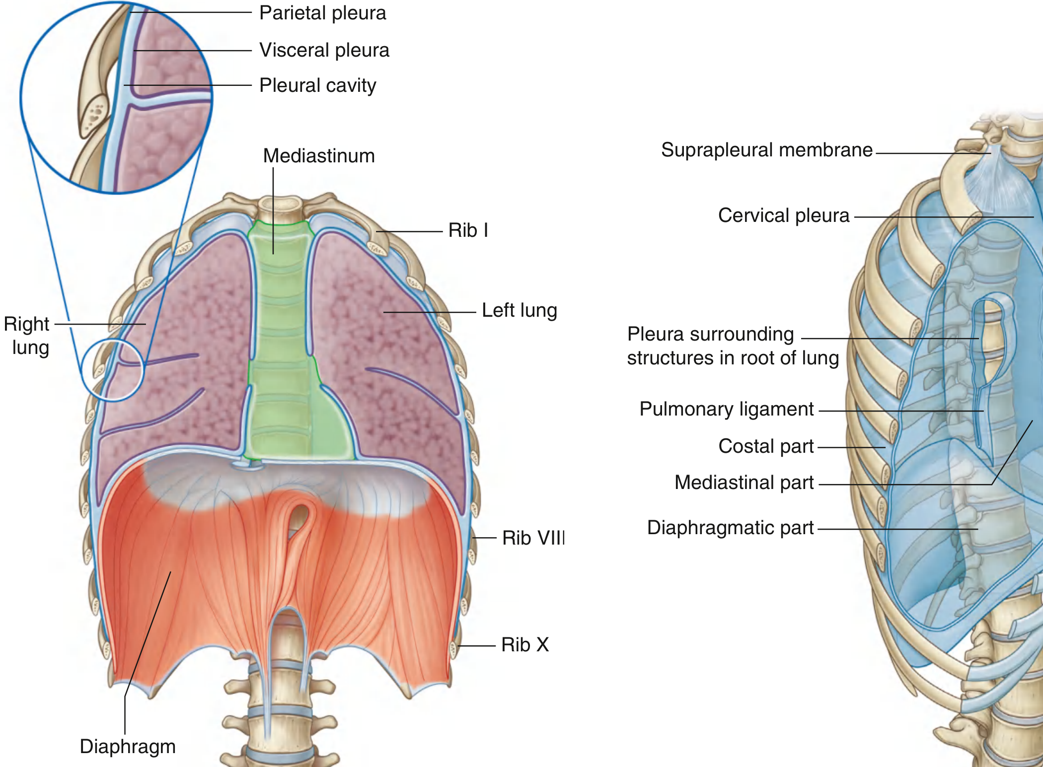
Layers
The pleura consists of a single layer of flat cells (mesothelium) supported by connective tissue. It is divided into two continuous layers:
1. Parietal Pleura
Lines the walls of the pleural cavity. It is further subdivided by location:
- Costal part - lines the inner surfaces of the ribs and intercostal spaces
- Diaphragmatic part - covers the superior surface of the diaphragm
- Mediastinal part - covers the lateral surface of the mediastinum
- Cervical pleura (pleural cupola) - dome-shaped portion projecting 3-4 cm above the first costal cartilage into the root of the neck; supported by the suprapleural membrane (Sibson's fascia)
2. Visceral Pleura (Pulmonary Pleura)
Directly covers the entire lung surface, including the interlobar fissures. It is continuous with the parietal pleura at the hilum. It cannot be stripped from the lung surface.
Between these two layers is the pleural cavity - a potential space containing only a thin capillary film of serous fluid (approximately 5-15 mL) that reduces friction during respiration.
- Gray's Anatomy for Students, p. 201; Color Atlas of Human Anatomy Vol. 2
Nerve Supply
| Layer | Nerve Supply | Nature of Pain |
|---|---|---|
| Parietal pleura | Somatic afferent nerves - well-innervated, pain-sensitive | Sharp, localised pain |
| - Costal part | Intercostal nerves (T1-T11) | Pain referred to the thoracic wall |
| - Diaphragmatic part (peripheral) | Intercostal nerves | Pain referred to the thoracic wall |
| - Diaphragmatic part (central dome) | Phrenic nerve (C3, C4, C5) | Pain referred to the shoulder tip (C4 dermatome) |
| - Mediastinal part | Phrenic nerve | Pain referred to the shoulder tip |
| Visceral pleura | Visceral afferent nerves accompanying bronchial vessels (autonomic) | Pain-insensitive - does not elicit pain when inflamed alone |
Key clinical point: Pleuritic chest pain (sharp, worsened by breathing) originates from parietal pleura stimulation, not the visceral pleura.
- Gray's Anatomy for Students, p. 201-202; Color Atlas of Human Anatomy Vol. 2, p. 224
c) Pleural Recesses - Enumeration and Clinical Importance
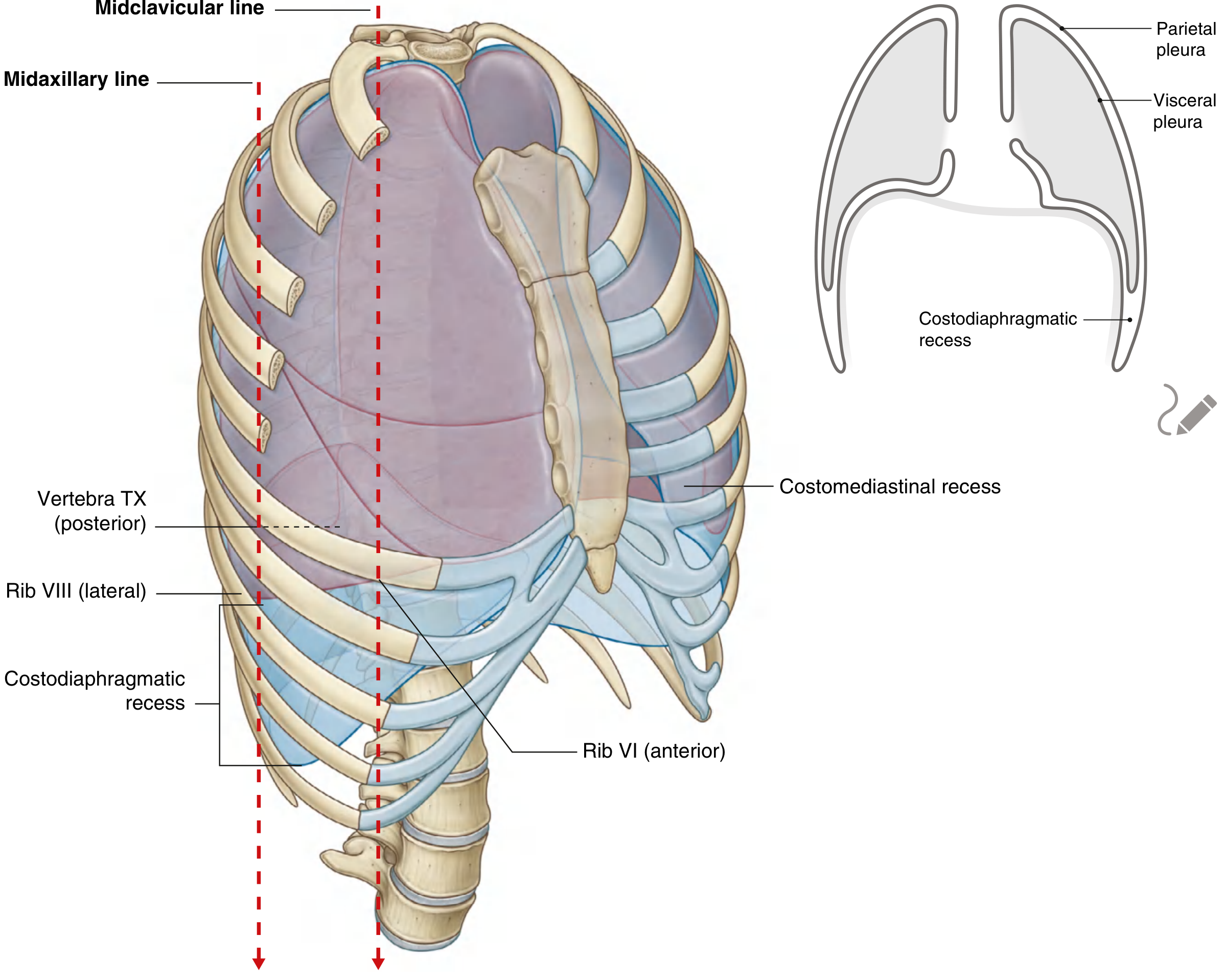
Pleural recesses are potential spaces where two layers of parietal pleura are apposed because the lung does not fully expand into these regions during quiet breathing.
1. Costodiaphragmatic Recess (Costodiaphragmatic Sinus)
- Located at the junction between the costal pleura and the diaphragmatic pleura, inferiorly on each side
- The largest and clinically most important recess
- Extends from the midclavicular line (rib VIII) to the midaxillary line (rib X) to the vertebral column (vertebra TXII)
- During quiet respiration, the inferior lung margin runs: rib VI (anterior) → rib VIII (lateral) → vertebra TX. The costodiaphragmatic recess occupies the gap between the lung edge and the lower pleural reflection.
- During expiration, the recess becomes larger as the lung rises.
Clinical Importance:
- This is the most dependent part of the pleural cavity - pleural fluid preferentially collects here first due to gravity
- Blunting of the costodiaphragmatic angle on chest X-ray (as seen in this patient) occurs when approximately 200-300 mL of fluid accumulates here
- It is the principal site for thoracocentesis (pleural aspiration)
- A needle inserted too low risks puncturing the diaphragm, liver (right) or spleen (left)
2. Costomediastinal Recess (Costomediastinal Sinus)
- Located anteriorly at the junction between the costal pleura and the mediastinal pleura
- Present on both sides, but larger on the left at the level of the cardiac notch (where the heart pushes the left lung away from the midline)
- During deep inspiration, the anterior border of the lung expands into this recess
Clinical Importance:
- Relevant during median sternotomy or anterior thoracic approaches
- The larger left costomediastinal recess corresponds to the "area of superficial cardiac dullness" on percussion
3. Vertebromediastinal Recess (minor)
-
Located posteriorly at the vertebral column where costal pleura meets mediastinal pleura
-
Smaller and less clinically significant
-
Gray's Anatomy for Students, p. 202-203; Color Atlas of Human Anatomy Vol. 2, p. 224
d) Definitions: Hydrothorax, Pneumothorax, and Chylothorax
Hydrothorax
Accumulation of non-inflammatory, serous (watery) fluid in the pleural cavity - i.e., a transudative pleural effusion. It occurs when systemic factors alter the balance of hydrostatic and oncotic pressures (e.g., heart failure, hepatic cirrhosis, nephrotic syndrome). The fluid has low protein content and low LDH. The term is sometimes used specifically for fluid accumulation from hepatic cirrhosis (hepatic hydrothorax).
Pneumothorax
Accumulation of gas or air within the pleural cavity. When air enters the pleural space, the natural elastic recoil of the lung parenchyma causes it to collapse. Types include:
- Spontaneous pneumothorax - occurs without an obvious cause (often in tall, young males with sub-pleural blebs)
- Traumatic pneumothorax - due to chest wall injury, rib fracture, or iatrogenic (e.g., central line insertion)
- Tension pneumothorax - a medical emergency: air accumulates under pressure, the mediastinum shifts to the opposite side, compressing the contralateral lung and great vessels; requires immediate needle decompression
Symptoms include sudden-onset chest pain, dyspnoea, and absent breath sounds on the affected side.
Chylothorax
Accumulation of chyle in the pleural space. Chyle is milky-white lymphatic fluid that drains from the intestines via the thoracic duct; it is rich in triglycerides, chylomicrons, and lymphocytes. Chylothorax results from disruption or obstruction of the thoracic duct at any point along its course. Common causes include:
- Trauma or iatrogenic injury (thoracic surgery, central line insertion)
- Malignancy (especially lymphoma compressing or invading the thoracic duct)
- Congenital abnormalities
The fluid is characteristically milky/turbid, with triglyceride levels >110 mg/dL and lymphocyte predominance.
- Gray's Anatomy for Students; Sabiston Textbook of Surgery; Murray & Nadel's Textbook of Respiratory Medicine
e) Thoracocentesis - Definition and Site
Definition
Thoracocentesis (also called pleural tap or thoracentesis) is a procedure in which a needle or catheter is inserted through the chest wall into the pleural space to aspirate pleural fluid (or air). It is performed both diagnostically (to analyse fluid and determine the cause of effusion) and therapeutically (to relieve symptoms by draining large effusions).
Indications
- Diagnostic: undiagnosed pleural effusion
- Therapeutic: large symptomatic effusions (relief of dyspnoea), empyema, haemothorax, hydrothorax, chylothorax, and malignant effusions
Site of Thoracocentesis
The needle is inserted into the costodiaphragmatic recess - the most dependent and accessible part of the pleural space where fluid collects.
Standard anatomical site:
- Patient position: Sitting upright, leaning slightly forward (arms resting on a table), which allows fluid to pool inferiorly and posteriorly
- Intercostal space: The 7th or 8th intercostal space posteriorly (or 5th-6th intercostal space in the midaxillary line for lateral approach)
- Vertical line: Posterior to the midaxillary line - typically the mid-scapular line or just lateral to the posterior angle of the scapula
Critical anatomical rule for needle insertion:
- The needle must be passed over the superior border of the lower rib (i.e., at the lower part of the intercostal space)
- This avoids the neurovascular bundle (intercostal vein, artery, and nerve) which runs in the groove on the inferior surface of each rib (subcostal groove)
- Inserting along the upper border of a rib = safe; inserting near the lower border of the rib above = risk of intercostal vessel/nerve injury
Ultrasound guidance is now routinely used to confirm the presence of fluid, assess loculations, and select the safest entry point before needle insertion.
Complications to avoid:
-
Too low: puncture of diaphragm, liver (right side), or spleen (left side)
-
Too medial: risk of intercostal vessels (larger near the sternum)
-
Injury to the intercostal nerve: post-procedural neuralgia
-
Gray's Anatomy for Students, p. 202-203 (Thoracostomy/chest tube insertion section)
Summary Table
| Term | Contents | Cause |
|---|---|---|
| Hydrothorax | Serous/watery fluid (transudate) | Heart failure, cirrhosis, nephrotic syndrome |
| Pneumothorax | Air/gas | Trauma, spontaneous, iatrogenic |
| Chylothorax | Chyle (milky, high triglycerides) | Thoracic duct disruption |
| Pleural effusion | Any excess fluid | Multiple (infection, malignancy, cardiac, hepatic) |
This is a shared conversation. Sign in to Orris to start your own chat.