
astroncutes
It looks like "astroncutes" might be a typo. Did you mean:
- Astronauts - people trained for spaceflight
- Astrocytes - star-shaped glial cells in the brain/spinal cord
- Asteronyx - a type of brittle star
- Something else entirely?
Could you clarify what you were looking for?
astrocytes
Astrocytes are star-shaped glial cells that are the most abundant cell type in the CNS. They provide structural and metabolic support to neurons, maintain the blood-brain barrier, regulate synaptic transmission, and respond to injury. They are derived from neuroectoderm and are identified by the marker GFAP (glial fibrillary acidic protein).
Searching sources...
Reading File
Finding Sources
Finding Sources
Searching PubMed
astrocytes functions CNS
Reading File
Reading File
Reading File
Reading File
Now let me fetch a couple of the key histology images to include in the answer:
Loading Image
Loading Image
Here is a full, textbook-sourced overview of astrocytes:
Astrocytes
What Are They?
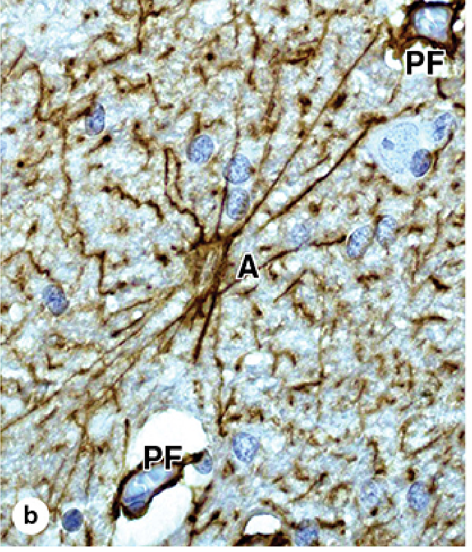
Astrocytes (from Greek astro- = star, kyotos = cell) are star-shaped glial cells and by far the most abundant cell type in the CNS - they constitute nearly half of all brain cells. They originate from progenitor cells in the embryonic neural tube (neuroectoderm). The defining molecular marker is GFAP (glial fibrillary acidic protein), an intermediate filament protein found in all astrocytic processes.
Types
| Type | Location | Processes |
|---|---|---|
| Protoplasmic | Gray matter | Numerous short, branching processes; closely associated with synapses |
| Fibrous (fibrillary) | White matter | Long, thin, relatively few processes; contact axons and nodes of Ranvier |
| Bergmann glia | Cerebellum | Specialized type, processes run parallel to Purkinje cell dendrites |
| Müller cells | Retina | Span entire retinal width |
Terminal processes of a single astrocyte can occupy a volume much larger than its cell body and associate with up to 2 million synaptic sites - Junqueira's Basic Histology, p. 435.
Key Functions
1. Blood-Brain Barrier (BBB) Support
Astrocytic perivascular end-feet completely envelop CNS capillaries, forming a critical component of the BBB. They regulate the passage of molecules and ions from blood into brain tissue. - Robbins Pathologic Basis of Disease, p. ~3373
2. K⁺ Spatial Buffering
When neurons fire action potentials, K⁺ floods the extracellular space. Astrocytes have high densities of K⁺ channels and act as spatial buffers - absorbing K⁺ near active synapses and releasing it at distant blood vessel contacts. This maintains ionic homeostasis needed for normal neuronal excitability. - Principles of Neural Science (Kandel), p. 201
3. Neurotransmitter Regulation
- Glutamate: High-affinity transporters on astrocytes rapidly clear glutamate from the synaptic cleft. Inside the astrocyte, glutamate → glutamine (via glutamine synthetase) → transferred back to neurons as a precursor. Failure of this system → excitotoxicity (neuronal death).
- Monoamines: Astrocytes degrade dopamine, norepinephrine, epinephrine, and serotonin.
- GABA: Astrocytes can also release GABA, reducing neuronal excitability.
- Principles of Neural Science (Kandel), p. 201
4. Metabolic Support - The Lactate Shuttle
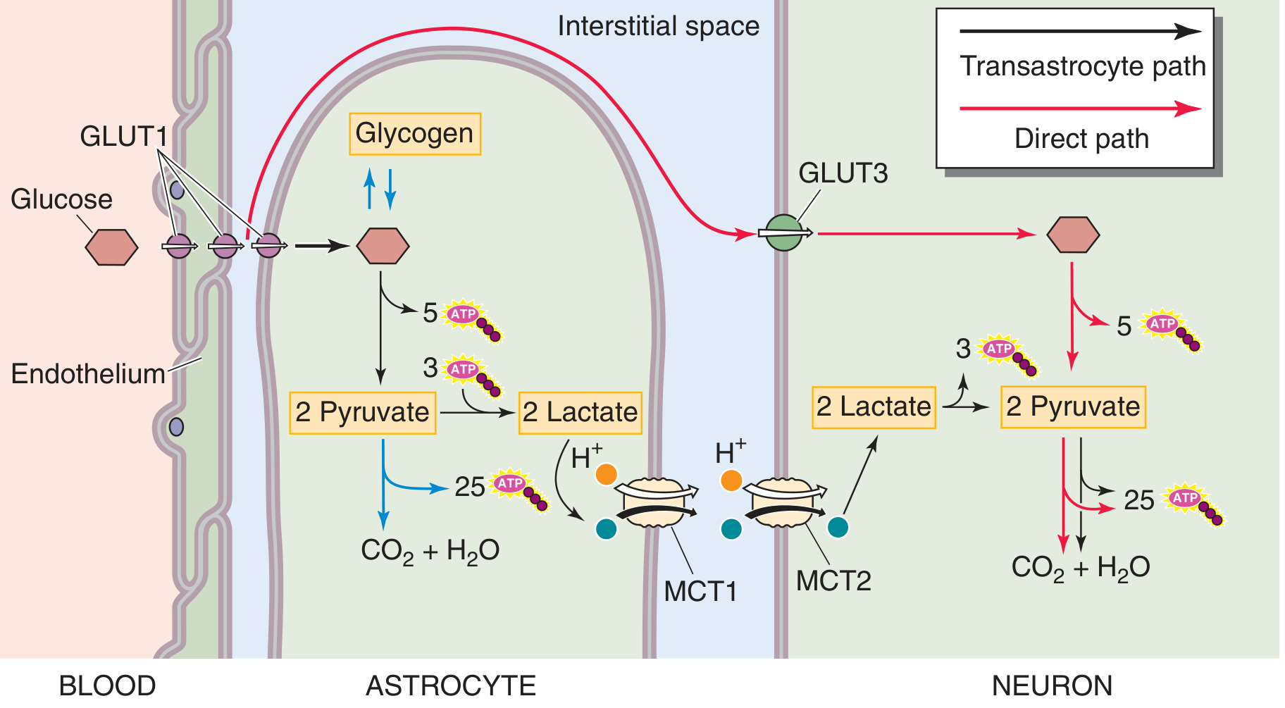
Astrocytes store virtually all the brain's glycogen. They break glucose/glycogen down to lactate, which is transferred via monocarboxylate transporters (MCT1/MCT2) to neurons for aerobic metabolism. This is especially important during periods of intense neuronal activity.
5. Synaptic Regulation - The Tripartite Synapse
Astrocytes are often described as forming a tripartite synapse with the pre- and postsynaptic neuron. They:
- Sense neurotransmitter release via their own receptor expression
- Modulate synapse formation and elimination (especially during development)
- Respond to glutamate binding with a rise in intracellular Ca²⁺, which propagates as a Ca²⁺ wave through neighboring astrocytes via gap junctions over hundreds of micrometers
- Neuroscience: Exploring the Brain (5e), p. ~2625
6. Trophic and Structural Support
Astrocytes secrete trophic factors that promote neuronal survival and synaptogenesis. They also communicate with each other through gap junctions, forming a large syncytium-like network. - Medical Physiology
Reactions to Injury - Gliosis
Any CNS injury triggers reactive gliosis - the most important histopathologic marker of CNS damage. Astrocytes undergo:
- Hypertrophy and hyperplasia
- Nuclear enlargement, vesicular chromatin, prominent nucleoli
- Bright pink cytoplasm due to increased GFAP expression
- Formation of gemistocytic (reactive) astrocytes
Special pathological forms include:
-
Alzheimer type II astrocyte: seen in hyperammonemia (chronic liver disease, urea cycle disorders); large pale nucleus with intranuclear glycogen droplet
-
Rosenthal fibers: thick eosinophilic inclusions in processes; seen in long-standing gliosis and pilocytic astrocytoma; contain αB-crystallin, HSP27, and ubiquitin
-
Corpora amylacea (polyglucosan bodies): round PAS-positive lamellated structures in subpial/perivascular zones; increase with age
-
Robbins Pathologic Basis of Disease, pp. 3373-3382
Clinical Relevance Summary
| Condition | Astrocyte Involvement |
|---|---|
| TBI / Hypoxia | Cellular swelling, reactive gliosis |
| Liver failure | Alzheimer type II astrocytes (hyperammonemia) |
| Pilocytic astrocytoma | Rosenthal fibers |
| Alexander disease | GFAP gene mutation → massive Rosenthal fiber accumulation |
| Excitotoxicity (stroke) | Failure of glutamate reuptake |
| Head injury edema | Astrocyte K⁺/water accumulation → cerebral edema |
This is a shared conversation. Sign in to Orris to start your own chat.