
PROM
Reading File
"PROM" has multiple meanings - let me clarify before diving in.
Asking for Preferences
What does PROM refer to in your question?
Searching sources...
Finding Sources
Searching PubMed
"premature rupture of membranes"[MeSH Terms]
Good results. Now let me read the most comprehensive sources - Creasy & Resnik's MFM and Tintinalli's for clinical management.
Reading File
Reading File
Reading File
Finding Sources
I have rich content. Let me read the management section more fully and also get the anesthesia perspective.
Reading File
Reading File
I now have comprehensive information to write a complete, well-structured answer on PROM. Let me compile the full response.
Premature Rupture of Membranes (PROM)
Definition and Classification
PROM is rupture of fetal membranes before the onset of labor. It complicates approximately 8-10% of all pregnancies and up to 35% of preterm deliveries.
Key classifications:
- PROM at term (≥37 weeks) - rupture before labor onset
- Preterm PROM (PPROM) - rupture before 37 weeks' gestation
- Previable PROM - rupture before approximately 22-23 weeks
- The latent period = time from membrane rupture to onset of labor
Predisposing Factors
- Short cervix
- Prior history of PROM or preterm delivery
- Genital tract infection (Chlamydia, gonorrhea, Trichomonas vaginalis, bacterial vaginosis)
- Non-genital infections (pyelonephritis, pneumonia, appendicitis, periodontal disease)
- Multiple gestation / polyhydramnios
- Uterine overdistention
- Smoking
- Placental abruption
Diagnosis
Avoid digital cervical examination - it shortens the latent period and increases infection risk. Use speculum examination instead.
Bedside testing (combined ~90% diagnostic accuracy):
| Test | Method | Positive Result |
|---|---|---|
| History | Gush/leakage of fluid | Ask patient to Valsalva during speculum exam |
| Nitrazine test | Paper applied to vaginal fluid | Orange → Blue (amniotic fluid pH >7.1; vaginal secretions pH <6) |
| Ferning test | Fluid dried on slide, view under microscope | Ferning pattern (arborization) |
| Pooling | Fluid visible in posterior fornix | Presence of fluid |
| Ultrasound | AFI measurement | Oligohydramnios (AFI <5 cm); not diagnostic alone |
Newer markers: IGFBP-1 (Actim PROM test) and PAMG-1 (AmniSure) have high sensitivity/specificity when nitrazine/fern are equivocal.
Complications
Maternal Complications
- Chorioamnionitis: 9% with term PROM; rises to 24% after >24 hours of rupture; 13-35% with PPROM remote from term; up to 25-77% with second-trimester PROM on conservative management
- Endometritis: 2-13%
- Placental abruption: 4-12%
- Maternal sepsis: 0.8% (death: 0.14%) - more likely with very early preterm PROM
Fetal Complications
- Umbilical cord compression (from oligohydramnios - FHR decelerations common)
- Umbilical cord prolapse (especially with malpresentation)
- Fetal death: ~1-2% with conservatively managed PROM; 12-71% (avg ~30%) with PROM <24 weeks
- Intrauterine infection
Neonatal Complications
- Gestational age at delivery is the primary determinant of severity
- Neonatal sepsis is twice as common after PPROM versus preterm labor
- Infection can manifest as congenital pneumonia, sepsis, meningitis
- Long-term: chronic lung disease, developmental disabilities, cerebral palsy, periventricular leukomalacia, visual/hearing deficits
- Long-term morbidities are uncommon with delivery after ~32 weeks
Management (Gestational Age-Based)
Management balances risk of infection against risk of fetal prematurity.
Term PROM (≥37 weeks)
- Induce labor - usually with oxytocin infusion
- No substantial fetal benefit to expectant management at term
- Reduces risk of chorioamnionitis (rises with prolonged rupture)
- Allow adequate time for latent phase; minimize digital vaginal exams until active phase
Preterm PROM at 34-36 weeks (Late Preterm)
- Conservative management prolongs pregnancy by only a few days (mean ~71-78 hours) while significantly increasing chorioamnionitis risk (16% vs 2%)
- PROMEXIL trials: conservative management doubled chorioamnionitis (5.6% vs 2.3%)
- Delivery is generally recommended - active management reduces RDS (5.2% vs 8.3%), ventilator need, and NICU stay
- Antenatal corticosteroids: administer if not previously given
Preterm PROM at <34 weeks (Remote from Term)
Conservative ("expectant") management with:
- Antibiotics - broad-spectrum (e.g., ampicillin + erythromycin; ACOG recommends GBS prophylaxis); prolong latency and reduce infection
- Antenatal corticosteroids - single course (betamethasone or dexamethasone) to accelerate fetal lung maturation
- Tocolytics - short-term, to allow time for corticosteroids to act; not for prolonged use
- Magnesium sulfate for neuroprotection - at 24-32 weeks to reduce risk of cerebral palsy
- GBS prophylaxis - if colonization status unknown or positive
- Continuous fetal monitoring - assess for cord compression, infection signs
- DVT prophylaxis - leg exercises, compression stockings (prolonged bed rest increases DVT risk)
Care should occur at a facility capable of emergent delivery and neonatal intensive care.
Previable PROM (<22-23 weeks)
- Particularly high fetal jeopardy
- Survival with conservative management at ≤24 weeks: ~44% overall (14% for PROM before 22 weeks; 58% for PROM at 22-24 weeks)
- Immediate delivery → neonatal death
- Requires detailed counseling; decisions individualized
Chorioamnionitis (Complication of PROM)
Diagnosis (clinical):
- Fever >38°C
- Maternal and fetal tachycardia
- Uterine tenderness
- Foul-smelling or purulent amniotic fluid
- Elevated CRP (>2 mg/dL); WBC unreliable (normally elevated in labor)
Treatment: IV broad-spectrum antibiotics + delivery (regardless of gestational age once diagnosis is established)
Anesthesia note: Regional anesthesia (epidural/spinal) is generally considered acceptable in chorioamnionitis if the patient has received antibiotics and there are no signs of overt septicemia, thrombocytopenia, or coagulopathy.
Summary Algorithm
Confirmed PROM
    ↓
Assess: gestational age, labor status, infection signs, fetal well-being
    ↓
≥37 weeks → Induce labor (oxytocin)
34-36 weeks → Active delivery (steroids if not given)
24-33 weeks → Conservative management:
              Antibiotics + Steroids + MgSO4 (24-32wks) + Tocolysis + Monitoring
<22-23 weeks → Individualized counseling; consider viability/prognosis
Sources: Creasy & Resnik's Maternal-Fetal Medicine, 9e; Tintinalli's Emergency Medicine; Morgan & Mikhail's Clinical Anesthesiology, 7e
Malpresentation
Searching sources...
Finding Sources
Finding Sources
Reading File
Reading File
Reading File
Finding Sources
Finding Sources
Reading File
Finding Sources
Finding Sources
Reading File
Reading File
Finding Sources
Reading File
Reading File
Reading File
I now have comprehensive information to compile a complete answer on malpresentation. Let me fetch the ECV diagram image as well.
Loading Image
Here is a comprehensive overview of malpresentation.
Malpresentation
Key Definitions
Lie = relationship of the fetal long axis to the uterine long axis (longitudinal, transverse, or oblique).
Presentation = the part of the fetus nearest the birth canal. All presentations except cephalic vertex are considered malpresentations.
Position = relationship of the presenting part to the maternal pelvis. The reference landmark is the occiput (cephalic), sacrum (breech), or mentum/chin (face).
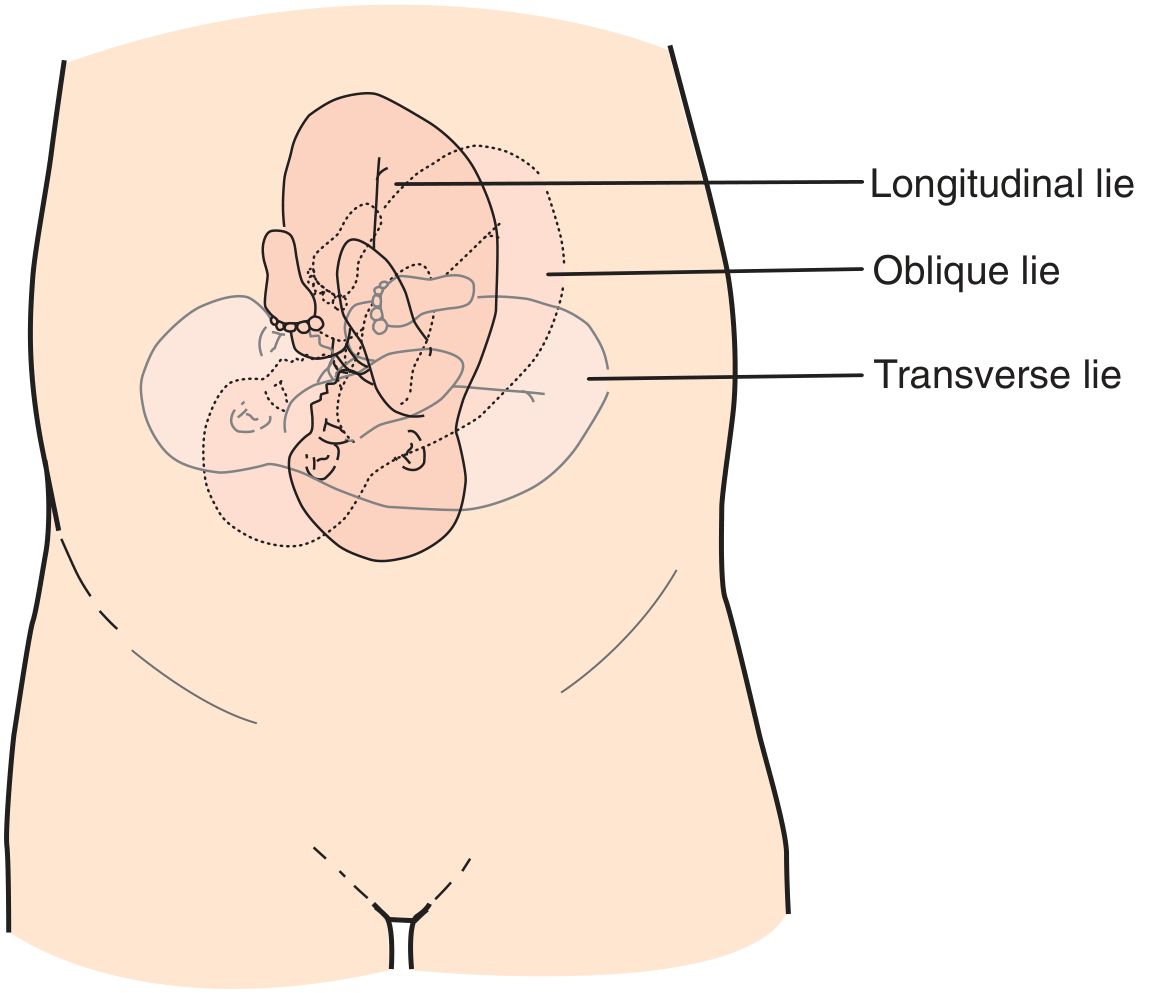
Fig: Longitudinal, oblique, and transverse lie (Roberts & Hedges' Clinical Procedures in Emergency Medicine)
Incidence (Order of Decreasing Frequency)
| Malpresentation | Incidence |
|---|---|
| Breech | 1 in 25 live births (~3-4%) |
| Shoulder dystocia | 1 in 300 live births |
| Face | 1 in 550 live births |
| Brow | 1 in 1400 live births |
| Transverse lie | ~1 in 300 deliveries |
1. Breech Presentation
Types
| Type | Description |
|---|---|
| Frank breech | Hips flexed, legs extended upward (most common, ~65%) |
| Complete breech | Hips and knees both flexed (fetus "sitting cross-legged") |
| Incomplete/Footling | One or both feet/knees are lowermost in canal |
Predisposing Factors
- Prematurity (24% at 18-22 wks; 8% at 28-30 wks; 3-4% at term)
- Fundal-cornual placental implantation
- Uterine anomalies / polyhydramnios
- Multiple gestation
- Fetal neuromuscular disorders (diminished motor tone/activity)
- Prior breech presentation
Risks During Labor & Delivery
- Umbilical cord prolapse - especially footling breech (incomplete cervical occlusion)
- Head entrapment - after-coming head trapped by incompletely dilated cervix (especially prematurity <1500 g or CPD)
- Trauma from extension of the head or nuchal arms
- Fetal asphyxia - ~1/3 of breech fetal deaths considered preventable
Diagnosis
- Leopold maneuvers (globular hard head palpable at fundus)
- Speculum: soft, irregular presenting part
- Ultrasound - confirms type and fetal position
Management
Preferred: Planned Cesarean Section
- ACOG recommends planned CS if ECV fails or is not possible
- The landmark Term Breech Trial (Hannah et al., 2000, n=2088) showed statistically significant increase in perinatal morbidity and mortality with planned vaginal delivery vs. planned CS
External Cephalic Version (ECV)
- Success rate ~60%; should be attempted at ≥36 weeks (before this, spontaneous version may still occur)
- Requires: ultrasound, electronic fetal monitoring, tocolytic agents, readiness for emergency CS
- Factors predicting failure: engaged breech, oligohydramnios, posterior placenta, fetal spine anterior, nulliparity
- Neuraxial anesthesia increases ECV success rate
- Contraindications: placenta previa, oligohydramnios, non-reassuring CTG, prior uterine scar (relative), multiple gestation
Criteria for Vaginal Breech Delivery Trial (selected cases only):
- Frank or complete breech (not footling)
- Estimated fetal weight 2000-3800 g
- Normal gynecoid pelvis with adequate measurements (AP inlet ≥11 cm, widest transverse ≥12 cm, interspinous ≥9 cm)
- Flexed fetal head
- Qualified obstetrician present
- Continuous fetal monitoring throughout labor
- Membranes kept intact as long as possible (hydraulic cord protection)
Key principle in vaginal breech delivery: Support but do NOT pull the presenting part - pulling the hips brings the shoulders through the OS, trapping the head.
2. Transverse Lie / Shoulder Presentation
- Fetal long axis perpendicular to uterine axis; presenting part is the shoulder
- Incidence: ~1 in 300 deliveries
- Common causes: prematurity (38%), high parity (87%), uterine anomaly, placenta previa, polyhydramnios, pelvic obstruction
- Oblique lie is unstable and typically converts to longitudinal or transverse during labor
Management
- <36-37 weeks, not in labor: await spontaneous version; do not attempt external version (risk of cord entanglement)
- ≥36-37 weeks, intact membranes, no placenta previa/CPD: external version → induction of labor at 38 weeks
- Unstable lie at ≥37 weeks: admit to hospital; evaluate for etiology; ECV + induction if no contraindication
- Awaiting spontaneous labor in unstable lie → high cord prolapse rate (10 cord prolapses and 4 perinatal deaths in 50 unmanaged cases vs. 1 cord prolapse and 0 deaths in 102 actively managed cases)
- Labor with transverse lie + ruptured membranes or placenta previa: Cesarean section mandatory
- Back-up transverse: low transverse uterine incision often adequate
- Back-down shoulder presentation: classical (vertical) uterine incision usually required
3. Face and Brow Presentations (Deflection Abnormalities)
Both represent degrees of cephalic deflection (failure of the fetal head to flex). They occur in approximately 1 in 500 deliveries each.
Engaging Diameters
- Vertex (normal): smallest - suboccipitobregmatic (~9.5 cm)
- Face: ~0.8 cm larger than vertex
- Brow: ~1.5 cm larger than vertex (mento-occipital diameter - the largest diameter of the fetal head)
Causes
- Cephalopelvic disproportion (CPD) - most important
- Increased parity, prematurity, PROM
- Anencephaly (almost always produces face presentation)
Face Presentation
- Reference landmark: chin (mentum)
- Mentum anterior: usually delivers vaginally
- Mentum transverse: frequently rotates to anterior
- Mentum posterior: cannot deliver vaginally at term → CS
- Clinical rule: "If a face presentation is progressing, leave it alone"
- 70-90% of face presentations result in spontaneous delivery
Brow Presentation
- Head partially extended; mento-occipital diameter must negotiate the pelvic inlet
- Most spontaneously convert to face or vertex (>50% of cases)
- If brow fails to convert → labor arrest → CS required
- Associated with CPD in ~10.9% (vs. 2.7% with vertex)
- If labor progresses normally without CPD, oxytocin augmentation may be cautiously tried
Management of Both
- Recognize abnormality on vaginal exam (inability to centrally palpate posterior fontanelle; palpation of anterior fontanelle, orbital ridges, or nose)
- Suspect if lambdoid sutures cannot be identified centrally
- If labor arrested + CPD suspected → Cesarean section
- Persistent mentum posterior (face) or failed conversion (brow) → Cesarean section
- Labor arrest + poor contractions + no CPD: cautious oxytocin augmentation
4. Compound Presentation
- An extremity (usually hand/arm) enters the birth canal alongside the head or breech
- Small/premature fetuses usually proceed to vaginal delivery without incident
- Do NOT attempt to reduce the hand/arm - manipulation increases cord prolapse risk
- Cord prolapse rate: 10-20% even without manipulation
- CS indicated for labor arrest or cord prolapse
Summary Table
| Malpresentation | Mechanism | Key Risk | Management |
|---|---|---|---|
| Breech | Non-vertex longitudinal | Cord prolapse, head entrapment | ECV → if fails, planned CS |
| Transverse lie | Perpendicular fetal axis | Cord prolapse, obstructed labor | ECV at term; CS if in labor/ruptured |
| Face | Full head extension | Mentum posterior = obstructed labor | Expectant if progressing; CS if mentum posterior |
| Brow | Partial head extension | Largest diameter, labor arrest | Expectant (usually converts); CS if persistent |
| Compound | Extremity + presenting part | Cord prolapse | No manipulation; CS if arrested/cord prolapse |
Sources: Creasy & Resnik's Maternal-Fetal Medicine, 9e; Rosen's Emergency Medicine; Roberts & Hedges' Clinical Procedures in Emergency Medicine; Pfenninger & Fowler's Procedures for Primary Care, 3e; Morgan & Mikhail's Clinical Anesthesiology, 7e
This is a shared conversation. Sign in to Orris to start your own chat.