
Role of nepafenac eye drop
nepafenac ophthalmic NSAID
PMID: 39936354
Nepafenac Eye Drops - Role in Ophthalmology
Drug Class & Formulation
Mechanism of Action
- Reduced intraocular prostaglandin synthesis
- Inhibition of the prostaglandin-mediated inflammatory cascade
- Reduction in vascular permeability (key to preventing macular edema)
Indications
1. Postoperative Pain and Inflammation after Cataract Surgery (Primary Indication)
2. Prevention and Treatment of Cystoid Macular Edema (CME)
- After cataract surgery, prostaglandins cause breakdown of the blood-retinal barrier, leading to CME (Irvine-Gass syndrome)
- Topical NSAIDs including nepafenac (3 times daily) reduce risk of CME in both non-diabetic and diabetic patients
- Beneficial even in established long-standing CME; treatment may be required for several months
- Used as nepafenac 0.3% once daily for CME management
3. Uveitis / Anterior Segment Inflammation
- Postoperative uveitis
- UGH syndrome (uveitis-glaucoma-hyphema) due to IOL irritation
- Iritis/anterior uveitis
4. Corneal Abrasions and UV Keratitis (Pain Relief)
- Traumatic corneal abrasions
- Ultraviolet (UV) photokeratitis
- Dosing: 1-2 drops, 2-4 times daily
5. Prevention of Intraoperative Miosis
Dosing Summary
| Indication | Formulation | Dosing |
|---|---|---|
| Post-cataract pain/inflammation | 0.1% suspension | 3 times daily, starting day before surgery |
| CME prevention/treatment | 0.3% suspension | Once daily |
| UV keratitis / corneal abrasion | Either | 1-2 drops, 2-4 times daily |
| Uveitis (adjunct) | Either | As prescribed alongside steroids |
Advantages Over Other Topical NSAIDs
| Feature | Nepafenac | Bromfenac | Ketorolac |
|---|---|---|---|
| Type | Prodrug | Active drug | Active drug |
| Penetration | High (prodrug design) | Moderate-high | Moderate |
| Dosing frequency | Once or 3x daily | Once-twice daily | 4x daily |
| Onset of action | Requires hydrolysis | Faster | Faster |
Adverse Effects
- Corneal toxicity (most important): Sterile corneal melt and perforation, particularly in patients with dry eye syndrome, autoimmune ocular surface disease, or elderly patients. All topical NSAIDs carry this risk.
- Transient burning/stinging on instillation
- Blurred vision (due to suspension formulation)
- Conjunctival hyperemia
- Rarely: delayed wound healing, foreign body sensation
Warning: Use with caution in patients with pre-existing ocular surface disease, dry eye, or rheumatologic conditions affecting the eye due to the risk of corneal melt.
Clinical Position
- Perioperative cataract surgery regimens
- Prevention of CME (especially in diabetic patients undergoing cataract surgery, who are at higher risk)
- Goodman & Gilman's Pharmacological Basis of Therapeutics, Nonsteroidal Anti-inflammatory Agents chapter
- Kanski's Clinical Ophthalmology (10th ed.), Cystoid Macular Oedema section
- Wills Eye Manual, CME and UGH Syndrome treatment sections
- Almasri et al. (2025), Eur J Ophthalmol - Systematic Review & Meta-analysis
What about use in 21 year old person having -7D lens number & having mild symptoms of blackness but normal looking retina
high myopia fundus retina myopic degeneration
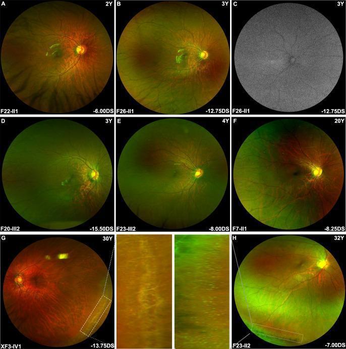
A composite of diagnostic ophthalmological images featuring wide-field fundus photography and fundus autofluorescence (FAF) in patients with early-onset high myopia (eoHM) related to ARR3 variants. Panels A-H display wide-field fundus photographs showing a predominantly tessellated fundus appearance, characterized by increased visibility of the large choroidal vessels due to thinning of the retinal pigment epithelium and choriocapillaris. In younger patients (A-F, ages 2-20Y), the peripheral retina appears relatively preserved. Panel C demonstrates a normal FAF scan with no conspicuous hyper- or hypo-autofluorescence, indicating stable metabolic activity of the retinal pigment epithelium. Panels G and H (ages 30Y and 32Y) include magnified insets of the peripheral retina, highlighting age-related progression marked by lattice degeneration and distinctive white dots. Clinical metadata indicates high myopic refractive errors ranging from -6.00DS to -15.50DS. The collection illustrates the spectrum of myopic maculopathy and peripheral retinal changes associated with X-linked female-limited inheritance in ARR3-related myopia.
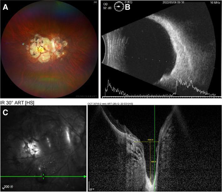
This composite diagnostic image illustrates pathological myopia in a 57-year-old patient. Panel A is a fundus photograph showing severe myopic retinal degeneration and chorioretinal atrophy at the posterior pole, exposing the underlying white sclera. A distinct grayish oval depression (macular scleral pit) is visible inferior to the macula. Panel B displays B-scan ultrasonography (top) and A-scan (bottom), revealing a significant posterior staphyloma characterized by an abnormal elongation and posterior bulging of the ocular wall. Panel C presents an Enhanced Depth Imaging Optical Coherence Tomography (EDI-OCT) cross-section, demonstrating a deep, crater-like pit measuring approximately 958 μm in depth. The OCT scan highlights extreme thinning of the neurosensory retina and partial loss of the retinal pigment epithelium and choroid within the pit area. These findings collectively demonstrate a macular scleral pit (MSP) associated with high axial myopia and posterior staphyloma.
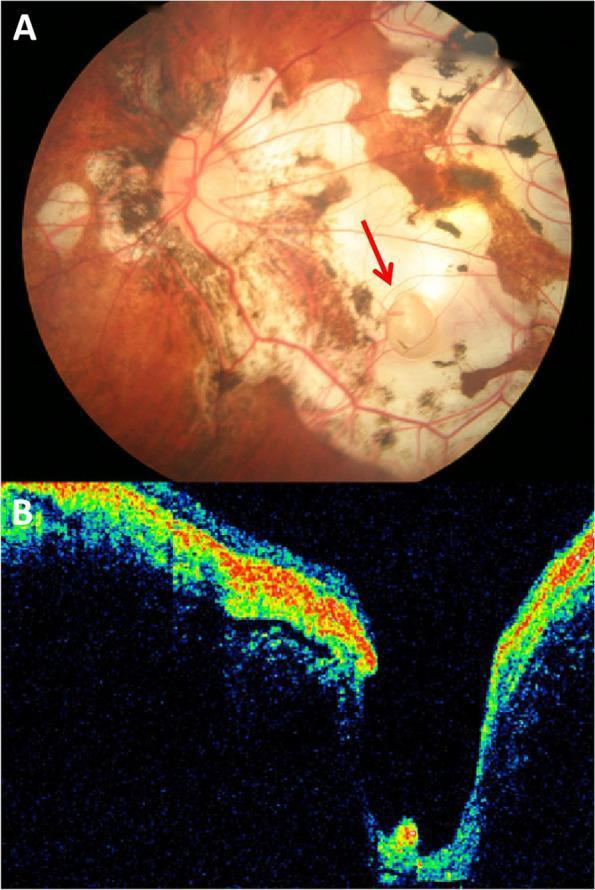
This medical image displays findings from a 62-year-old patient with pathological myopia and metamorphopsia. Panel A is a color fundus photograph showing severe myopic retinal degeneration. Key features include extensive chorioretinal atrophy, choroidal lacquer cracks, and marked retinal thinning, exposing the white underlying sclera. A red arrow identifies a Macular Scleral Pit (MSP) located inferotemporally to the macula. This MSP is a well-demarcated, oval, gray-white lesion approximately one optic disc diameter in size, with linear retinal vessels visible across its base. Panel B is a cross-sectional Optical Coherence Tomography (OCT) scan corresponding to the lesion. It reveals a deep, crater-like excavation that extends through the thinned retina and choroid into the sclera. The OCT shows a sharp depression located approximately 30 ̀̀m from the fovea centralis. There is a focal area of hyperintensity at the base of the pit beneath the neurosensory retina, though no active neurosensory detachment or defects are observed. These findings illustrate the structural complications associated with axial elongation in high myopia.
Clinical Scenario Analysis: 21-year-old, -7D Myopia, "Blackness," Normal Retina
Step 1 - What Does "Blackness" Mean Here?
| Symptom | Description | Likely Cause |
|---|---|---|
| Floaters | Dark spots/threads/cobwebs drifting in vision | Vitreous syneresis/liquefaction (most common) |
| Scotoma | Fixed dark patch | Retinal or optic nerve issue |
| Momentary blackouts | Transient vision darkening | Vascular (amaurosis fugax) or optic nerve |
| Shadow/curtain | Peripheral darkness | Early retinal detachment (urgent) |
Step 2 - Why the Retina Looks Normal
- Lattice degeneration in the periphery
- Early lacquer cracks (not always visible without detailed fundus exam)
- Early posterior vitreous detachment (PVD) not yet causing a retinal tear
Step 3 - Pathologic Myopia Context (-7D)
- Posterior staphyloma (abnormal posterior bulging of sclera)
- Myopic crescent adjacent to the optic disc
- Macular pigmentary abnormalities and Fuchs spots
- Lacquer cracks (breaks in Bruch's membrane)
- Peripheral lattice degeneration
- Vitreal degeneration/syneresis
-
Rhegmatogenous retinal detachment (RRD) - vitreous traction can tear the thinned retina
-
Choroidal neovascularization (CNV)
-
Macular schisis or macular hole (from staphyloma traction)
-
Glaucoma (IOP measurement may be underestimated by standard tonometry in highly myopic eyes)
-
Wills Eye Manual, Section 11.22 - Pathologic/Degenerative Myopia
Step 4 - Role of Nepafenac in THIS Patient
- Vitreous degeneration or floaters
- Vitreoretinal traction
- Myopia-related peripheral retinal thinning
- Prevention of retinal detachment
Step 5 - What SHOULD Be Done for This Patient
Immediate Workup:
- Dilated fundus examination with indirect ophthalmoscopy + scleral indentation - to look for peripheral retinal breaks, lattice degeneration, or early detachment (the "normal retina" may have been assessed without full dilation/indentation)
- Slit lamp biomicroscopy with 90D lens - assess macula for schisis, CNV, lacquer cracks
- B-scan ultrasonography if the media is not clear - assess posterior staphyloma and vitreous
- OCT macula - to detect foveal schisis, sub-retinal fluid, early CNV that is not visible clinically
- Visual fields - to detect any scotoma pattern
- Applanation tonometry (note: standard Schiotz/Tono-pen may underestimate IOP in high myopia; use Goldmann)
Management Based on Findings:
| Finding | Action |
|---|---|
| Vitreous floaters only, no retinal pathology | Reassurance, observation, return if acute floaters/flashes/curtain |
| Lattice degeneration | Prophylactic laser photocoagulation (controversial - done if symptomatic) |
| Retinal break/tear | Urgent laser/cryotherapy |
| Retinal detachment | Urgent surgical repair (scleral buckle / vitrectomy) |
| CNV detected on OCT/IVFA | Anti-VEGF (bevacizumab/ranibizumab) |
| Macular schisis | Monitor; vitrectomy if progressive |
| Glaucoma | IOP-lowering therapy |
Lifestyle Advice:
- Wear polycarbonate safety goggles for sports - high myopia significantly increases choroidal rupture risk from minor trauma
- Return urgently if: sudden increase in floaters, flashes of light (photopsia), or a curtain/shadow in vision - these are red flag signs of retinal tear or detachment
- Follow-up every 6-12 months even if asymptomatic
Summary
| Question | Answer |
|---|---|
| Is nepafenac indicated? | No - not for vitreous floaters or myopic degeneration |
| What is the likely cause of "blackness"? | Vitreous syneresis / floaters from high myopia |
| Is the "normal retina" finding sufficient? | Not without a full dilated exam with scleral indentation |
| What is the priority? | Rule out retinal breaks, detachment, and early macular complications with full workup |
| When would nepafenac ever be used in myopia? | Only if concurrent CME/uveitis/post-surgical inflammation is present |