
Management of low r gi bleed with evidence
Reading File
Lower GI bleeding (LGIB) management involves initial resuscitation, risk stratification, colonoscopy as the primary diagnostic-therapeutic tool, and targeted hemostasis. Now let me search the medical library and recent evidence simultaneously.
Searching sources...
Finding Sources
Finding Sources
Searching PubMed
"lower gastrointestinal bleeding"[MeSH Terms] AND management
Reading File
Reading File
Reading File
Searching PubMed
"lower gastrointestinal bleeding" AND management
Reading File
Reading File
Reading Article
PMID: 36735555
Reading File
Reading File
Reading File
Finding Sources
Searching PubMed
"lower gastrointestinal bleeding" AND "colonoscopy" AND "CTA"
Reading File
Reading File
Reading File
Reading File
I now have comprehensive information from the textbooks and the 2023 ACG guideline. Let me compile the full evidence-based answer.
Loading Image
Here is a comprehensive, evidence-based overview of the management of lower GI bleeding (LGIB).
Management of Lower GI Bleeding (LGIB) - Evidence-Based
Definition and Epidemiology
LGIB generally signifies bleeding from the colon or anorectum (distal to the ligament of Treitz). Annual incidence is approximately 20 cases/100,000 population, predominantly affecting adults over 70 years. Most patients present with painless hematochezia. If orthostasis accompanies hematochezia, a brisk upper GI source must be excluded - approximately 15% of severe hematochezia in non-cirrhotic patients originates from an upper GI source.
- Sleisenger and Fordtran's GI and Liver Disease, p. 320
Common Causes
| Cause | Frequency (%) |
|---|---|
| Diverticulosis | ~30% |
| Internal hemorrhoids | ~20% |
| Colon cancer/polyps | ~20% |
| Colitis (ischemic, IBD, infectious) | ~17% |
| Angioectasia | ~6-7% |
| Post-polypectomy bleeding | ~6-8% |
| Rectal ulcer / anorectal | ~8-10% |
| Radiation colitis | ~2% |
- Sleisenger and Fordtran's, Table 20.8
Step 1 - Resuscitation
All patients with LGIB should be resuscitated first:
- Two large-bore IV lines, IV crystalloid fluid resuscitation
- Blood transfusion: The 2023 ACG Guideline recommends a restrictive transfusion threshold (transfuse at Hb <7 g/dL; Hb <8 g/dL in patients with cardiovascular disease) - identical to the evidence base in upper GI bleeding
- Coagulopathy correction: For patients on vitamin K antagonists with life-threatening LGIB, the ACG 2023 guideline recommends reversal (4-factor PCC preferred over FFP). For patients on direct oral anticoagulants (DOACs) with life-threatening LGIB, specific reversal agents (andexanet alfa for Xa inhibitors, idarucizumab for dabigatran) are recommended
- ICU monitoring for hemodynamically unstable patients
Step 2 - Risk Stratification
Oakland Score (validated tool, ACG 2023 recommended)
The Oakland score uses age, sex, prior LGIB admission, rectal examination findings, heart rate, systolic BP, and hemoglobin to stratify risk:
-
Oakland score ≤8: Minor self-limited bleeding - may be discharged safely from the ED and undergo outpatient colonoscopy within 2 weeks
-
Oakland score >8: Requires inpatient management
-
Mulholland and Greenfield's Surgery, p. 3213
Strate Clinical Prediction Score (for severe LGI bleeding)
Risk points (1 point each):
- Aspirin use
-
2 comorbid illnesses
- Heart rate ≥100/min
- Nontender abdomen
- Rectal bleeding within first 4 hours
- Syncope
- Systolic BP ≤115 mmHg
| Score | Risk of Severe Bleeding | Mortality |
|---|---|---|
| 0 | 6% | 0% |
| 1-3 | 43% | 2.9% |
| ≥4 | 79% | 9.6% |
- Sleisenger and Fordtran's, Table 20.9
Step 3 - Diagnostic and Management Algorithm
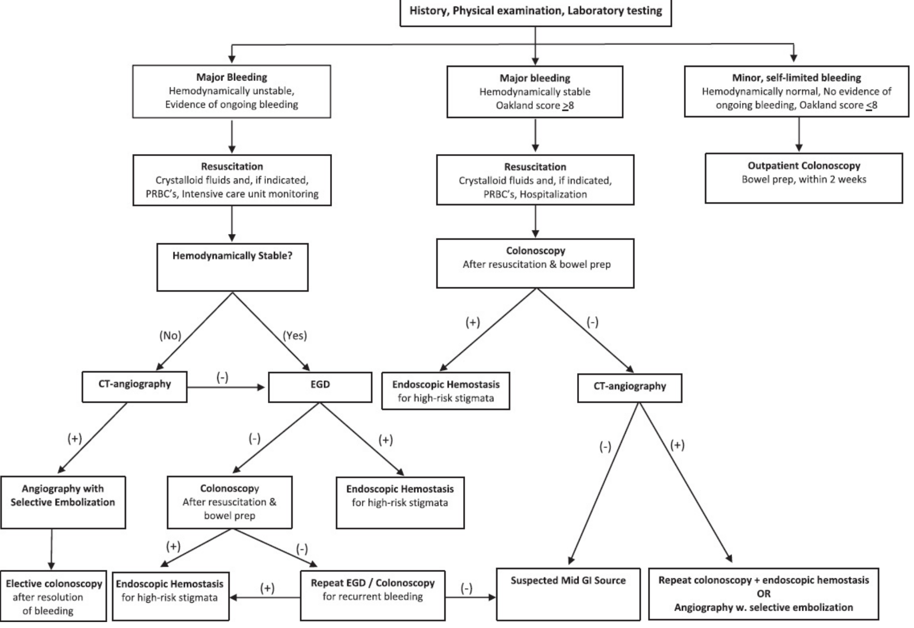
Figure: Diagnostic algorithm for managing LGIB - Mulholland and Greenfield's Surgery
A) Hemodynamically Unstable - Ongoing Bleeding
- Urgent CT Angiography (CTA) - first-line investigation
- Detects bleeding rates as low as 0.3 mL/min
- Sensitivity 85%, specificity 92% in active bleeders
- These patients cannot tolerate bowel prep or colonoscopy
- If CTA positive: Proceed to visceral angiography + superselective embolization (achieves hemostasis in ~80%)
- If CTA negative: Perform EGD (upper GI source in ~15% of unstable patients with hematochezia)
B) Hemodynamically Stable - Major Bleeding (Oakland >8)
-
Colonoscopy after resuscitation and bowel prep (PEG purge)
- Bowel prep improves diagnostic yield
- For cirrhotic patients, recent melena/hematemesis, or PUD history: panendoscopy (EGD + colonoscopy) recommended first
- Urgent colonoscopy diagnoses the source in 70-95% of cases
-
Timing of colonoscopy - ACG 2023 Update:
- Two systematic reviews showed colonoscopy within 24 hours improved bleeding localization and reduced hospital stay
- However, a recent multicenter RCT showed no difference in rebleeding rates between urgent (<24 hrs) and elective colonoscopy
- ACG 2023 recommends NON-URGENT colonoscopy for most patients (i.e., within the admission but not necessarily within 24 hours), as urgent colonoscopy has not been shown to improve outcomes like rebleeding
Key evidence: ACG 2023 LGIB Guideline (Sengupta N et al., Am J Gastroenterol 2023; PMID 36735555) -
If colonoscopy negative: CT angiography for a possible missed bleeding source or mid-GI source
C) Minor, Self-Limited Bleeding (Oakland ≤8)
- Outpatient colonoscopy within 2 weeks
- No need for inpatient admission
Step 4 - Endoscopic Hemostasis
When high-risk stigmata are found (active bleeding, non-bleeding visible vessel, adherent clot), endoscopic hemostasis is indicated:
| Technique | Notes |
|---|---|
| Hemoclips (with or without epinephrine injection) | Preferred for right colon diverticula (thin-walled - avoids transmural injury from thermal devices). 92% hemostasis success rate in 137 patients |
| Epinephrine injection (1:20,000 in saline, 4-quadrant, 1 mL/quadrant) | Always combine with a second modality |
| MPEC (multipolar electrocoagulation) | Low power (10-15 W), 1-second pulses |
| Argon Plasma Coagulation (APC) | Invaluable for angioectasia/vascular ectasia |
| Rubber band ligation / endoloops | Diverticula, hemorrhoids |
| Fibrin glue | Adjunct |
After successful colonoscopic hemostasis of diverticular bleeding, the UCLA CURE group reported:
- Rebleeding rate: 0% vs 53% in historical controls without hemostasis
- Emergency hemicolectomy: 0% vs 35% in controls
- In a later series of 63 patients: rebleeding rate 4.8% with endoscopic treatment
A submucosal tattoo should be placed around the treated lesion for future identification.
- Sleisenger and Fordtran's, p. 323
Step 5 - Angiographic Management
Indications:
- Active bleeding on CTA in hemodynamically unstable patients
- Failure of endoscopic hemostasis
- Patient cannot tolerate colonoscopy
Selective vasopressin infusion: Controls bleeding in up to 80% but recurrence rate is as high as 50%. Contraindicated in coronary artery disease (risk of MI).
Superselective embolization (3 Fr coaxial catheter system, microcoils/polyvinyl alcohol/gelatin foam):
-
First-line angiographic therapy - preferred over vasopressin
-
Initial hemostasis success: 85-90%
-
Reduced bowel infarction risk with superselective technique
-
Detects bleeding at 0.5-1.0 mL/min (requires active extravasation)
-
Yamada's Textbook of Gastroenterology, p. 2833
Step 6 - Surgical Management
Indications for surgery (rare):
- Bleeding unresponsive to endoscopic hemostasis AND angiographic embolization
- Recurrent bleeding refractory to endoscopic or angiographic therapy
- Massive hemorrhage unresponsive to resuscitation
Principles:
-
Preoperative localization is essential - allows partial (segmental) colectomy
-
Subtotal colectomy reserved for when the bleeding site cannot be localized
- Recurrent bleeding rate after subtotal colectomy: <4%
- Perioperative mortality: 2-6%
-
Blind subtotal colectomy should be avoided when possible
-
Mulholland and Greenfield's Surgery, p. 3231
Step 7 - Specific Cause-Based Management
Diverticular Bleeding
- Stops spontaneously in 75-80% of cases
- Risk factors for recurrence: age >70, NSAID/aspirin/anticoagulant use, pancolonic disease
- Recurrence incidence: 3,626/100,000 person-years; risk after second bleed: 11% annually
- Treatment: hemoclips ± epinephrine injection (preferred in thin-walled right colon)
Hemorrhoids
- Anoscopy should be the initial diagnostic study in ALL LGIB patients
- Mild bleeding: dietary fiber + lifestyle modification (avoids straining)
- Rubber band ligation: effective in >90% of patients
- Severe/grade III-IV: operative hemorrhoidectomy
Ischemic Colitis
- Usually resolves spontaneously in a few days; antibiotic therapy generally not required
- ~10% have a focal ulcer with major stigmata on colonoscopy - treat with epinephrine + hemoclip
- Large-vessel mesenteric ischemia: worse outcomes, higher rates of surgery/perforation/death
Post-Polypectomy Bleeding
- Occurs in ~1% of polypectomies, typically 5-7 days post-procedure
- 50-75% require transfusion; usually self-limited
- Treatment: hemoclip ± epinephrine injection (avoids thermal injury at polypectomy site)
Angioectasia
- APC is the treatment of choice
- If active extravasation: selective arterial embolization
IBD-related Bleeding
- Most cases managed medically
- Surgery (colectomy) for refractory cases
Step 8 - Antithrombotic Medications After Discharge
Per ACG 2023 Guideline:
- In patients on aspirin for secondary prevention: resume aspirin as soon as hemostasis is achieved (do not stop)
- In patients on dual antiplatelet therapy (aspirin + P2Y12 inhibitor):
- High cardiothrombotic risk: continue at least aspirin; restart P2Y12 inhibitor within 5 days
- Lower cardiothrombotic risk: continue aspirin; restart P2Y12 within 5 days of hemostasis
- Anticoagulants: Restart after 7-10 days in most cases after hemostasis is confirmed; decision based on thrombotic vs. bleeding risk
Summary Flow
LGIB presentation
│
├─ Oakland ≤8 → Outpatient colonoscopy (2 weeks)
│
├─ Stable + Oakland >8 → Resuscitate → Colonoscopy (non-urgent per ACG 2023)
│   ├─ High-risk stigmata found → Endoscopic hemostasis (clips ± epinephrine)
│   └─ No source on colonoscopy → CTA → angiography/embolization if positive
│
└─ Unstable + ongoing bleeding → Urgent CTA
    ├─ CTA positive → Angiography + superselective embolization
    ├─ CTA negative → EGD (rule out UGI source)
    └─ Failure of all above → Surgery (segmental or subtotal colectomy)
Key Evidence References
| Source | Key Recommendation |
|---|---|
| ACG 2023 Guideline (PMID 36735555) | Non-urgent colonoscopy for most inpatients; Oakland score for triage; CTA role in severe LGIB; DOAC reversal guidance |
| Sleisenger and Fordtran's GI and Liver Disease | Strate prediction score; endoscopic hemostasis techniques; mortality data |
| Mulholland and Greenfield's Surgery (7e) | Diagnostic algorithm; Oakland score; surgical indications |
| Yamada's Textbook of Gastroenterology (7e) | Angiographic techniques; vasopressin vs embolization |
| Fischer's Mastery of Surgery (8e) | Diverticular bleeding epidemiology and treatment outcomes |
Management of ugi bleed
Upper GI bleeding (UGIB) management centers on early resuscitation, risk stratification (Glasgow-Blatchford or Rockall score), PPI therapy, and urgent EGD within 24 hours (within 12 hours for high-risk patients). Variceal and non-variceal causes have distinct pathways.
Searching sources...
Finding Sources
Searching PubMed
"upper gastrointestinal bleeding" AND management
Reading File
Finding Sources
Searching PubMed
"nonvariceal upper gastrointestinal bleeding" AND endoscopy
Reading File
Finding Sources
Finding Sources
Reading File
Reading File
Finding Sources
Reading File
Reading File
Reading File
Reading File
Finding Sources
Reading File
Reading File
Reading File
Loading Image
I now have comprehensive data from multiple authoritative textbooks and current guidelines. Let me compile the full answer.
Management of Upper Gastrointestinal Bleeding (UGIB) - Evidence-Based
Definition and Causes
UGIB is bleeding proximal to the ligament of Treitz, presenting as hematemesis, coffee-ground emesis, melena (black tarry stool), or occasionally hematochezia (in massive bleeding).
Common causes:
| Cause | Notes |
|---|---|
| Peptic ulcer disease | Most common (~50%); duodenal > gastric |
| Esophagogastric varices | Cirrhosis-related; high mortality |
| Mallory-Weiss tear | Forceful vomiting, usually self-limited |
| Arteriovenous malformation / angioectasia | |
| Dieulafoy lesion | Submucosal artery, can be massive |
| Gastric cancer / tumor | |
| Aortoenteric fistula | Prior aortic surgery - life-threatening |
- Sabiston Textbook of Surgery, Table 98.1
Management Algorithm
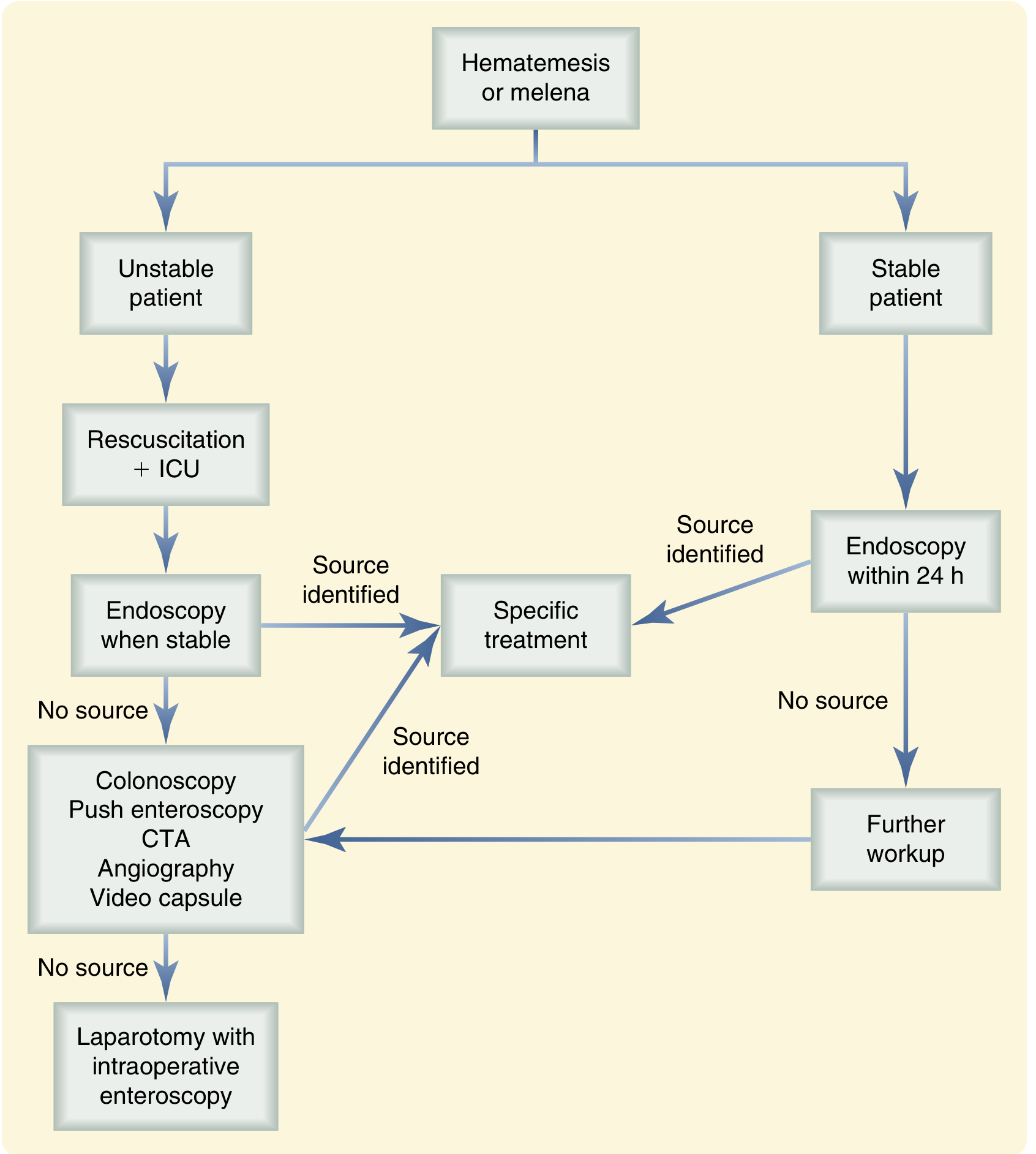
Step 1 - Resuscitation (ABCDE)
Airway
- Patients with hematemesis, altered consciousness, or inability to protect their airway require endotracheal intubation
- Prophylactic intubation is NOT routinely indicated - it is associated with cardiopulmonary complications including pneumonia
- Intubation is mandatory before balloon tamponade (Sengstaken-Blakemore tube)
Circulation - IV Access
- Two large-bore IV catheters (≥18G) in antecubital veins, OR large-bore single-lumen central cordis
- Begin crystalloid resuscitation immediately
Transfusion Strategy
- Restrictive strategy (transfuse at Hb <7 g/dL) - shown by multiple RCTs and guidelines to reduce all-cause mortality and rebleeding vs. liberal strategy
- Higher threshold (Hb <8 g/dL) for patients with ischemic cardiac disease
- For severe/ongoing massive hemorrhage: balanced resuscitation 1:1:1 ratio of packed RBCs : fresh frozen plasma : platelets (as in trauma)
- In active brisk bleeding, do not wait for lab results - transfuse clinically
Acid Suppression - PPI
- High-dose IV PPI is a cornerstone of UGIB management:
- Active bleeding / hemodynamic instability: Esomeprazole or Pantoprazole 80 mg IV bolus
- No active bleeding: 40 mg IV
- If EGD delayed beyond 12 hours: repeat 40 mg IV dose
- IV PPI infusion (80 mg bolus + 8 mg/hr continuous) after endoscopic hemostasis in high-risk ulcers significantly reduces rebleeding, need for surgery, and hospital stay
- H2-receptor antagonists are NOT adequate for acute ulcer bleeding (not shown to reduce rebleeding)
- Pre-endoscopy PPI: A 2022 Cochrane systematic review (PMID 34995368) found that pre-endoscopy PPI reduces the proportion of patients with high-risk stigmata at endoscopy and decreases the need for endoscopic treatment, but does not reduce mortality, rebleeding, or surgery rates
Vasoactive Drugs (for suspected variceal bleeding)
- Octreotide: 50 mcg IV bolus + 50 mcg/hr continuous infusion for up to 5 days
- Causes selective splanchnic vasoconstriction
- Meta-analysis: as effective as sclerotherapy for controlling variceal bleeding, with fewer adverse events
- Preferred over vasopressin due to safety (no cardiac complications)
- Terlipressin: 2 mg IV q4-6h (not available in USA) - shown to reduce mortality in variceal bleeding
- Somatostatin: 250 mcg bolus + 250 mcg/hr infusion
Prokinetics (pre-endoscopy)
- Erythromycin 250 mg IV over 20-30 minutes, 20-90 minutes before endoscopy
- Motilin receptor agonist - clears stomach of blood, clots, food
- Improves gastric visualization (77% vs 51%; OR 4.14, 95% CI 2.01-8.53)
- Reduces need for second-look endoscopy (15% vs 26%; OR 0.51)
- Shorter hospital stay (mean -1.75 days)
- Monitor QTc prolongation; check CYP3A4 drug interactions
- Metoclopramide: alternative if erythromycin contraindicated
Antibiotics (cirrhotic patients only)
- Up to 20% of cirrhotic patients admitted with GIB already have bacterial infection; up to 50% develop infection during admission - associated with increased mortality
- Meta-analyses show prophylactic antibiotics reduce both infectious complications and possibly mortality in cirrhotic patients with variceal bleeding
- Regimen: IV ceftriaxone 1 g q24h × 7 days (preferred; superior to fluoroquinolones in areas of high quinolone resistance); alternatives: IV ciprofloxacin 400 mg q12h or IV levofloxacin 500 mg q24h
Nasogastric Tube
- NOT routinely recommended - no benefit on clinical outcomes in RCTs
- May be used to confirm UGI source if unclear, or to clear stomach before endoscopy as an alternative to erythromycin
- Nasogastric aspirate with non-bloody bilious fluid suggests pylorus is open and no active UGI bleeding distal to the pylorus
Step 2 - Risk Stratification
Glasgow-Blatchford Score (GBS) - Pre-endoscopy
| Variable | Points |
|---|---|
| Systolic BP 100-109 | 1 |
| Systolic BP 90-99 | 2 |
| Systolic BP <90 | 3 |
| Heart rate ≥100/min | 1 |
| Hb (men) 12-13 g/dL | 1 |
| Hb (men) 10-12 g/dL | 3 |
| Hb (men) <10 g/dL | 6 |
| BUN elevated | 2-6 |
| Melena | 1 |
| Syncope | 2 |
| Hepatic disease | 2 |
| Cardiac disease | 2 |
- GBS = 0: Very low risk - safe discharge and outpatient management
- GBS ≥7: Predicts need for endoscopic control of bleeding
- GBS is superior to Rockall score for predicting need for intervention - endorsed by ESGE, International Consensus Group, Asia-Pacific Working Group
Rockall Score - Post-endoscopy (predicts mortality and rebleeding)
Combines clinical variables (age, shock, comorbidities) with endoscopic findings (diagnosis, stigmata of recent hemorrhage):
- Score ≥3: Significant mortality/rebleeding risk
- Score 0-1: Low risk
AIMS65 Score
Uses Albumin, INR, Mental status, Systolic BP, Age ≥65 - predicts in-hospital mortality
- Mulholland and Greenfield's Surgery, p. 3204-3205
Step 3 - Endoscopy (EGD)
EGD is the diagnostic and therapeutic modality of choice for acute UGIB.
Timing
-
Most patients: EGD within 24 hours of presentation - improves outcomes, allows safe discharge of low-risk patients
-
Suspected variceal bleeding or cirrhosis: EGD within 12 hours
-
Hemodynamically unstable despite resuscitation: EGD within 12 hours after stabilization
-
A 2023 meta-analysis (PMID 36578195) confirmed early colonoscopy improves outcomes, but for non-variceal UGIB, very urgent (<6 hours) endoscopy has NOT been shown to improve mortality
-
Upper GI barium studies are contraindicated in acute UGIB (interfere with subsequent endoscopy, angiography, or surgery)
-
Routine second-look endoscopy is NOT recommended
Step 4 - Forrest Classification and Endoscopic Hemostasis Decisions
Forrest Classification for Peptic Ulcer Bleeding
| Class | Finding | Prevalence | Rebleeding Risk (without treatment) |
|---|---|---|---|
| Ia | Active arterial spurting | 12% (Ia+Ib) | 55% |
| Ib | Active oozing | ||
| IIa | Non-bleeding visible vessel | 8% | 43% |
| IIb | Adherent clot | 8% | 22% |
| IIc | Flat pigmented spot | 16% | 10% |
| III | Clean ulcer base | 55% | 5% |
Endoscopic hemostasis is indicated for Forrest Ia, Ib, IIa, and IIb (high-risk stigmata).
Forrest IIc and III: medical management alone (PPI + H. pylori eradication); early discharge safe.
- Sabiston Textbook of Surgery, Table 98.2
Endoscopic Hemostasis Techniques
Non-variceal bleeding (peptic ulcer, Dieulafoy, Mallory-Weiss):
| Technique | Notes |
|---|---|
| Injection therapy (epinephrine 1:10,000 - 1:20,000) | Always combine with a second modality; epi alone insufficient |
| Thermal coagulation (MPEC / heater probe / APC) | Contact coagulation preferred for visible vessels |
| Hemoclips | Mechanical - preferred where thermal risk is high (thin wall) |
| Hemostatic powder (TC-325 / Hemospray) | Bridging measure; useful in diffuse/tumor bleeding |
| Over-the-scope clips (OTSC) | For refractory/recurrent bleeding; large vessel tamponade |
| Combination therapy (epi + thermal or clip) | Standard of care for high-risk lesions; reduces rebleeding vs monotherapy |
Variceal bleeding - Esophageal:
-
Endoscopic band ligation (EBL) - preferred over sclerotherapy
- Initial hemostasis: 80-85%; rebleeding: 25-30%
- Meta-analysis: EBL reduces rebleeding, overall mortality, and death from bleeding vs sclerotherapy, with fewer complications (especially esophageal strictures)
- Technique: 2 bands per variceal column - 1 at the GEJ, 1 about 4-6 cm proximally
-
Endoscopic variceal sclerotherapy (EVS) - alternative if EBL not feasible
- Sclerosants: ethanolamine oleate, sodium tetradecyl sulfate, sodium morrhuate
- Hemostasis in 85-95% of cases; rebleeding 25-30%
- Complications: esophageal ulcers/perforation, strictures, mediastinitis
-
Sleisenger and Fordtran's, p. 318
Gastric variceal bleeding:
- Cyanoacrylate (tissue glue) injection - more effective than band ligation for fundal varices (GOV2/IGV1)
- EBL can be used for type 1 gastroesophageal varices (GOV1)
Step 5 - Second-Line Interventions (Failed Endoscopy)
Interventional Radiology
- CT Angiography (CTA): Detects bleeding at ≥0.3 mL/min; sensitivity 85%, specificity 92%
- Useful when EGD fails to identify source, or patient cannot undergo endoscopy
- Useful for deep small bowel lesions beyond endoscopic reach
- Visceral angiography + transcatheter arterial embolization (TAE):
- Detects active bleeding at 0.5-1.0 mL/min
- Achieves hemostasis in ~80% when bleeding site identified
- If bleeding has stopped/is intermittent, angiography may be non-diagnostic
TIPS (Transjugular Intrahepatic Portosystemic Shunt):
- For variceal bleeding refractory to endoscopic + pharmacologic therapy (~10-20% of cases)
- Rescue TIPS (emergency TIPS within 72 hours) is now preferred over surgical shunt in Child-Pugh B/C patients with refractory variceal bleeding
- Preferred over surgery because operative mortality is high in cirrhotic patients
Balloon Tamponade (Sengstaken-Blakemore / Minnesota / Linton-Nachlas tube)
-
Used only as a temporizing bridge to definitive therapy in massive variceal hemorrhage
-
Achieves initial control in 85-98% of cases
-
Rebleeding recurs in 21-60% after deflation
-
30% rate of serious complications: aspiration pneumonia, esophageal rupture, airway obstruction
-
Endotracheal intubation is mandatory before placement
-
Should NOT be used as definitive therapy
-
Sleisenger and Fordtran's, p. 318
Step 6 - Surgical Management
Indications (rare - last resort when all above fail):
- Ongoing hemorrhage unresponsive to endoscopic + angiographic therapy
- Hemodynamic instability refractory to resuscitation
Bleeding Duodenal Ulcer
- Anterior longitudinal duodenotomy extending across the pylorus
- Figure-of-eight sutures to ligate the gastroduodenal artery at the top and bottom of ulcer crater + U-stitch for transverse pancreatic branches
- Close incision vertically as Heineke-Mikulicz pyloroplasty
- Truncal vagotomy added if patient hemodynamically stable (reduces recurrent ulceration)
Bleeding Gastric Ulcer
- Wedge resection and primary closure for greater curvature, antrum, body lesions
- Distal gastrectomy (Billroth I or II or Roux-en-Y) for lesser curvature ulcers at the incisura
- Anterior gastrotomy with biopsy + oversewing for GEJ ulcers (avoids GEJ compromise)
- Important: 4-5% of benign-appearing gastric ulcers are malignant - always biopsy/excise
Esophageal Varices - Failed TIPS/Endoscopy
-
Surgical portosystemic shunts (total or selective):
- Total/nonselective: end-to-side or side-to-side portocaval shunt (decompresses entire portal system but increases encephalopathy)
- Selective: distal splenorenal shunt (Warren shunt) - preserves hepatic portal flow while selectively decompressing gastroesophageal varices
-
Devascularization procedures (Sugiura procedure): extensive paraesophageal devascularization + esophageal transection + splenectomy + vagotomy + pyloroplasty
- Rarely used emergently (operative mortality up to 100% in Child C)
- 5-year survival: 44% (Child A), 22.5% (Child B), 0% (Child C)
-
Sabiston Textbook of Surgery, p. 2187-2188
Step 7 - Antithrombotic Drug Management
| Patient | Recommendation |
|---|---|
| Warfarin + life-threatening bleeding | 4-factor PCC + vitamin K (FFP if PCC unavailable); do not delay endoscopy |
| Warfarin + high thromboembolic risk (e.g., mechanical mitral valve) | Resume warfarin once hemostasis achieved; bridge with UFH |
| DOAC (apixaban, rivaroxaban, edoxaban) + bleeding | Hold DOAC; "time is the most important antidote" (short half-life); do not delay endoscopy |
| Dabigatran + life-threatening bleeding | Idarucizumab (specific reversal agent) |
| Aspirin (primary prevention) | Hold; consider permanent discontinuation |
| Aspirin (secondary prevention) | Continue or restart within 72 hours of hemostasis |
| Dual antiplatelet (aspirin + P2Y12) + high thrombotic risk | Continue both |
| Dual antiplatelet + low thrombotic risk, severe bleeding | Continue aspirin; restart P2Y12 inhibitor within 5 days of hemostasis |
- Mulholland and Greenfield's Surgery, p. 3210-3211
Summary Algorithm
UGIB (hematemesis / melena / coffee-ground emesis)
│
├─ GBS = 0 → Safe discharge + outpatient EGD
│
├─ Hemodynamically STABLE
│   ├─ Resuscitate (IV access, fluids, blood if Hb <7)
│   ├─ PPI 40-80 mg IV + erythromycin 250 mg IV
│   ├─ Octreotide if suspected varices; ceftriaxone if cirrhotic
│   └─ EGD within 24 hrs (within 12 hrs if suspected varices)
│       ├─ High-risk stigmata (Forrest Ia/Ib/IIa/IIb) → Endoscopic hemostasis
│       │   (combo therapy: injection + thermal/clips) → IV PPI infusion post-EGD
│       └─ Low-risk (Forrest IIc/III) → PPI + H. pylori test and treat → early discharge
│
└─ Hemodynamically UNSTABLE (persistent despite resuscitation)
    ├─ ICU admission + airway protection
    ├─ EGD when stabilized (within 12 hrs)
    ├─ If EGD fails → CTA → angiography + TAE (~80% success)
    ├─ Variceal: rescue TIPS if EBL + octreotide fails
    ├─ Balloon tamponade (SB tube) as bridge to TIPS
    └─ Surgery if all above fail (last resort)
Key Evidence References
| Source | Key Contribution |
|---|---|
| Cochrane SR 2022 (PMID 34995368) | Pre-endoscopy PPI reduces high-risk stigmata at EGD but not mortality/rebleeding |
| Meta-analysis Shung & Laine 2024 (PMID 38517201) | Updated evidence on UGIB management |
| Japan Guideline 2025 (PMID 40114631) | Endoscopic management of non-variceal UGIB (2nd edition) |
| CAG Guideline 2025 (PMID 40778886) | Non-peptic non-variceal UGIB endoscopic management |
| Sabiston Textbook of Surgery (21e) | Surgical management, resuscitation protocols, variceal surgery |
| Sleisenger and Fordtran's GI & Liver Disease | Variceal pharmacotherapy, balloon tamponade, EBL vs sclerotherapy |
| Mulholland and Greenfield's Surgery (7e) | Risk scores, antithrombotic management, resuscitation |
This is a shared conversation. Sign in to Orris to start your own chat.