
Translation in eukaryotic cells
Reading File
Translation in eukaryotes is the process by which ribosomes decode mRNA into protein. It involves four stages: initiation, elongation, termination, and ribosome recycling. Let me pull detailed content from the medical textbooks now.
Searching sources...
Finding Sources
Finding Sources
Good - I found relevant sections in Harper's Biochemistry and Lippincott. Let me read these in parallel.
Reading File
Reading File
Good content. Let me now read more of the Lippincott translation section (elongation, termination) and the Harper's initiation section in parallel.
Reading File
Reading File
Excellent content from both sources. Let me read the continuation for Harper's elongation/termination section.
Reading File
Reading File
Reading File
I now have comprehensive content. Let me also fetch the key diagram images to include in the response.
Loading Image
Loading Image
I have comprehensive information from the textbooks. Now I'll compose a thorough, well-structured answer.
Translation in Eukaryotic Cells
Translation is the process by which the nucleotide sequence of an mRNA is decoded into the amino acid sequence of a protein. In eukaryotes, translation occurs in the cytoplasm on 80S ribosomes and is divided into three main stages: initiation, elongation, and termination, followed by post-translational modifications.
Components Required
Before translation begins, the following must be present:
- 80S ribosomes (40S small subunit + 60S large subunit), each containing three tRNA-binding sites: A (aminoacyl), P (peptidyl), and E (exit)
- mRNA (monocistronic in eukaryotes - one coding region per mRNA, unlike prokaryotes)
- Aminoacyl-tRNAs - tRNAs charged with their specific amino acids by aminoacyl-tRNA synthetases
- Eukaryotic initiation factors (eIFs), elongation factors (eEFs), and release factors (eRF)
- GTP and ATP as energy sources
The Genetic Code & tRNA
Each codon is a three-nucleotide sequence on mRNA read 5'→3'. Of 64 possible codons, 61 specify amino acids and 3 are stop codons (UAA, UAG, UGA). The code is degenerate (multiple codons can specify the same amino acid).
tRNA links codons to amino acids. The anticodon region (7 nucleotides) base-pairs with the mRNA codon in an antiparallel fashion. The wobble hypothesis explains how the 5' nucleotide of the anticodon can form non-Watson-Crick base pairs with the 3' base of the codon, allowing a single tRNA species to recognize more than one codon.
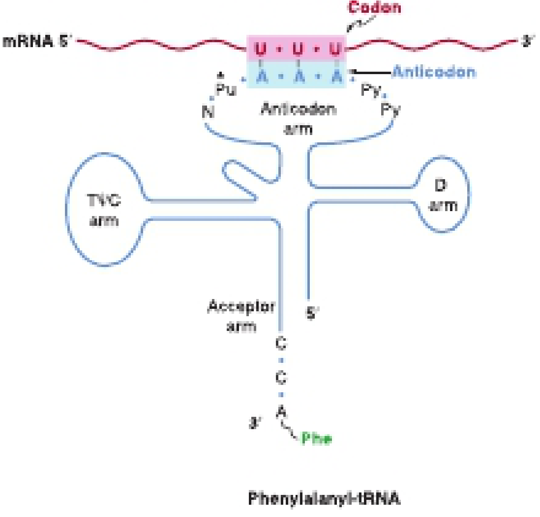
Aminoacyl-tRNA synthetases catalyze tRNA charging in a two-step reaction: (1) amino acid activation with ATP to form aminoacyl-AMP-enzyme complex, then (2) transfer of the amino acid to the 3'-OH of the tRNA. The error rate is extremely low (< 1 in 10^6 events). - Harper's Illustrated Biochemistry, 32nd Ed.
Stage 1: Initiation
Initiation assembles the full ribosome on the start codon. It involves at least 10 eukaryotic initiation factors (eIFs) and requires both GTP and ATP.
Step 1 - Ribosomal Dissociation: The 80S ribosome dissociates into 40S and 60S subunits at the end of each translation cycle. eIF-3, eIF-1, and eIF-1A bind the 40S subunit and prevent premature reassociation with the 60S subunit. - Harper's Illustrated Biochemistry, 32nd Ed.
Step 2 - 43S Preinitiation Complex: eIF-2 binds GTP, then recruits the initiator methionyl-tRNAi (Met-tRNAi) - a special tRNA used only at the start codon AUG (distinct from the tRNA used at internal AUG codons). This ternary complex (eIF-2•GTP•Met-tRNAi) joins the 40S subunit to form the 43S preinitiation complex, stabilized by eIF-3, eIF-1A, and eIF-5.
Step 3 - mRNA Binding and Scanning (48S complex): Unlike prokaryotes, eukaryotes lack a Shine-Dalgarno sequence. Instead, the small subunit (aided by eIF-4 family proteins) binds near the 5' cap structure (m7G cap) of the mRNA and scans 5'→3' until it encounters the first AUG codon. This scanning requires ATP hydrolysis. Interactions between cap-binding eIF-4 proteins and poly-A tail-binding proteins cause mRNA circularization, likely preventing use of incompletely processed mRNA. - Biochemistry, Lippincott 8th Ed.
Cap-independent initiation can also occur via binding of the 40S subunit to an Internal Ribosome Entry Site (IRES) near the start codon.
Step 4 - 80S Initiation Complex: Once the AUG is located, the 60S subunit joins the 48S complex (aided by eIF-5 and eIF-5B with GTP hydrolysis) to form the 80S initiation complex. The initiator Met-tRNAi occupies the P site (not the A site, unlike all subsequent aminoacyl-tRNAs). The A site is now empty and ready to accept the next aminoacyl-tRNA.
Key eukaryotic vs. prokaryotic differences: Eukaryotes use methionine as the initiating amino acid (prokaryotes use N-formylmethionine). The initiating codon is AUG in both.
Stage 2: Elongation
Elongation adds amino acids one at a time to the carboxyl end of the growing peptide. Each cycle has three steps:
1. Decoding (Aminoacyl-tRNA delivery to the A site):
The aminoacyl-tRNA matching the next codon in the A site is delivered by eEF-1α•GTP (equivalent to prokaryotic EF-Tu•GTP). When the correct anticodon base-pairs with the mRNA codon, GTP is hydrolyzed, eEF-1α•GDP is released, and the aminoacyl-tRNA is accommodated into the A site. The exchange factor eEF-1γ (EF-Ts analog) regenerates eEF-1α•GTP.
2. Peptide Bond Formation (Transpeptidation):
Peptidyl transferase - a ribozyme activity intrinsic to the 28S rRNA of the 60S subunit - catalyzes peptide bond formation between the α-carboxyl group of the amino acid in the P site and the α-amino group of the aminoacyl-tRNA in the A site. The growing peptide chain is transferred from the P-site tRNA to the A-site tRNA (transpeptidation), leaving an uncharged tRNA in the P site.
3. Translocation:
The ribosome moves exactly 3 nucleotides (one codon) toward the 3' end of the mRNA, facilitated by eEF-2•GTP (equivalent to prokaryotic EF-G) with GTP hydrolysis. This moves:
- The uncharged tRNA from P site → E site (then released)
- The peptidyl-tRNA from A site → P site
- The next codon into the A site
The cycle repeats until a stop codon enters the A site. Because mRNAs are long, many ribosomes can simultaneously translate a single mRNA, forming a polysome (polyribosome). - Biochemistry, Lippincott 8th Ed.
Stage 3: Termination
When one of the three stop codons (UAA, UAG, or UGA) moves into the A site, no aminoacyl-tRNA recognizes it. Instead:
- eRF-1 (a single eukaryotic release factor) recognizes all three stop codons
- eRF-1 triggers hydrolysis of the bond between the completed polypeptide and the P-site tRNA, releasing the protein
- eRF-3•GTP (analogous to prokaryotic RF-3) functions with eRF-1 and assists in release of eRF-1 upon GTP hydrolysis
- The ribosomal subunits, mRNA, and tRNA are released and recycled (requiring eRF and ATP hydrolysis in eukaryotes)
In prokaryotes, two release factors (RF-1 and RF-2) are needed; RF-1 recognizes UAA and UAG, RF-2 recognizes UGA and UAA.
Regulation of Translation
eIF-2 Phosphorylation (Key Control Point)
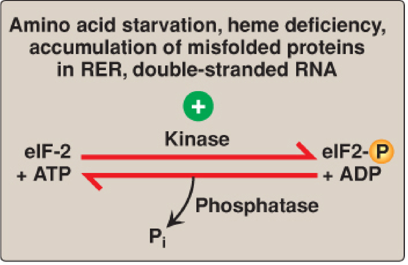
Under cellular stress conditions (amino acid starvation, heme deficiency, misfolded proteins in RER, viral infection with double-stranded RNA), specific kinases phosphorylate eIF-2α at Serine 51:
| Kinase | Trigger |
|---|---|
| HCR | Heme deficiency |
| PKR | Viral dsRNA |
| PERK | ER stress / misfolded proteins |
| GCN2 | Amino acid / glucose starvation |
Phosphorylated eIF-2α binds tightly to eIF-2B (the GEF that recycles GDP→GTP on eIF-2), inactivating it. This prevents formation of the 43S preinitiation complex and globally shuts down protein synthesis. This is a protective mechanism - PKR in particular provides antiviral defense by halting viral protein synthesis.
mTOR and eIF-4E-BP1
The mTOR pathway also regulates translation initiation. When active, mTOR phosphorylates 4E-BP1 (eIF-4E-binding protein), causing it to release eIF-4E. Free eIF-4E can then bind the 5' cap and initiate translation. When mTOR is inactive, 4E-BP1 sequesters eIF-4E, blocking translation of specific mRNAs.
Post-Translational Processing
After translation, proteins may undergo:
- Cleavage - removal of signal sequences or propeptides
- Covalent modifications - phosphorylation, glycosylation, acetylation, hydroxylation, etc.
- Folding - assisted by chaperone proteins
- Targeting - signal sequences direct proteins to organelles or for secretion
Summary Comparison: Eukaryotes vs. Prokaryotes
| Feature | Eukaryotes | Prokaryotes |
|---|---|---|
| Ribosome | 80S (40S + 60S) | 70S (30S + 50S) |
| mRNA type | Monocistronic | Polycistronic |
| Start amino acid | Methionine | N-formylmethionine |
| AUG recognition | 5' cap + scanning | Shine-Dalgarno sequence |
| Initiation factors | ≥10 eIFs (complex) | 3 IFs (simpler) |
| Elongation factors | eEF-1α, eEF-1γ, eEF-2 | EF-Tu, EF-Ts, EF-G |
| Release factors | eRF-1 (recognizes all 3 stops) | RF-1, RF-2 (separate) |
| Coupling of transcription/translation | No (nuclear membrane separates them) | Yes (simultaneous) |
| Energy for initiation | GTP + ATP | GTP |
Sources: Biochemistry, Lippincott Illustrated Reviews 8th Ed., pp. 1254-1270; Harper's Illustrated Biochemistry 32nd Ed., pp. 418-440
This is a shared conversation. Sign in to Orris to start your own chat.