
Is pulse rate of 55 normal in a 55 y/o male whose BP is 110/72 and no other co morbidity? He has recently experienced a 20 second chest pain at night which resulted in spontaneous resolution. Following which he hasn't experienced any symptoms of breathlessness or fatigue . He usually c
Is pulse rate of 55 normal in a 55 y/o male whose BP is 110/72 and no other co morbidity? He has recently experienced a 20 second chest pain at night which resulted in spontaneous resolution. Following which he hasn't experienced any symptoms of breathlessness or fatigue . He also swims for 30 mins 5 days a week . No episode of fatigue or tiredness during swimming. However he is always anxious about his day to day activities and is very impatient. After the episode of chest pain his chest sounds were normal along with normal S1 and S2 no added sounds . Pulse was regular in volume and no wall stiffness or missed beats . Now is it anything to worry about ?
sinus bradycardia athletes vagal tone clinical significance
Clinical Assessment: 55-Year-Old Male Swimmer
1. Is a Pulse of 55 Normal?
2. The Chest Pain Episode - Should It Be Dismissed?
- Single episode, 20 seconds, at night (at rest)
- Spontaneous resolution
- No recurrence
- No dyspnoea, no fatigue, no exertional component
Most Likely: Vasospastic (Prinzmetal/Variant) Angina
"The chest discomfort occurs predominantly at rest... There is a predilection for the pain to wake the patient in the early morning hours when sympathetic activity is increasing. The syndrome is often cyclical; periods of exacerbation with repetitive episodes of chest pain may persist only for seconds..."
Other Differentials
- Oesophageal spasm - also occurs at rest/night, brief, self-resolving; very common mimicker of cardiac pain
- Musculoskeletal chest pain - though typically positional/reproducible on palpation
- Benign cardiac arrhythmia - a brief run of SVT or even a transient sinus pause can cause a sense of chest discomfort
3. The Reassuring Clinical Picture
- Normal S1, S2, no added sounds
- Regular pulse, normal volume
- No pulse wall stiffness or missed beats
- No ongoing symptoms
4. What Should Be Done?
- Vasospastic angina can cause life-threatening ventricular arrhythmias during the spasm
- A 55-year-old male has background cardiovascular risk that is not zero
- The episode was brief and self-limiting precisely because spasm or ischaemia resolved - it does not mean the underlying substrate is benign
| Investigation | Reason |
|---|---|
| Resting 12-lead ECG | Baseline; may show ST changes, Brugada pattern, QTc, conduction defects |
| 24-48 hour Holter monitor | Captures any nocturnal arrhythmia or transient ST changes |
| Fasting lipid panel + blood glucose + HbA1c | Standard CV risk assessment at 55 |
| Thyroid function (TSH) | Hypothyroidism can cause bradycardia + chest discomfort |
| Echo (if clinically indicated) | Assess LV function, wall motion, valves - not mandatory if fully asymptomatic afterward |
5. The Anxiety/Impatience Factor
- Psychological stress is a recognized trigger for coronary vasospasm and can raise sympathetic tone
- Anxiety disorder itself can cause atypical chest pain
Bottom Line
| Question | Answer |
|---|---|
| Is HR 55 normal? | Yes - athletic bradycardia from regular swimming |
| Is BP 110/72 normal? | Yes - optimal |
| Is the chest pain dangerous? | Uncertain, needs investigation |
| Should he panic? | No |
| Should he ignore it? | No |
Ecg pattern of prinzmetal angina . And it's treatment
Prinzmetal variant angina ECG ST elevation coronary spasm
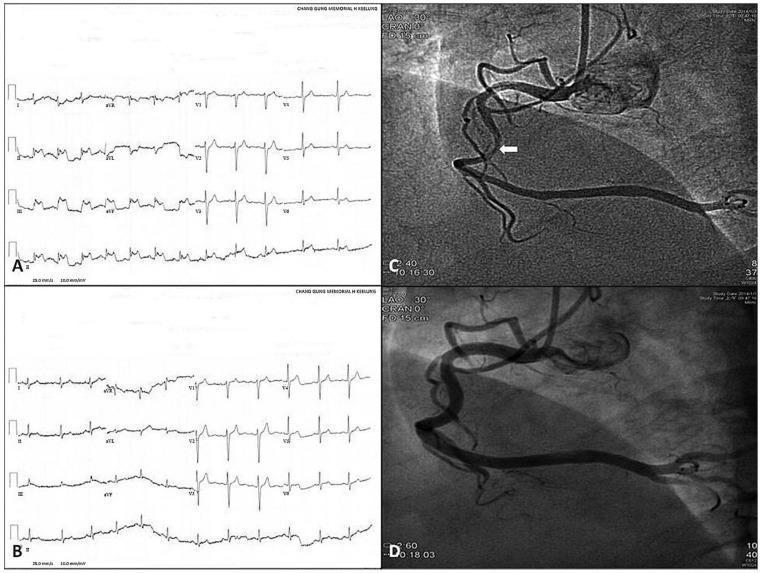
A multi-panel medical image illustrating variant (Prinzmetal) angina diagnostic findings. Panels A and B display 12-lead electrocardiograms (ECG). Panel A shows significant ST-segment elevation in inferior leads II, III, and aVF during a symptomatic angina attack. Panel B shows the resolution of these ST-segment elevations following the administration of sublingual nitroglycerin. Panels C and D are coronary angiograms of the right coronary artery (RCA). Panel C reveals a focal, severe narrowing or vasospasm in the mid-portion of the RCA (indicated by a white arrow) induced by methylergonovine. Panel D demonstrates the complete relief of this coronary artery spasm (CAS) after treatment with intracoronary nitroglycerin, resulting in restored vessel patency and normal caliber. The composite image serves as a clinical comparison for medical students and clinicians to recognize the reversible nature of vasospastic angina through both electrical (ECG) and anatomical (angiography) evidence.
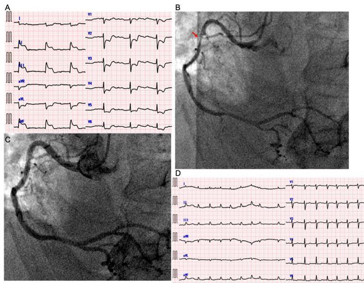
This composite medical image illustrates a clinical case of variant angina. Panel A shows an initial 12-lead electrocardiogram (ECG) with significant ST-segment elevation in the inferior leads (II, III, aVF) and an escape rhythm, indicative of acute inferior myocardial ischemia. Panel B is a coronary angiogram of the right coronary artery (RCA) showing a focal, severe stenosis in the proximal segment (indicated by a red arrow) caused by coronary vasospasm. Panel C shows the same RCA after the administration of intracoronary nitroglycerin, demonstrating the complete resolution of the stenosis and restoration of normal vessel diameter. Panel D shows a follow-up ECG with the resolution of the ST-segment elevation in leads II, III, and aVF, now displaying atrial fibrillation. The set of images effectively demonstrates the diagnostic hallmarks of variant (Prinzmetal) angina: reversible ST-elevation on ECG associated with angiographic evidence of coronary artery spasm that responds to vasodilators.
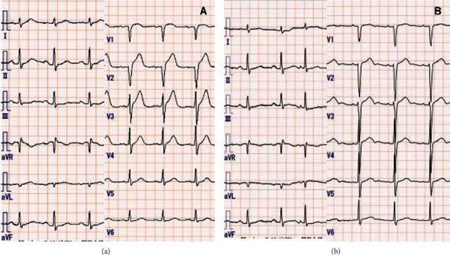
This comparison chart displays two 12-lead electrocardiograms (ECG) labeled (a) and (b), demonstrating the transient nature of vasospastic angina. ECG (a) shows significant ST-segment elevation in the anterior precordial leads, specifically V2, V3, and V4, consistent with an acute transmural myocardial injury pattern. Reciprocal ST-segment depressions are subtly visible in limb leads I and aVL. ECG (b) illustrates the same patient's cardiac rhythm following sublingual nitroglycerin administration, showing a complete resolution of the ST-segment elevations and a return to a baseline isoelectric ST-segment morphology in all leads. The educational focus of this image is the recognition of Prinzmetal (vasospastic) angina, where coronary artery spasm causes reversible ST elevation. Key clinical features include the dynamic shift from an injury pattern during symptomatic episodes to a normal or baseline ECG after vasodilator therapy. The material is suitable for medical students and clinicians studying cardiovascular diagnostics and acute coronary syndromes.
ECG Pattern of Prinzmetal (Variant/Vasospastic) Angina
The Hallmark: Transient ST-Segment Elevation
ECG Features - Detailed Breakdown
| Feature | Detail |
|---|---|
| During attack | Transient ST-segment elevation in leads overlying the ischemic territory |
| Territory | Inferior (II, III, aVF) if RCA spasm; anterior (V1-V4) if LAD spasm; lateral (I, aVL, V5-V6) if LCx spasm |
| Mechanism | Total occlusive spasm causes transmural ischemia - hence elevation, not depression |
| After resolution | ECG reverts completely to normal - no persistent changes, no Q waves |
| Between attacks | ECG is typically completely normal |
| Other ECG changes | T-wave inversion, negative U-waves, and occasionally ST depression (in opposite leads - reciprocal changes) |
| Arrhythmias during spasm | Premature ventricular ectopics, ventricular bigeminy, complete AV block, sinus bradycardia <45 bpm - in ~19% of episodes |
| Life-threatening arrhythmias | Ventricular tachycardia/fibrillation possible during spasm - this is why vasospasm carries risk even when brief |
Diagnosis
- Nitrate-responsive chest pain
- Ischemic ST-segment changes on ECG during the episode
- ≥90% spasm of the coronary artery on acetylcholine provocation testing
Treatment of Prinzmetal Angina
The Core Principle
First-Line: Calcium Channel Blockers (CCBs)
| Drug Class | Examples | Mechanism |
|---|---|---|
| Dihydropyridines (preferred) | Amlodipine 5-10 mg/day, Nifedipine | Vascular smooth muscle relaxation, prevents coronary spasm |
| Non-dihydropyridines | Diltiazem, Verapamil | Also effective; diltiazem has additional HR-lowering effect |
Second-Line: Nitrates
- Sublingual nitroglycerin for acute attacks - first-line rescue medication
- Long-acting nitrates (isosorbide mononitrate/dinitrate) for prophylaxis, though recent data from Fuster's The Heart suggests long-term nitrate use has a neutral or potentially adverse effect on MACE, so they are used judiciously - bedtime dosing preferred if nocturnal episodes predominate
- Nicorandil (a nitrate + K⁺-channel opener) has shown a neutral effect on MACE and is an alternative in some guidelines
AVOID: Beta-Blockers
Other Considerations
| Intervention | Evidence |
|---|---|
| Aspirin | Neutral in pure vasospastic angina without atherosclerosis; use cautiously - may worsen spasm via thromboxane A₂ inhibition paradox at high doses |
| Statins | Reduce 5-year MACE in vasospastic angina (Fuster's Heart, 15th Ed - HR 0.11 for MACE in statin users) |
| Avoid triggers | Smoking cessation (smoking is the strongest modifiable trigger), cold exposure, cocaine, ergot-containing medications, emotional stress |
| PCI/CABG | Not suitable - high risk of stent failure due to ongoing vasospasm; CABG not routinely recommended per Fuster's Heart |
Summary Treatment Algorithm
Acute attack:         Sublingual nitroglycerin → rapid relief
Ongoing prevention:   Calcium channel blocker (amlodipine 5-10 mg)
                      ± long-acting nitrate (bedtime dosing)
Lifestyle:            Stop smoking, reduce stress, avoid cold
Avoid:                Beta-blockers, high-dose aspirin
Monitoring:           24-hr Holter for arrhythmias
Refractory:           Add second CCB (diltiazem + amlodipine)