
I am a 2nd year medical student i want you to make histlogy notes for digestive system also provide evey histlogy slide
digestive system histology slide overview layers
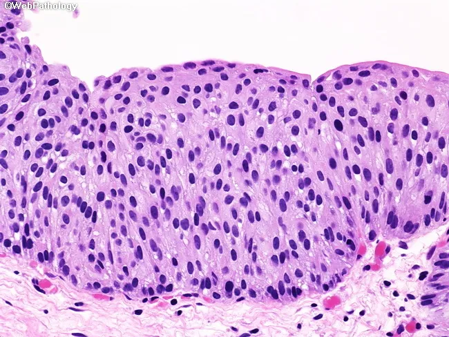
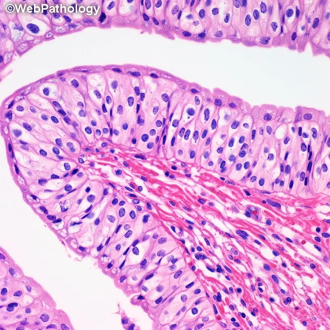
Histology slide of urothelium from urinary bladder mucosa, stained with Hematoxylin and Eosin, viewed under bright-field microscopy (paraffin-embedded, 4 µm sections; objective magnification approximately 40x). The urothelial lining is markedly thickened, comprising 12–15 cell layers. Basal cells appear prominent with preserved architecture, but there is loss of apical polarity accompanied by mild nuclear pleomorphism, nuclear enlargement, and hyperchromasia. Mitotic activity is not increased. No inflammatory infiltrate or infectious/reactive changes account for these cytologic abnormalities, and there is no evidence of stromal invasion or tumor formation in this field. Taken together, these features fall short of carcinoma in situ (CIS) and are most consistent with urothelial dysplasia (a premalignant lesion). Diagnostic significance includes recognition of precancerous change with potential progression to CIS or invasive urothelial carcinoma if unchecked. Differential considerations comprise CIS, high-grade urothelial neoplasm without invasion, and reactive/ regenerative atypia, though the latter lacks cytologic severity. Clinical correlation is essential, including cystoscopic assessment, urine cytology, and surveillance strategy; management may involve interval rebiopsy and risk-stratified follow-up. This image serves educational purposes for grading urothelial intraepithelial lesions and distinguishing dysplasia from CIS. Correlation with patient history and carcinogen exposure can further refine prognosis and monitoring. This image exemplifies urothelial dysplasia criteria.
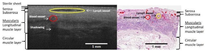
This dual-panel medical graphic provides a direct comparison between an optical coherence tomography (OCT) B-scan (left) and an H&E stained histology slide (right) of human gastric fundus tissue. Both panels illustrate the layered architecture of the gastric wall with corresponding labels. The tissue layers identified from the surface downward include the serosa and subserosa, followed by the muscularis propria, which is subdivided into the longitudinal muscle layer and the circular muscle layer. In the OCT B-scan, microvascular structures are visible, including a lymph vessel appearing as a low-scattering (dark) horizontal void and a blood vessel marked with a red circle. Vertical shadowing is noted beneath the blood vessel due to signal attenuation. In the histopathology slide, blood vessels are highlighted in red and lymph vessels in yellow, primarily localized within the serosa and subserosa layers. A sterile sheet artifact is visible on the OCT surface. Scale bars in both panels represent 1 mm, emphasizing the microscale resolution used to assess microvasculature and tissue morphology at the end of a gastric tube.
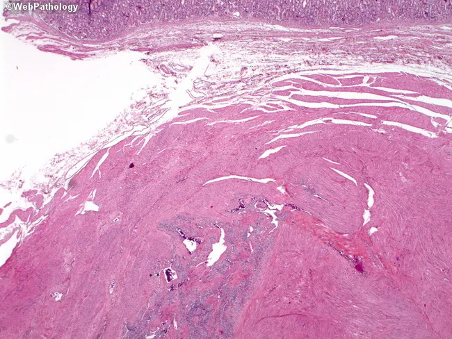
This histology slide displays gastric wall tissue with mucosa at the superior aspect and deeper muscularis propria beneath. A discrete neoplastic lesion resides within the muscular layers, visible near the bottom center as basophilic aggregates embedded in smooth muscle bundles. The lesion morphology is compatible with a glomus tumor (glomangioma) of the stomach, characterized by nests or sheets of uniform, round to polygonal cells with pale to eosinophilic cytoplasm surrounding slit- or vesicular vascular channels. The surrounding stroma is fibromuscular with conspicuous vasculature; the tumor shows a well-circumscribed architecture without frank pleomorphism, high-grade atypia, or conspicuous mitotic figures in this field. The overlying mucosa is intact. In this setting, immunohistochemistry is helpful: glomus tumor cells typically express smooth muscle actin (SMA) and vimentin, and are negative for desmin and CD117 (c-KIT) to distinguish from GIST or smooth muscle tumors. The differential for a submucosal gastric mass includes GIST, leiomyoma, hemangioma, paraganglioma, and neuroendocrine tumor; however, the perivascular arrangement and uniform cell population favor glomus tumor.
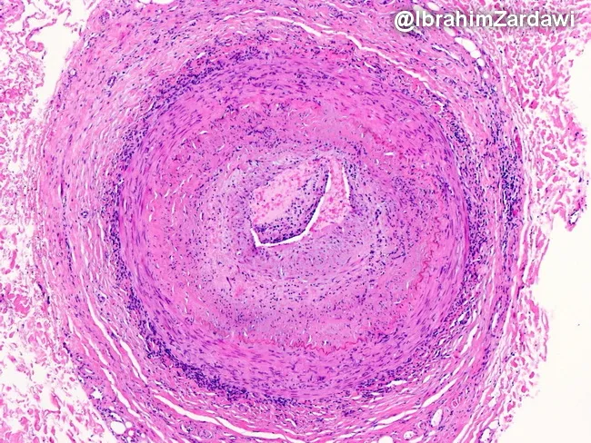
This histology image captures a cross-sectional temporal artery biopsy illustrating the hallmarks of giant cell arteritis. Prepared as a paraffin-embedded tissue section and stained with Hematoxylin and Eosin, the slide reveals a transmural inflammatory infiltrate that traverses all arterial wall layers. At the center, a narrowed lumen is surrounded by concentric intimal hyperplasia with marked medial disruption. Multinucleated giant cells and mononuclear macrophage clusters are present within the media, accompanied by lymphocytic infiltrate along the vessel wall. The elastic lamina appears disrupted, and fragmentation of the media is evident, consistent with robust granulomatous vasculitis. These features produce near-occlusive narrowing of the artery, explaining the ischemic risk associated with temporal arteritis. Clinically, such histopathology confirms a diagnosis of GCA and helps differentiate from atherosclerotic disease, infectious vasculitis, or other large-vessel vasculitides. The image underscores the systemic nature of the disease, as inflammation can extend from cranial vessels to the aorta. The diagnostic significance lies in correlating histology with patient presentation, including age-related cranial symptoms and risk of vision loss; timely immunosuppressive therapy can prevent irreversible ischemic complications. This specimen exemplifies classic angiitis with granulomatous inflammation and intimal hyperplasia driving luminal compromise.
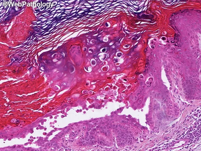
Histopathology slide of a cutaneous lesion stained with Hematoxylin and Eosin (H&E) under light microscopy, depicting classic Darier disease features. The epidermis shows marked hyperkeratosis with irregular, papillated acanthosis and prominent suprabasal clefting due to focal autoimmune-like detachment of squamous cells. Within the spinous and granular layers, numerous dyskeratotic cells are present, including the hallmark corps ronds (round, eosinophilic dyskeratotic cells with pyknotic nuclei) and grains (smaller, flattened dyskeratotic cells) situated near the keratin layer. The basal layer remains relatively preserved, whereas keratinocyte cohesion is variably disrupted. The dermis displays a mild, mixed inflammatory infiltrate without overt dermal necrosis. The composite histology—hyperkeratosis, acantholysis with suprabasal clefts, and dyskeratosis with corps ronds and grains—is highly characteristic for keratosis follicularis (Darier disease), and closely correlates with ATP2A2 gene mutations. Clinically, these findings support a diagnosis in patients with seborrheic-like papules and nail/ mucocutaneous involvement. Differential considerations include Hailey-Hailey disease (acantholysis without prominent corps ronds or grains) and Grover disease, though the dyskeratotic patterns are distinguishing. Overall, histologic pattern recognition aids diagnostic confirmation, genetic counseling, and therapeutic planning, including retinoid-based strategies and monitoring of disease progression in dermatology practice and educational settings. This image exemplifies canonical diagnostic criteria for educational and clinical use.
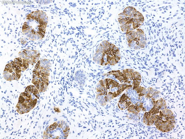
This histopathology slide depicts a formalin-fixed, paraffin-embedded prostatic needle biopsy specimen subjected to immunohistochemical staining. The tissue showcases prostatic glandular units with a preserved, continuous basal cell layer; gland lumina are variably sized and lined by two cell layers in many foci. The basal cell population exhibits diffuse, strong positivity for high molecular weight cytokeratin (HMWCK) with a brown chromogen, outlining the acinar architecture. Adjacent stromal and epithelial cells show non-specific blue hematoxylin counterstain. The staining pattern contrasts with adenocarcinoma, where the basal layer is typically disrupted or absent; here, the intact basal cell layer argues against neoplastic invasion and favors basal cell hyperplasia. p63 immunoreactivity is present in basal cells, reinforcing benign histology. In differential, prostatic hyperplasia with epithelial budding may mimic carcinoma on H&E, but the IHC demonstrates basal cells per gland. Clinically relevant use: helps distinguish benign prostatic hyperplasia with basal cell hyperplasia from prostatic adenocarcinoma in suspicious needle biopsy cores, reducing overtreatment and guiding management. The image demonstrates an educational example of diagnostic IHC utility, where basal cell markers are essential in challenging cases. The technique is commonly employed in uropathology practice to confirm benign versus malignant prostatic lesions. Correlation with morphology improves diagnostic confidence significantly.
esophagus histology cross section stratified squamous epithelium slide
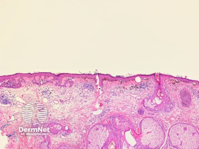
This is a light-microscopy histology image of human skin obtained from a skin biopsy. The section presents a cross-sectional view through the epidermis and dermis, highlighting classic skin architecture suitable for educational comparison. The epidermis shows stratified squamous epithelium with orderly keratinocyte maturation and a clearly visible keratin layer at the surface (stratum corneum). The epidermal-dermal junction appears intact. In the underlying dermis, dense collagenous connective tissue is intermingled with looser fibrous stroma, and adnexal elements such as hair follicles, sebaceous glands, and eccrine glands may be present depending on the field illuminated by the section. Hematoxylin provides purple nuclear detail, while eosin stains the cytoplasm and extracellular matrix pink, creating the familiar purple-pink contrast that delineates structures. No obvious atypia, dysplasia, or malignant invasion is discernible in this field, though assessment is limited to a single plane and context is clinical. The image serves as a reference for normal or near-normal skin histology and can assist in recognizing deviations arising from inflammatory dermatoses, hyperplasia, keratinization disorders, or benign adnexal lesions when correlated with patient history and additional levels. It is relevant for dermatology training, histopathology practice, and educational case discussions.
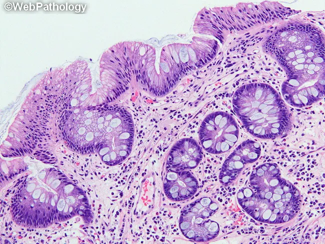
This histology slide demonstrates Barrett esophagus at the distal esophagus with intestinal metaplasia characterized by replacement of native squamous epithelium by columnar mucosa containing goblet cells. The specimen is prepared as a standard paraffin-embedded tissue section and stained with Hematoxylin and Eosin for routine morphological assessment. Under light microscopy, the mucosal surface reveals columnar epithelium arranged in irregular glands and foci of goblet cells with mucin-filled cytoplasm and pale staining. Goblet cells appear as rounded to oval cells with clear lumens, embedded within a fibromuscular lamina propria that contains a chronic inflammatory infiltrate. The underlying basal cell layer shows preserved architecture with minimal dysplasia in this field; there is no overt invasion or ulceration. The transition zone between squamous mucosa and intestinal-type mucosa is evident, consistent with Barrett esophagus. Clinically, BE is a premalignant condition linked to an increased risk of low- to high-grade dysplasia and esophageal adenocarcinoma, making histologic confirmation of goblet cell–containing metaplasia critical for diagnosis in many regions (e.g., United States, Germany) where goblet cells define specialized intestinal mucosa. This image is foundational for pathology education, differential diagnosis with reflux esophagitis, GERD-associated mucosal changes, and BE surveillance planning.
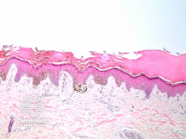
Imaging modality and technique: Light microscopy of a hematoxylin and eosin stained skin biopsy section. Specimen originates from cutaneous tissue, comprising epidermis with stratified squamous epithelium and underlying dermis. Anatomical context: integumentary system; skin organ; epidermis–dermis junction; documented with low magnification cross‑sectional view. Visual features: intact stratified squamous epithelium with multiple keratinocyte layers, prominent stratum corneum, rete ridges descending into the dermis, and a loose papillary dermis with collagenous stroma. The dermoepidermal interface is undisturbed; hair follicle and eccrine structures may be outside image field. No inflammatory cells, necrosis, dysplasia, or malignant features are evident. Staining highlights nuclei in blue (hematoxylin) and cytoplasmic/matrix components in pink (eosin), providing clear delineation of epidermal layers and dermal connective tissue. Diagnostic significance: representative normal skin histology used as a reference for dermatopathology, educational use in anatomy/gross pathology correlation, and comparative assessment in inflammatory dermatoses, autoimmune conditions, or neoplasia. Clinical relevance: baseline atlas image for teaching, slide review, and digital pathology databases; assists students and clinicians in recognizing epidermal stratification, keratinization, and dermal architecture. Clinical scenarios include dermatology education, histology labs, and virtual microscopy datasets, with contrasting examples (psoriasis, eczema, dermatitis, basal cell carcinoma) to illustrate epidermal thickening, rete peg morphology, and inflammatory patterns. Baseline image aids recognition of epidermal thickness and adnexal structures.
stomach histology gastric glands mucosa slide H&E
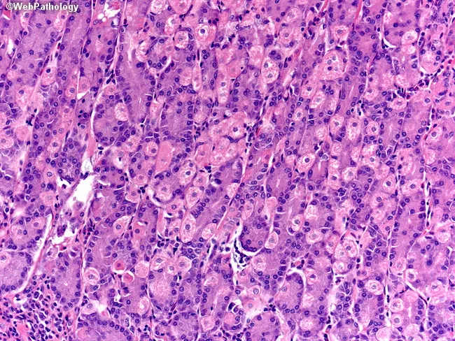
Gastric mucosal histology viewed by bright-field light microscopy on a Hematoxylin and Eosin (H&E) stained section of mucosa from the stomach, typically the oxyntic/fundic region. The glands are tubular and packed with two principal cell types: parietal (oxyntic) cells and chief (peptic) cells. Parietal cells display abundant eosinophilic (pink) cytoplasm and a central or slightly eccentric nucleus, giving a characteristic fried-egg appearance; they contribute acid secretion via gastric H+/K+-ATPase. Chief cells have basophilic (purple) cytoplasm with basal nuclei and apical zymogen granules, reflecting pepsinogen production. Neuroendocrine cells are present in minute numbers and are usually inconspicuous on routine H&E sections. Stem cells are scarce and not readily visible without special markers. The overall architecture shows intact gastric fundic glands with uniform cell density, minimal cytologic atypia, and preserved mucosal layering. The image emphasizes contrasts between cytoplasmic staining: eosinophilic parietal cells versus basophilic chief cells, as well as the densely staining nuclei. This morphology is essential for recognizing normal gastric mucosa, distinguishing parietal cell-rich areas, and identifying early metaplastic changes or inflammatory patterns in gastritis. Clinically, such images support reports of gastric biopsy evaluation, autoimmune gastritis assessment, and correlating acid-secreting cell distribution with disorders of digestion and nutrition and metabolic balance.
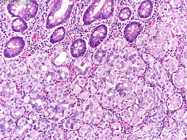
This is a high-magnification light microscopy image of gastric fundic mucosa (oxyntic glands) prepared with Hematoxylin and Eosin (H&E). The specimen represents stomach body/fundus mucosa; the image shows well-organized gastric tubular glands with prominent parietal (oxyntic) cells and abundant chief (zymogen) cells. Parietal cells appear as large, round to pyramidal cells with eosinophilic cytoplasm and intracellular canaliculi, often with a central or slightly eccentric nucleus, reflecting acid-secreting activity. Adjacent chief cells exhibit basophilic cytoplasm and basal nuclei; they contain apical zymogen granules. The glandular architecture demonstrates alternating parietal-rich regions and chief cell-rich zones within a single fundic unit, with supporting mucous cells and scattered enteroendocrine cells. The lamina propria shows a loose vascular stroma with scattered lymphocytes and capillaries; no acute or chronic inflammatory infiltrates are evident, and there is no dysplasia or metaplasia. This image captures histology suitable for educational references or diagnostic context as a normal comparator in gastritis, metaplasia, or neoplastic processes. Clinically, recognition of oxyntic glands and parietal/chief cell morphology under brightfield microscopy supports assessments of gastric acid secretion potential and helps differentiate fundic mucosa from antrum-type glands. The slide is useful for medical student teaching, histology atlases, and research focused on gastric gland physiology.
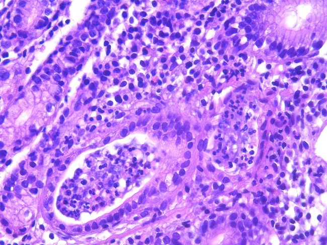
This histopathology image shows gastric mucosa with chronic active gastritis. Hematoxylin and eosin (H&E) staining reveals dense lymphoplasmacytic infiltrate in the lamina propria surrounding gastric glands, with crypt abscess formation and neutrophilic cryptitis. Epithelial architecture is variably preserved, but focal glandular distortion and inflammatory debris are evident. There is a prominent inflammatory milieu dominated by mature lymphocytes and plasma cells, consistent with a chronic inflammatory process, and occasional eosinophils are present. The image is contextually linked to Helicobacter pylori infection, which is identified in a subsequent Giemsa-stained preparation in most cases and is a key driver of acquired MALT in the stomach. Gastric mucosa in H. pylori-associated gastritis may harbor mucosa-associated lymphoid tissue (MALT) and can evolve toward MALT lymphoma in rare cases. Clinically, such findings warrant H. pylori testing and eradication therapy, since antibiotic treatment often induces remission in gastric MALT lymphoma and reduces cancer risk. This field illustrates the histologic substrate for chronic inflammation and B-cell-mediated lymphoid responses; correlation with clinical data, endoscopy, and ancillary studies (immunohistochemistry, molecular tests) is essential for accurate diagnosis and management decisions. The image is suitable for teaching histology of gastritis, differential diagnosis of gastric ulcers, and evaluation of H. pylori-related MALT pathology in practice.
small intestine villi histology duodenum jejunum slide H&E
This is a histopathology image obtained by light microscopy of an H&E-stained small intestinal mucosa biopsy. The specimen shows finger-like villi projecting from the mucosal surface, consistent with the mucosa of the small intestine (duodenum/jejunum). The epithelium comprises tall absorptive enterocytes with basally oriented nuclei and interspersed goblet cells. The brush border is variably visible at higher magnification. The lamina propria beneath the epithelium contains loose connective tissue with capillaries and scattered inflammatory cells, and the muscularis mucosae is not clearly seen in this field. The villi retain architecture with uniform height; crypts are not prominent in this field; no villous blunting or crypt hyperplasia is evident. Overall, the mucosal architecture appears preserved without acute inflammatory infiltrates, edema, or necrosis. These features are typical of normal or non-inflammatory small intestinal mucosa. Clinically, such images are used to illustrate baseline histology in education, to compare with pathologies (celiac disease with villous atrophy, inflammatory enteropathy, Giardia infection). Diagnostic significance: confirms normal mucosal histology when clinical suspicion of malabsorption or inflammatory disease is low. Potential use cases include teaching, quality control of biopsy processing, and reference for comparison in gastroenterology research. This image serves as a reference standard for educational and diagnostic practice.
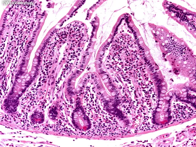
Imaging modality: Light microscopy of a hematoxylin and eosin (H&E) stained paraffin-embedded biopsy of small intestinal mucosa. The specimen reveals finger‑like villi projecting into the lumen, lined by tall simple columnar enterocytes with basally oriented nuclei and interspersed goblet cells. Crypts of Lieberkühn lie at the bases of the villi, forming closely packed glands within a fibrous lamina propria. The epithelium and lamina propria display preserved architecture without overt villous blunting, atrophy, or dysplasia. Nuclei are dark purple on hematoxylin; cytoplasm and extracellular matrix appear various shades of pink with eosin. Overall morphology is consistent with normal-appearing small intestinal mucosa, though subtle inflammatory cells may be present in the lamina propria depending on stage. This image demonstrates typical mucosal histology of the duodenum/jejunum region and is useful for assessing absorptive surface integrity and mucosal inflammation. Clinical relevance includes evaluation of malabsorption syndromes (celiac disease, tropical sprue, Giardia infection), inflammatory bowel disease involving the small intestine, and drug- or toxin-induced enteropathy. Diagnostic significance lies in verifying villous architecture and epithelial integrity, supporting differential diagnoses such as normal mucosa, chronic nonspecific enteritis, or villous atrophy when correlating with serology and symptoms. Educational value spans pathology, gastroenterology, and medical training.
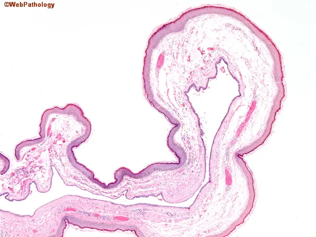
This histopathology image depicts a paraffin-embedded small intestinal mucosa section stained with Hematoxylin and Eosin (H&E), captured at a low-to-mid magnification to emphasize overall villous architecture. The tissue shows finger-like villi emanating from the lamina propria, with closely apposed, tall mucosal folds consistent with villous mucosa of the small intestine. The surface epithelium appears as simple columnar enterocytes with scattered goblet cells, and the absorptive cell brush border is not resolved at this scale. The lamina propria contains loose connective tissue with a sparse vascular network, and the muscularis mucosae is present as a thin boundary beneath the mucosal layer. No overt architectural distortion, crypt branching, or dysplastic glands are evident in the visible fields; there is no clear inflammatory infiltrate or edema highlighted at this magnification. The image emphasizes normal or near-normal mucosal pattern, suitable as a baseline reference for comparison with samples showing villous atrophy, blunting, or neoplastic villiform growth. This slide is relevant for routine evaluation of malabsorption syndromes, celiac disease workups, polyps with villous features, or educational demonstrations of normal intestinal histology. Accurate interpretation requires correlating endoscopy findings, patient history, and additional sections to differentiate normal variants from pathology.
large intestine colon histology crypts goblet cells slide
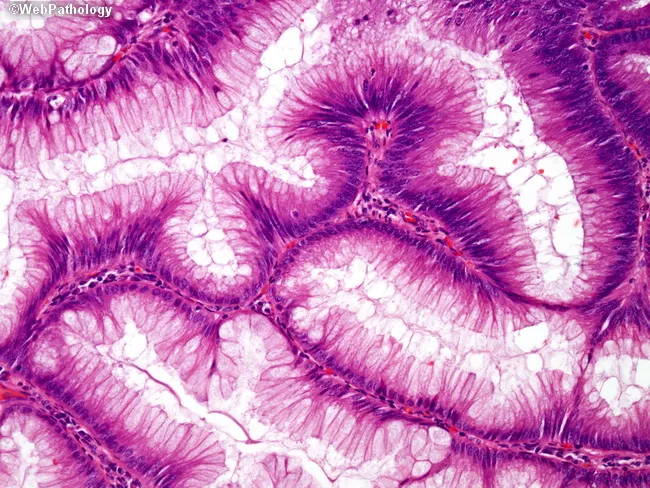
Imaging modality: brightfield light microscopy of a hematoxylin & eosin stained histology section from colorectal mucosa. Location: colorectal crypts within the mucosal layer of the large intestine (colon/rectum). The image shows high-magnification view (~400x) of tubular glands lined by tall columnar epithelial cells with basally located hyperchromatic nuclei. Nuclei appear elongated and densely staining (hyperchromasia) with preserved polarity in some regions and mild crowding in others. Goblet cells produce mucin and appear with pale cytoplasm interspersed among the epithelium. The gland lumina are irregularly shaped and show infolding and slight branching consistent with normal mucosal architecture, not overt neoplasia, though hyperchromasia can be a nonspecific reactive feature. The lamina propria contains minimal inflammatory cells; no frank desmoplasia, invasion, or significant mitotic activity is evident. These features are critical for diagnosing mucosal pathology, including evaluating for dysplasia or adenocarcinoma when clinical suspicion arises. Diagnostic significance: basally oriented nuclei are typical of healthy colorectal mucosa, but hyperchromasia and crowding require correlation with clinical data and additional sections. Potential clinical use cases include screening for colorectal neoplasia, assessment of mucosal inflammation, and educational reference for histopathology of the gastrointestinal tract. Additional note: correlate with endoscopic findings and patient history for definitive interpretation. Ultimately.
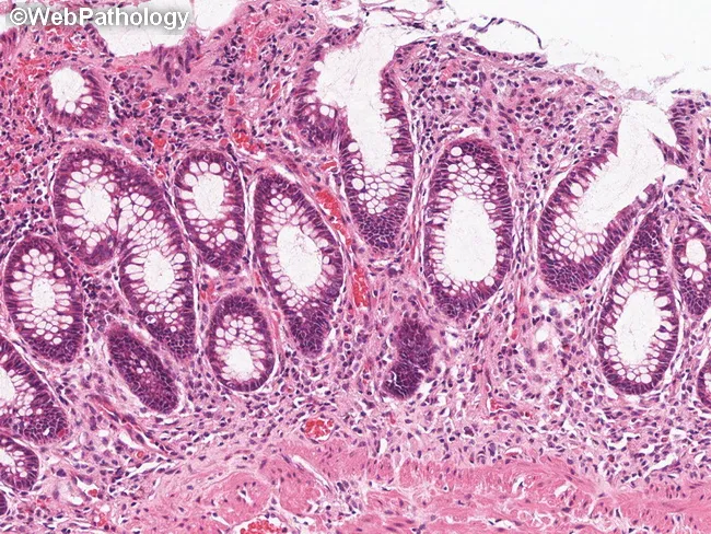
Imaging modality and technique: Light microscopy of a formalin-fixed colon mucosa biopsy (4–5 μm) stained with Hematoxylin and Eosin (H&E). Anatomical site: colorectal mucosa, large intestine, colonic lamina propria with crypts. Visual features: intact goblet cell–rich tubular glands with tall columnar epithelium; crypts preserved; lamina propria shows mild cellular inflammatory infiltrate, composed predominantly of lymphocytes and plasma cells; no active cryptitis, crypt abscess, or mucosal erosions evident. In this section, there is no obvious mast cell infiltrate or mast cell clusters near the muscularis mucosae; thus not diagnostic for GI involvement by systemic mastocytosis on this field. Differential considerations include inflammatory bowel disease patterns (ulcerative colitis or Crohn’s disease) versus reactive mucosal inflammation; other mimickers include infectious colitis and medication-related changes. Diagnostic significance: In SM, GI involvement is typically demonstrated by small compact MC infiltrates in the deep lamina propria near muscularis mucosae; their absence here does not exclude SM if clinical suspicion remains; immunohistochemical confirmation using tryptase, CD117 (c-KIT) staining or flow cytometry on adjacent tissue could identify mast cells. Clinical utility: correlate with flushing and mediator-related GI complaints; guide therapy with antihistamines and mast cell stabilizers; immunohistochemistry for mast cells (tryptase, CD117) may aid diagnosis; this image illustrates SM-related GI histology considerations for pathology and GI practice.
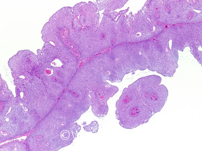
This is a hematoxylin and eosin stained histology slide of colonic mucosa examined under light microscopy in brightfield illumination. The specimen appears to show mucosal tissue from the large intestine with papillary or villiform epithelial protrusions, consistent with a mucosal polyp. The epithelium forms elongated, back-to-back glands lined by columnar cells with basophilic nuclei and moderate mucin-producing goblet cells; the lamina propria contains sparse inflammatory cells and a fibrous stroma in the core. There is no obvious invasion into submucosa. The architecture suggests benign polypoid proliferations such as a hyperplastic polyp or inflammatory polyp; however, small adenomatous changes cannot be excluded without higher magnification assessment for dysplasia, architectural complexity, and cytologic atypia. The slide emphasizes colonic mucosa without overt carcinoma. The differential includes hyperplastic polyp, inflammatory mucosal polyp, tubular adenoma, and serrated pathway lesions. Clinically, such images support histopathological diagnosis on colonoscopic biopsy and guide surveillance intervals. Potential use cases include educational training in gastrointestinal pathology, image-based teaching on mucosal polyps, and research into colorectal neoplasia progression. This image is suitable for algorithmic pattern recognition of mucosal polyp morphology and comparative histology studies. Metadata-ready keywords: histology, colon, mucosa, polyp, H&E, brightfield, microscopy, pathology training for clinical education and digital database.
liver histology hepatocytes portal triad sinusoids slide
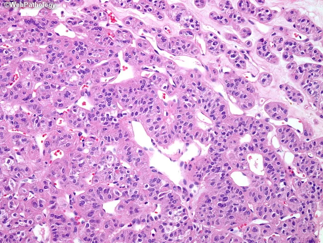
Low-power brightfield histology image of a paraffin-embedded liver biopsy stained with hematoxylin and eosin (H&E). Hepatic parenchyma demonstrates cords of polygonal hepatocytes separated by sinusoids. Cells show round to oval nuclei with finely dispersed chromatin and prominent nucleoli in a subset of nuclei. Cytoplasm is eosinophilic with variable intensity. Occasional binucleated hepatocytes are present. The plate architecture appears relatively preserved with intact sinusoidal networks; no widespread steatosis or bile stasis is evident at this field. No obvious portal triad congestion or inflammatory infiltration is conspicuous; however, a few small portal tracts appear in the economy of the field. The image emphasizes cellular morphology rather than a discrete neoplastic lesion, making definitive diagnosis of hepatocellular carcinoma or benign mimics challenging without immunohistochemistry and clinical data. This slide is suitable for educational purposes to illustrate hepatocyte cytology, binucleation, and sinusoidal arrangement, and to discuss differential diagnoses in hepatocellular lesions. Clinically, if a focal hepatic mass or chronic liver disease is suspected, this histology would be integrated with radiologic imaging and serology to determine whether a malignant tumor, benign lesion, or regenerative nodular disease is present. Further immunostains and clinical correlation are recommended for definitive classification and management planning in suspected tumors. Notes.
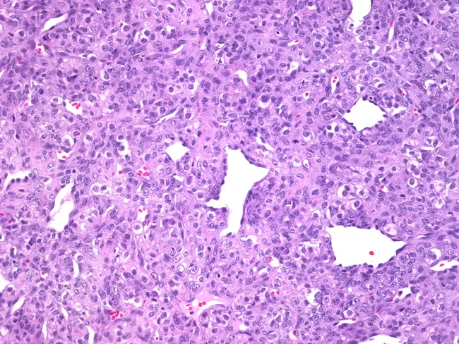
This image is a bright-field histology slide of liver parenchyma prepared by routine hematoxylin and eosin (H&E) staining and examined under light microscopy at high magnification. The specimen represents a biopsy tissue section oriented through hepatic lobules, showing cords of hepatocytes arranged in plate-like rows separated by narrow sinusoidal spaces. Hepatocytes are predominantly polygonal with round to oval nuclei and moderately basophilic cytoplasm; occasional binucleated cells are visible, consistent with normal hepatic turnover. The nuclear chromatin is evenly dispersed with visible nucleoli in a subset of hepatocytes. Intervening sinusoids appear as slender, pale vascular channels containing sparse red blood cells; there is no overt cholestasis. The overall architecture appears preserved without obvious disarray of cords, portal tract expansion, or focal necrosis in this field. No conspicuous inflammatory infiltrate or fibrotic bands are evident within the captured region. In this limited field, there is no clear evidence of steatosis, steatohepatitis, or malignant transformation, although small sampling areas may not reflect the entire organ. This image can serve as a reference for normal hepatic histology, educational demonstrations of hepatocyte morphology, and comparison with pathologic liver specimens. It has utility in training, research datasets, and radiology-pathology correlation exercises for hepatology and gastroenterology education.
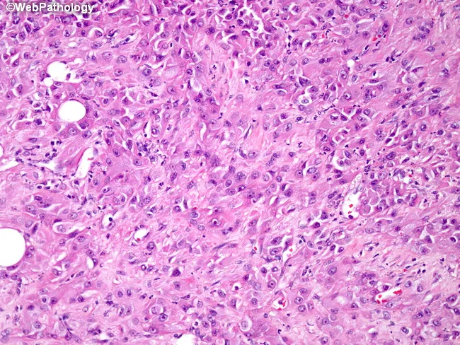
This histopathology slide depicts liver parenchyma prepared for light microscopy and stained with hematoxylin and eosin (H&E). The tissue shows polygonal hepatocytes arranged in an orderly plate-like architecture with cords separated by sinusoids. The cytoplasm is eosinophilic and mildly granular; nuclei are round or oval with inconspicuous nucleoli. Several large, clear vacuolar spaces within hepatocytes are compatible with macrovesicular fatty change (steatosis). The overall architecture appears preserved, but occasional cytoplasmic ballooning and mild cytoplasmic rarefaction may reflect early hepatocellular stress. Ductal structures and portal tracts are not the dominant features in this field and appear limited to scattered portal elements. There is no conspicuous necrosis, active inflammation, or significant fibrosis visible at this magnification, though sampling bias may obscure subtle changes. The image emphasizes hepatocellular morphology, lipid accumulation, and cellular detail that are essential for assessing fatty liver disease, steatohepatitis, or metabolic hepatopathy. Clinically, these findings can correlate with nonalcoholic fatty liver disease (NAFLD), alcoholic hepatopathy, or other causes of hepatic steatosis. Differential considerations include steatosis without inflammation, steatohepatitis, drug-induced liver injury with fatty change, or minimal chronic hepatitis. The slide is suitable for teaching hepatic histology, fatty change assessment, and correlating histologic pattern with clinical liver function abnormalities.
pancreas exocrine endocrine islets of Langerhans acini histology
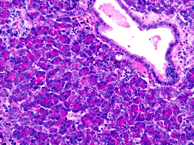
Histology image of ectopic pancreas within the gastric wall. Acinar cells with granular eosinophilic cytoplasm form pancreatic acini adjacent to ductal structures; islets of Langerhans appear as lighter, rounded cell clusters. The specimen demonstrates Heinrich type I ectopia, containing all pancreatic elements (acini, ducts, islets) embedded in gastric tissue, with preserved lobular organization and intervening gastric mucosa. The micrograph is stained with Hematoxylin and Eosin, highlighting basophilic nuclei and eosinophilic cytoplasm; acini display zymogen granules, ductal epithelium lines small ducts, and endocrine islets are dispersed in the exocrine pancreas. The lesion is a congenital anomaly presenting in the stomach, usually incidental but clinically relevant for differential diagnosis of submucosal gastric lesions. Diagnostic significance lies in recognizing heterotopic pancreatic tissue to avoid misdiagnosis as gastric neoplasm or inflammatory process. Potential clinical use cases include educational reference for surgical pathology, gastroenterology differential diagnosis, and radiologic-pathologic correlation. This image illustrates the histomorphology of ectopic pancreas, enabling recognition of all pancreatic components within gastric tissue and supports discussion of Heinrich classification, ectopic pancreas prevalence, and associated clinical scenarios such as pancreatitis or obstruction when symptomatic. The image thereby serves as a reference for teaching, diagnostic reasoning, and histopathological correlation in gastrointestinal pathology and education.
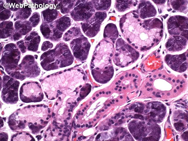
Histology micrograph of human pancreatic tissue analyzed by brightfield light microscopy after Hematoxylin and Eosin staining. The specimen shows classic lobular exocrine pancreas with densely packed acinar cells and intervening islets of Langerhans. Acinar cells display basophilic cytoplasm, basally located round nuclei, and granular eosinophilic zymogen content, contributing to the characteristic purple-blue cellular nests on the slide. The lobular architecture is preserved, with clear separation by connective septa. Within the ducts, cuboidal to low columnar epithelium lines the ductal lumina; small interlobular ducts are visible as white-staining channels. Vascular structures are present, containing erythrocytes. Adipose tissue is variably present at the periphery, consistent with normal pancreatic parenchyma. Notable features include the pale-staining islets that contrast with the intensely staining acinar lobules, providing a focal example of endocrine-exocrine juxtaposition. No overt inflammatory infiltrate, edema, necrosis, or fibrotic changes are evident in this field. Clinically, this image serves as a reference for normal pancreatic histology and as a teaching aid in pathology education. Potential diagnostic uses include benchmarking against pancreatopathic states such as pancreatitis or neoplasia, and facilitating differential diagnosis discussions in exam questions and research reviews. This image supports multiple learning modalities, including atlas reference, slide review, and examination preparation resources.
salivary gland histology serous mucous acini parotid submandibular
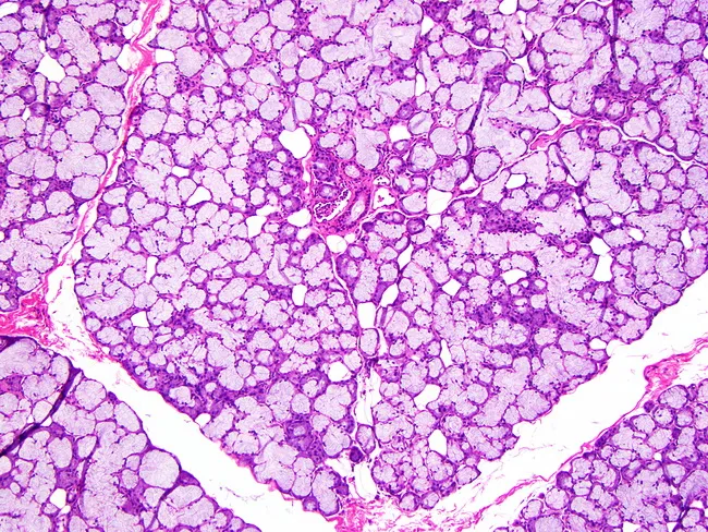
This histology image depicts sublingual salivary gland tissue examined by light microscopy after routine hematoxylin and eosin staining. The mucous acini predominate, appearing as large, pale-staining units with mucus-filled cytoplasm and relatively flattened basal nuclei; serous demilunes are minimal or absent in this field. Acini are arranged in a lobular architecture embedded in a connective tissue stroma that contains scattered capillaries and occasional adipose elements at the periphery. The excretory duct system is relatively inconspicuous compared with serous-rich glands such as the parotid or mixed glands like the submandibular; ducts are small, inconspicuous channels coursing between acini and encased in slender septa. The overall pattern reflects the mucous-dominant phenotype characteristic of the sublingual gland, with larger mucous acini than those observed in the submandibular counterpart. The image emphasizes glandular polarity with secretory cells facing a lumen and myoepithelial cells surrounding acini and ducts. Clinically, this histology supports identification of sublingual gland tissue in surgical specimens and serves as a reference for comparisons in glandular pathology, including evaluation for chronic sialadenitis, mucous retention phenomena, or rare mucous-secreting neoplasms. Overall, the slide demonstrates classic duct-acinar organization, mucous-rich cytoplasm, and a subtle ductal network within a loose connective tissue capsule.
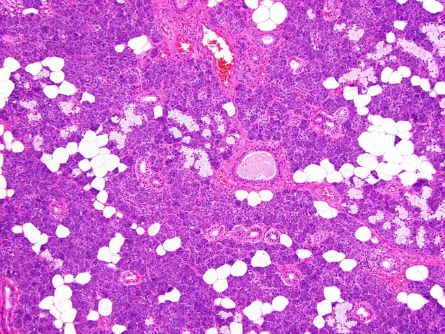
This histology image shows a hematoxylin and eosin stained paraffin-embedded section of human submandibular gland tissue, examined by light microscopy at high power. The gland demonstrates mixed seromucinous acini in which mucous cells form the central portions of the acini and serous cells lie at the periphery, with serous demilunes sometimes evident at mucous borders. Excretory and intercalated ducts are readily visible within the lobular architecture, appearing as columnar to cuboidal ductal elements with clear lumens. The acini are small to medium in size, and the surrounding stroma contains adipose tissue along with delicate connective tissue septa that partition the lobules. The overall pattern reflects normal salivary gland histology without overt cytologic atypia, notable inflammatory infiltrate, or neoplastic change in this field. Clinically, this image is pertinent for education on glandular microanatomy, including acinar polarity (serous periphery vs mucous center), ductal morphology, and the relationship between acini and excretory ducts. It supports differential considerations such as normal variant anatomy, sialadenitis, and salivary gland neoplasms in comparative studies. The image is a valuable teaching tool for pathologists, residents, and students to recognize typical histologic landmarks and enhance reproducibility in diagnostic reporting.
🔬 Histology of the Digestive System
Complete Notes for 2nd Year Medical Students
📌 OVERVIEW OF THE DIGESTIVE SYSTEM
- Transport of food and water along the canal
- Secretion of fluids, electrolytes, and digestive enzymes
- Digestion and absorption of nutrients
- Excretion of indigestible material
🔷 PART 1: GENERAL STRUCTURE OF THE GI TRACT WALL
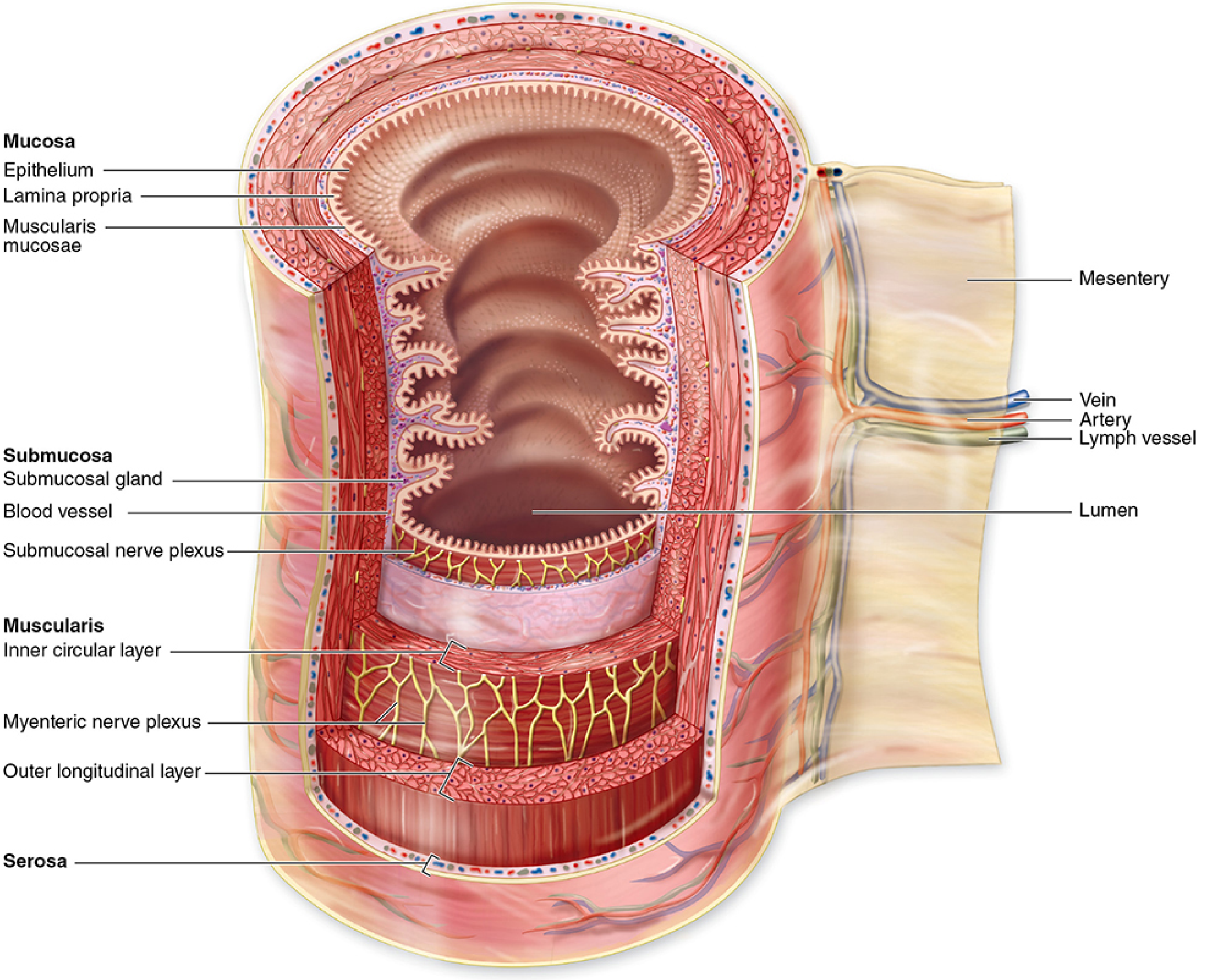
1. MUCOSA (innermost)
| Sub-layer | Description |
|---|---|
| Epithelium | Varies by region (stratified squamous, simple columnar, etc.) |
| Lamina propria | Loose connective tissue; contains vessels, lymphocytes, small glands |
| Muscularis mucosae | Thin smooth muscle; allows local mucosal movements |
2. SUBMUCOSA
- Dense connective tissue with larger blood/lymph vessels
- Contains Meissner's (submucosal) plexus - autonomic nerves
- May contain glands (esophageal glands, Brunner's glands in duodenum)
- Contains significant lymphoid tissue (MALT)
3. MUSCULARIS EXTERNA
- Inner circular layer (closer to lumen)
- Outer longitudinal layer
- Between layers: Auerbach's (myenteric) plexus - coordinates peristalsis
- Exception: Stomach has a 3rd oblique inner layer
4. SEROSA / ADVENTITIA
- Serosa = mesothelium (simple squamous) + loose connective tissue - present where organs are suspended by mesentery (stomach, intestines)
- Adventitia = only connective tissue, no mesothelium - present where organ is fixed (esophagus in mediastinum)
🔷 PART 2: ORAL CAVITY
Mucosa
- Lining epithelium: non-keratinized stratified squamous in most regions
- Keratinized on hard palate and gingiva (areas of mechanical stress)
Tongue
| Papilla | Epithelium | Taste Buds? | Notes |
|---|---|---|---|
| Filiform | Keratinized | ❌ | Most numerous; mechanical function |
| Fungiform | Non-keratinized | ✅ | Mushroom-shaped; scattered |
| Vallate (circumvallate) | Non-keratinized | ✅ | Largest; form a V-row; surrounded by a moat |
| Foliate | Non-keratinized | ✅ | Leaf-like folds on lateral tongue margins |
🔷 PART 3: ESOPHAGUS
Key Histological Features
| Feature | Details |
|---|---|
| Epithelium | Non-keratinized stratified squamous |
| Lamina propria | Contains mucous glands at the cardiac end |
| Submucosa | Contains esophageal glands (mucous) |
| Muscularis externa | Upper 1/3: striated muscle; Middle 1/3: mixed; Lower 1/3: smooth muscle |
| Outer layer | Adventitia (not serosa - it's fixed to mediastinum) |
🔷 PART 4: STOMACH
Regions (4 major):
- Cardia - short region near esophageal junction
- Fundus - dome above cardia
- Body - large central region (fundus + body are histologically similar)
- Pylorus - distal portion connects to duodenum
Mucosa of Fundus/Body
- Surface lined by surface mucous cells secreting a thick viscous mucus + bicarbonate (protection)
- Mucosa pitted with gastric pits (foveolae) leading to gastric glands
Cell Types in Gastric Glands (Fundus/Body)
| Cell Type | Location in Gland | Appearance | Function |
|---|---|---|---|
| Surface mucous cells | Surface + pit | Pale cytoplasm, basal nucleus | Mucus + HCO₃⁻ secretion (protection) |
| Mucous neck cells | Neck region | Irregular, mucus-filled | Immature precursors + mucus |
| Parietal (oxyntic) cells | Upper/mid gland | Large, eosinophilic (pink), central nucleus, "fried egg" appearance | HCl secretion via H⁺/K⁺-ATPase; Intrinsic factor (vitamin B12 absorption) |
| Chief (zymogenic) cells | Lower half of gland | Basophilic (purple) cytoplasm, basal nucleus, apical zymogen granules | Pepsinogen (→ pepsin in acid) secretion |
| Enteroendocrine cells | Scattered throughout | Pale, inconspicuous on H&E | Hormones: gastrin (G-cells), somatostatin, histamine |
| Stem cells | Neck region | Undifferentiated | Regenerate all gland epithelial cells |
Histology Slide - Gastric Fundus (Normal)
Cardia & Pyloric Mucosa
- Glands consist almost entirely of mucous cells
- Lack parietal and chief cells
- Pyloric glands are more branched and deeper
- Pylorus has a well-developed pyloric sphincter (thick circular smooth muscle)
🔷 PART 5: SMALL INTESTINE
Structural Adaptations to Increase Surface Area
- Plicae circulares (valves of Kerckring) - permanent circular folds of mucosa + submucosa
- Villi - finger-like projections of mucosa into the lumen
- Microvilli - on surface of each enterocyte → form the brush border (glycocalyx)
Cell Types of Small Intestinal Epithelium
| Cell Type | Description | Function |
|---|---|---|
| Enterocytes (absorptive cells) | Tall columnar, basal nucleus, apical brush border | Nutrient absorption; final digestion at glycocalyx |
| Goblet cells | Pale mucin-filled cytoplasm, basal nucleus squeezed | Mucus secretion (lubrication + protection) |
| Paneth cells | Deep in crypts; eosinophilic granules at apex | Defensins (antimicrobial); innate immunity |
| Enteroendocrine cells | Scattered, inconspicuous on H&E | Hormones: secretin, CCK, GIP, motilin |
| Stem cells | Base of crypts of Lieberkühn | Generate all epithelial cell types |
| M cells | Over Peyer's patches (ileum) | Antigen sampling; transport to lymphoid tissue |
Lamina Propria of Villi
- Loose connective tissue + capillaries + central lacteal (lymphatic vessel)
- Lipids absorbed as chylomicrons are taken up by the lacteal
- Sugars and amino acids enter capillaries → portal system
Intestinal Glands (Crypts of Lieberkühn)
- Simple tubular glands between villi
- Contain stem cells, Paneth cells, goblet cells
- Stem cells proliferate continuously → cells migrate up onto villi
Regional Differences in Small Intestine
| Feature | Duodenum | Jejunum | Ileum |
|---|---|---|---|
| Villi shape | Leaf/plate-like | Tall finger-like | Shorter, finger-like |
| Special feature | Brunner's glands in submucosa (alkaline mucus to neutralize acid chyme) | Tallest villi; most plicae | Peyer's patches (large lymphoid nodules in mucosa + submucosa) |
| Goblet cells | Fewer | Moderate | Most numerous |
Histology Slides - Small Intestine
🔷 PART 6: LARGE INTESTINE
General Features
- No villi (key difference from small intestine!)
- No plicae circulares
- Abundant simple tubular intestinal glands (crypts)
- Very large number of goblet cells (dominant cell type)
- Colonocytes (absorptive cells) for water and electrolyte reabsorption
Regions: Cecum → Ascending → Transverse → Descending → Sigmoid colon → Rectum + Appendix
Distinguishing Structural Feature
- Outer longitudinal muscle layer condensed into 3 bands = Teniae coli (colon only)
- Between teniae: haustra (sacculations)
Appendix
- Similar structure to large intestine
- Extremely abundant lymphoid tissue in submucosa (large lymphoid nodules)
- Very prominent MALT
Anal Canal
- Transition: simple columnar epithelium (rectum) → stratified squamous epithelium (anus)
- Circular muscle layer forms internal anal sphincter (smooth muscle)
- Striated muscle forms external anal sphincter
Histology Slides - Large Intestine
🔷 PART 7: SALIVARY GLANDS
Secretory Units (Acini)
| Type | Appearance | Secretion |
|---|---|---|
| Serous acini | Round/oval; dark basophilic cytoplasm; zymogen granules | Watery, protein-rich (enzymes like amylase) |
| Mucous tubules | Elongated; pale cytoplasm; flat basal nuclei | Viscous mucin |
| Mixed (serous demilunes) | Mucous tubule capped by serous cells at one end | Both watery + mucous |
Duct System (from smallest to largest)
| Duct | Lining Epithelium | Special Feature |
|---|---|---|
| Intercalated | Simple cuboidal | Drains acini; myoepithelial cells present |
| Striated | Simple columnar | Basolateral folds with mitochondria = electrolyte reabsorption; striated appearance |
| Interlobular | Stratified cuboidal/columnar | Merges into main excretory duct |
Comparison of Major Salivary Glands
| Gland | Type | Serous:Mucous | Key Feature |
|---|---|---|---|
| Parotid | Pure serous | 100% serous | Largest; only serous acini; rich in amylase |
| Submandibular | Mixed (mainly serous) | ~75% serous | Serous + mucous with demilunes; main serous |
| Sublingual | Mixed (mainly mucous) | ~75% mucous | Primarily mucous; some serous demilunes |
Histology Slides - Salivary Glands
🔷 PART 8: PANCREAS
Two functional components:
A. Exocrine Pancreas (~99% of tissue)
- Serous acini - pyramidal cells with:
- Apical end (narrow): zymogen (secretory) granules - eosinophilic
- Basal end (wide): large nucleus, rough ER, Golgi - basophilic
- Secretes digestive enzymes (trypsinogen, lipase, amylase, etc.)
B. Endocrine Pancreas (Islets of Langerhans)
- Pale-staining cell clusters scattered among acini
- Richly vascularized
| Cell Type | % in Islet | Secretion |
|---|---|---|
| B (beta) cells | ~70% | Insulin (lowers blood glucose) |
| A (alpha) cells | ~20% | Glucagon (raises blood glucose) |
| D (delta) cells | ~5% | Somatostatin (inhibits A + B cells) |
| PP cells | ~5% | Pancreatic polypeptide |
Histology Slide - Pancreas
🔷 PART 9: LIVER
Hepatic Lobule (Classical Lobule)
- Central vein at center
- Portal tracts (triads) at 6 corners
- Hepatocyte plates radiate from center to periphery
- Blood flows FROM portal triads → sinusoids → central vein (periportal to centrilobular)
Portal Triad - Contains THREE structures:
- Portal venule (branch of portal vein) - thin wall, large lumen, irregular shape
- Hepatic arteriole (branch of hepatic artery) - thick wall, small lumen, round
- Bile ductule - simple cuboidal epithelium; bile flows in OPPOSITE direction to blood
Hepatocytes
- Large polygonal cells with large central nuclei
- Often binucleated (polyploidy is common)
- Much smooth ER + rough ER; many Golgi complexes
- Functions: plasma protein synthesis, bile secretion, glycogen storage, detoxification
Hepatic Sinusoids
- Irregular channels between hepatocyte plates
- Discontinuous, fenestrated endothelium (allows plasma-hepatocyte exchange)
- Space of Disse (perisinusoidal space) - between endothelium and hepatocytes; site of exchange
- Kupffer cells - specialized stellate macrophages within sinusoid wall; phagocytose old erythrocytes, pathogens, debris
- Ito cells (hepatic stellate cells) - in Space of Disse; store vitamin A; become fibroblasts in cirrhosis
Zones of the Hepatic Lobule (Rappaport's Acinus)
| Zone | Location | Characteristics |
|---|---|---|
| Zone 1 (periportal) | Near portal triad | Most oxygenated; first to see nutrients; last to die in ischemia |
| Zone 2 | Middle | Intermediate |
| Zone 3 (centrilobular) | Near central vein | Least oxygenated; most metabolically active (CYP450, lipogenesis); first to die in ischemia and toxic injury |
Bile Canaliculi
- Narrow channels formed between adjacent hepatocytes (not separate structures)
- Bile flows from canaliculi → Canal of Hering → Bile ductules → Bile ducts in portal tracts
Histology Slides - Liver
🔷 PART 10: GALLBLADDER
Key Histological Features
| Layer | Description |
|---|---|
| Mucosa | Simple columnar epithelium with microvilli; highly folded (gives honeycombed appearance); NO muscularis mucosae |
| Lamina propria | Loose connective tissue with many capillaries |
| Muscularis | Irregularly arranged smooth muscle (no distinct layers) |
| Perimuscular connective tissue | Dense connective tissue; contains vessels and nerves |
| Serosa/Adventitia | Serosa on free surface; adventitia on hepatic surface |
📊 QUICK COMPARISON TABLE: KEY HISTOLOGICAL IDENTIFIERS
| Organ | Epithelium | Special Structures | Key Cells |
|---|---|---|---|
| Esophagus | Non-keratinized stratified squamous | Esophageal mucous glands; mixed muscle | - |
| Stomach (fundus) | Simple columnar; gastric pits | Gastric glands | Parietal cells (pink), Chief cells (purple) |
| Duodenum | Simple columnar; villi | Brunner's glands in submucosa | Enterocytes, goblet cells, Paneth cells |
| Jejunum | Simple columnar; tallest villi | Plicae circulares prominent | Enterocytes, goblet cells |
| Ileum | Simple columnar; villi | Peyer's patches; M cells | Goblet cells (↑), Paneth cells |
| Colon | Simple columnar; NO villi | Teniae coli; haustra | Colonocytes, goblet cells (↑↑) |
| Parotid | Serous acini only | Intercalated + striated ducts | Serous cells |
| Submandibular | Mixed (mainly serous) | Serous demilunes | Serous + mucous cells |
| Pancreas | Serous acini + islets | Centroacinar cells; zymogen granules | Acinar cells, A/B/D islet cells |
| Liver | Hepatocyte plates | Sinusoids; Space of Disse; portal triads | Hepatocytes, Kupffer cells, Ito cells |
| Gallbladder | Simple columnar (no muscularis mucosae) | Rokitansky-Aschoff sinuses; no villi | Columnar absorptive cells |
⭐ EXAM HIGH-YIELD POINTS
- Parietal cells = eosinophilic (pink), "fried egg" look → secrete HCl + intrinsic factor
- Chief cells = basophilic (purple), basal nucleus → secrete pepsinogen
- Brunner's glands = ONLY in duodenum submucosa (the one submucosal gland of the small intestine)
- Paneth cells = deep in crypts, apical eosinophilic granules → defensins (antimicrobial)
- Peyer's patches = ONLY in ileum mucosa/submucosa → M cells overlying them
- No villi = large intestine (key differentiator from small intestine)
- Centroacinar cells = ONLY in pancreas (pale cells sitting inside acinar lumen)
- Kupffer cells = liver macrophages in sinusoids
- Space of Disse = between fenestrated sinusoidal endothelium and hepatocytes
- Bile flows opposite to blood in liver (blood: portal → central vein; bile: canaliculi → portal tract)
- Zone 3 (centrilobular) = most damaged in ischemia and toxins (alcohol, acetaminophen)
- Esophagus has adventitia (not serosa) - it's fixed in mediastinum
- Parotid = only pure serous gland among the three major salivary glands
- Teniae coli = 3 longitudinal muscle bands unique to colon