
Stroma
stroma parenchyma histology
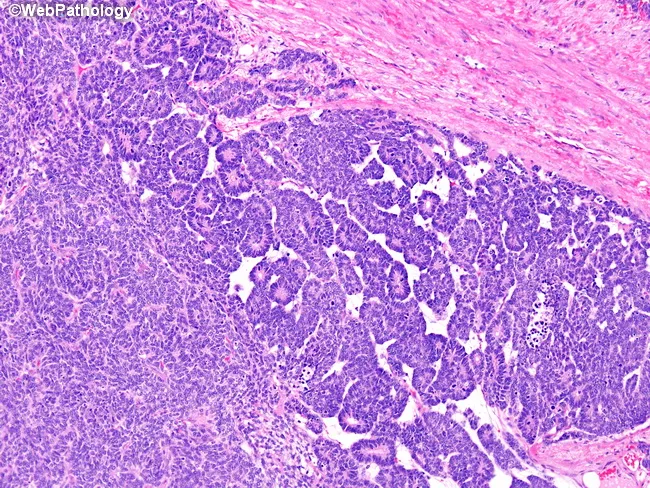
Imaging modality: light microscopy of a formalin-fixed, paraffin-embedded kidney tumor section stained with Hematoxylin and Eosin. Anatomic site: renal parenchyma within a pediatric nephroblastoma (Wilms tumor); imaging perspective: histology micrograph. The tumor exhibits classic triphasic histology with an epithelial component forming papillary structures, a prominent blastemal population arranged in sheets on the lower left, and a primitive fibroblastic stroma occupying the upper right. Epithelial elements resemble abortive tubules and papillae embedded in a cellular, small round blue cell background; blastemal cells are hyperchromatic with scant cytoplasm and high nuclear-to-cytoplasmic ratio. The stroma is variably fibrous, containing loose, spindle-shaped mesenchymal cells. The tumor–stroma interface is relatively well circumscribed in this field, with focal invasion not evident. Immunohistochemistry is not provided; in Wilms tumor, WT1 expression may be variable across components, and morphological context is essential for diagnosis. Clinically, this histology supports nephroblastoma, guiding pediatric management with nephrectomy and multimodal therapy (chemotherapy, and occasionally radiotherapy). Differential considerations include other pediatric renal neoplasms with epithelial differentiation or blastemal-predominant variants. Recognizing triphasic architecture and cellular morphology is critical for diagnostic accuracy, risk stratification, and treatment planning in pediatric renal oncology. These features confirm Wilms tumor.
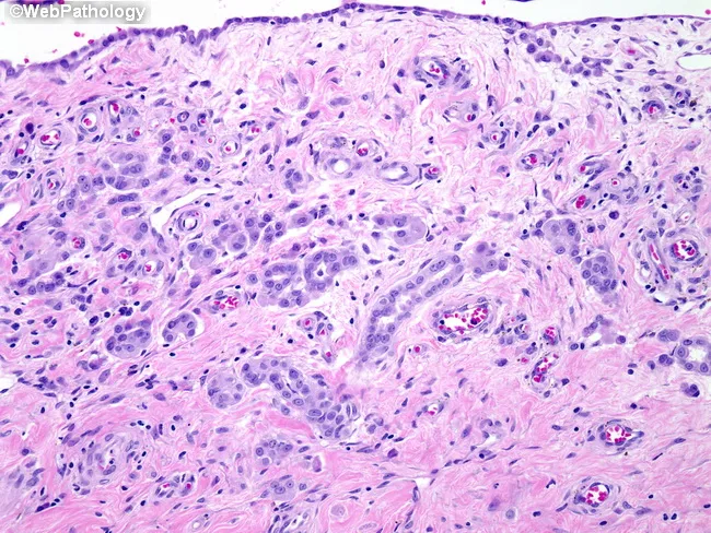
This histology slide shows reactive mesothelial hyperplasia in an orchiectomy specimen. The tissue section includes tunica vaginalis with submesothelial stroma; within this stroma, clusters of glandular-like structures are evident, lined by single to multi-layered mesothelial cells. The glands appear well-formed, with luminal spaces lined by cuboidal to flattened epithelial cells that demonstrate bland, uniform nuclei and minimal cytologic atypia. There is no cellular pleomorphism, mitotic activity, necrosis, or angular infiltration into surrounding stroma. The underlying stroma is fibrous and mildly edematous with chronic inflammatory infiltrate; no appreciable invasion of adjacent parenchyma by malignant cells is seen. The overall pattern is reactive, corresponding to chronic epididymo-orchitis as patient history; conspicuously, if the image is taken out of context, this could be mistaken for invasive carcinoma or mesothelioma due to gland-like arrangements of mesothelial cells. The complete absence of nuclear anaplasia and lack of a discrete mass favor a benign reactive process. Clinically, this finding underscores the importance of correlating histology with inflammation history and, if needed, ancillary immunohistochemistry (calretinin, WT1, CK5/6 positive for mesothelial origin; Ber-EP4, MOC31 negative or variable; PAX8 negative) to avoid misdiagnosis. This case illustrates the diagnostic challenge of mesothelial proliferations in testicular structures and guides management toward conservative interpretation.
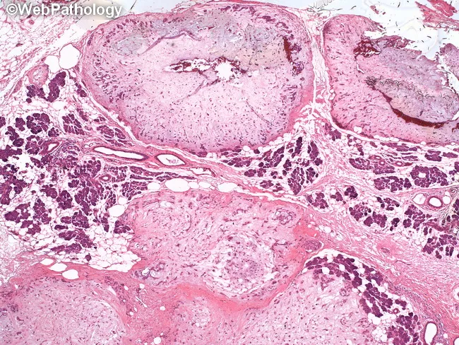
Histopathology of a recurrent multinodular pleomorphic adenoma of salivary gland demonstrates a biphasic tumor with epithelial ductal structures embedded in a variably myxoid to chondroid stroma. The architectural pattern shown is multinodular, with several discrete nodules of tumor separated by thin fibrous septa and by intervening normal salivary gland parenchyma. The stroma is predominantly myxochondroid, ranging from loose myxoid to cartilaginous-like areas, often containing chondroid droplets and cartilaginous differentiation. Ductal and myoepithelial cell populations form spindle to plasmacytoid shapes, with occasional plasmacytoid cells lining ducts. There is no overt cytologic atypia or increased mitotic activity, and perineural and vascular invasion are not evident in this field, consistent with benign histology. The presence of satellite nodules and interposed normal tissue explains the recurrent multinodular growth pattern despite benign behavior. Clinically, this histology supports a diagnosis of pleomorphic adenoma with recurrence, highlighting the importance of complete excision with wide margins to reduce residual disease. The differential diagnosis includes canalicular adenoma, basal cell adenoma, mucoepidermoid carcinoma (in areas of concern for malignant transformation), and adenoid cystic carcinoma (consider if perineural invasion emerges in other sections). Overall, findings correlate with a benign salivary gland tumor showing characteristic myxochondroid stroma.
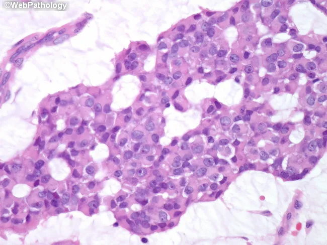
High-magnification bright-field histology image of breast parenchyma showing invasive mucinous (colloid) carcinoma with a mixture of solid and cribriform growth patterns. The tumor displays abundant extracellular mucin pools that create lakes in which clusters and occasional cords of neoplastic cells are dispersed. The neoplastic cells are relatively uniform, with small to medium-sized nuclei, round to oval contours, few conspicuous nucleoli, and minimal pleomorphism, consistent with well-differentiated histology. Mitotic activity is low and necrosis is not evident. Cribriform nests intersect the mucinous stroma, while solid foci demonstrate cohesive tumor islands embedded in mucinous matrix. In the surrounding breast parenchyma, fibrous stroma and adipose tissue may be present with mild stromal reaction but no extensive desmoplasia. The overall pattern aligns with mucin-producing adenocarcinoma of the breast, colloid type, which generally carries a favorable prognosis relative to conventional ductal carcinoma. Clinically relevant features include ER/PR positivity in many cases and a relatively indolent course; radiologic correlation may show a well-circumscribed, mucin-rich mass mimicking benign lesions. This image is valuable for pathology education, differential diagnosis with other mucinous and ductal carcinomas, and training in recognizing low-grade mucinous patterns at high magnification. Overall, this image captures characteristic mucin pools and uniform cellularity.
"tumor microenvironment"[MeSH Terms] AND stroma
Quick answer
- Cells: fibroblasts, myofibroblasts, pericytes, adipocytes, and immune cells such as macrophages and mast cells.
- Extracellular matrix: collagen, elastin, proteoglycans and glycoproteins.
- Blood vessels, lymphatics and nerves that run through the supporting tissue.
- Provides mechanical support and organ shape.
- Carries the blood supply, lymphatic drainage and innervation to the parenchyma.
- Signals to parenchymal cells through growth factors and matrix cues.
- Helps repair tissue, and can become fibrotic when injury is chronic.
- Cornea: the stroma is about 90% of corneal thickness. It consists of orderly lamellae of collagen fibrils, which keeps the cornea transparent.
- Ovary: the stroma is the connective tissue and cells surrounding the follicles.
- Lymphoid organs: the stroma is a reticular fibre network that supports the lymphocytes.
- Chloroplast (botany and cell biology): the stroma is the fluid-filled space where the Calvin cycle takes place.
- Bone marrow: the stroma comprises mesenchymal stromal cells and the supporting matrix that supports haematopoiesis.
- Tumours: the stroma is part of the tumour microenvironment. It includes cancer-associated fibroblasts, matrix, immune cells and vessels.
- Desmoplasia is a dense, fibrous stromal reaction, seen for example in pancreatic and breast carcinoma.
- Stromal signalling affects tumour growth, invasion and response to therapy.
- Biphasic tumours: some tumours have both epithelial and stromal components. Examples are Wilms tumour (blastemal, epithelial and stromal elements), phyllodes tumour, and pleomorphic adenoma, which has a myxochondroid stroma.
- Stromal tumours: GISTs and ovarian sex cord-stromal tumours arise from stromal cells themselves.
- Fibrosis: chronic injury makes the stroma over-produce matrix, as in cirrhosis, pulmonary fibrosis and keloid.
Stroma surgery in detail prosure
Which stromal surgery do you want the step-by-step procedure for? · What level of detail do you need?