
Which stain use in CSF examination
The main stains used in CSF (cerebrospinal fluid) examination are:
- India Ink (Nigrosin) - for Cryptococcus neoformans (capsule appears as clear halo)
- Gram stain - for bacterial meningitis organisms
- Ziehl-Neelsen (ZN) stain - for Mycobacterium tuberculosis (AFB)
- Giemsa stain - for parasites (Trypanosoma, Plasmodium) and malignant cells
Searching sources...
Reading File
Finding Sources
Searching the Web
stains used in CSF examination microbiology
Finding Sources
Finding Sources
Reading File
Reading File
Loading Image
Loading Image
Finding Sources
Here is a thorough, textbook-backed summary of all the stains used in CSF examination:
Stains Used in CSF (Cerebrospinal Fluid) Examination
1. Gram Stain - Most Important Routine Stain
The Gram stain is the cornerstone of CSF microbiology. All CSF specimens should be concentrated by centrifugation before staining. Sensitivity ranges from 60-90% depending on organism type and concentration:
- Gram-positive: Streptococcus pneumoniae, Listeria monocytogenes, Group B strep
- Gram-negative diplococci: Neisseria meningitidis
- Gram-negative bacilli: E. coli, H. influenzae
Note: Sensitivity for Listeria and gram-negative bacilli can be as low as 50% or less.
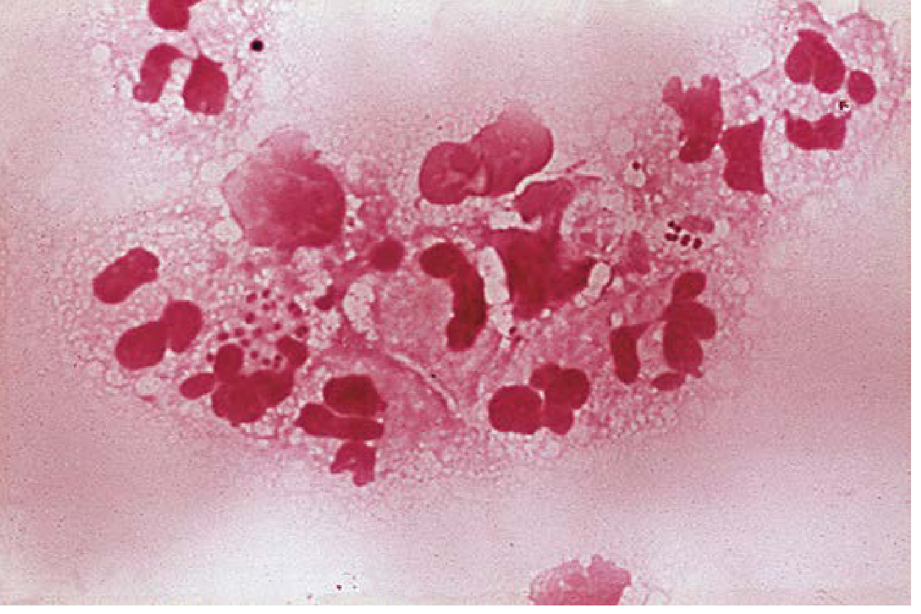
Fig. 30.10 - CSF Gram stain showing gram-negative diplococci characteristic of N. meningitidis (Henry's Clinical Diagnosis, p. 193)
2. India Ink (Negative Stain) - For Cryptococcal Meningitis
Used to detect Cryptococcus neoformans/C. gattii in CSF. India ink particles cannot penetrate the polysaccharide capsule, so the capsule appears as a clear halo around the yeast cell against a dark background. Sensitivity is less than 50% (especially in non-HIV patients), so the cryptococcal antigen (CrAg) test has largely replaced it - but India ink remains useful when antigen testing is unavailable.
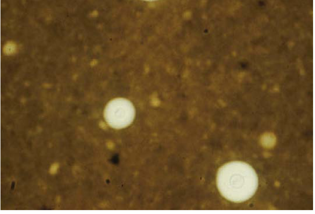
Fig. 60.14 - India ink preparation of CSF with C. neoformans (Henry's Clinical Diagnosis, p. 2114)
3. Acridine Orange Stain - Fluorescent/Supplementary
A fluorescent stain used when Gram stain is negative but clinical suspicion remains high (e.g., many PMNs but no organisms seen). More sensitive than Gram stain for:
- Bacteria and fungi (appear bright orange/red under fluorescence)
- Gram-negative organisms
- Differentiates amoebas (brick red) from leukocytes (bright green) in primary amoebic meningoencephalitis (Naegleria fowleri)
4. Ziehl-Neelsen (ZN) / AFB Stain - For TB Meningitis
Used when tuberculous meningitis is suspected. Detects Mycobacterium tuberculosis as acid-fast bacilli (appear red on blue background). Sensitivity in CSF is low (10-40%), so culture and PCR are also essential.
5. Giemsa / Wright-Giemsa Stain
Used for:
- Parasites: Trypanosoma species (African sleeping sickness), Naegleria, Acanthamoeba
- Malignant cells in CSF cytology
- Also helps identify cell morphology in the CSF differential
6. KOH (Potassium Hydroxide) Preparation
Used for fungal elements - clears cellular debris and highlights fungal hyphae and yeast. Can be combined with Calcofluor white (fluorescent) for enhanced visibility.
Summary Table
| Stain | Target Organism/Purpose | Key Feature |
|---|---|---|
| Gram stain | Bacteria (meningitis) | 60-90% sensitivity; most routine |
| India ink | Cryptococcus (fungi) | Negative stain; halo capsule; <50% sensitivity |
| Acridine orange | Bacteria, fungi, amoeba | Fluorescent; more sensitive than Gram |
| ZN / AFB stain | Mycobacterium tuberculosis | Acid-fast; low CSF sensitivity |
| Giemsa/Wright | Parasites, malignant cells | Cell morphology, trypanosomes |
| KOH ± Calcofluor white | Fungal elements | Clears debris; shows hyphae/yeast |
- Henry's Clinical Diagnosis and Management by Laboratory Methods, pp. 193-195, 2114-2135
This is a shared conversation. Sign in to Orris to start your own chat.