
Allman classification of clavicle fracture in detail
Allman classification clavicle fracture diagram
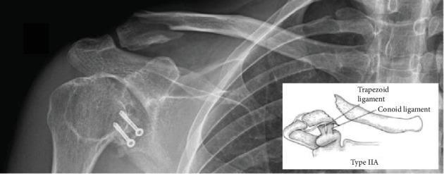
Anteroposterior (AP) radiograph of the right shoulder and clavicle demonstrating a distal clavicle fracture, classified as Neer type IIA. The fracture is located medial to the coracoclavicular (CC) ligaments, resulting in a characteristic superior displacement of the proximal clavicle fragment due to the pull of the sternocleidomastoid muscle, while the distal fragment remains attached to the acromion. Adjacent to the glenohumeral joint, two metallic cortical screws with washers are visible, originating from a previous Latarjet procedure; these are positioned within the coracoid process remnant and appear stable. An inset anatomical diagram illustrates the Neer type IIA classification, specifically highlighting the relationship between the fracture line and the conoid and trapezoid components of the CC ligament. The image serves as an educational tool for identifying unstable distal clavicle fractures and assessing postoperative orthopedic hardware in the context of complex shoulder trauma history.
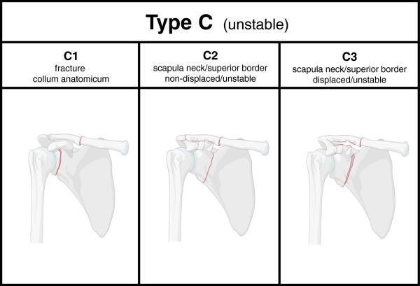
This medical illustration presents a diagnostic classification chart for Type C scapular fractures, characterized as unstable injuries or 'floating shoulder' configurations. The diagram is divided into three panels (C1, C2, C3) showing posterior-anterior views of the right shoulder girdle, highlighting fracture lines in red. Panel C1 illustrates a fracture of the collum anatomicum (anatomical neck) of the scapula. Panel C2 depicts a non-displaced but unstable fracture involving both the scapular neck/superior border and the mid-shaft clavicle. Panel C3 demonstrates a displaced and unstable fracture pattern involving the scapular neck/superior border and the clavicle, with visible misalignment of the bone fragments. The infographic serves as an educational tool for orthopedic trauma, focusing on the Superior Shoulder Suspensory Complex (SSSC) and distinguishing between injury types based on anatomical location and the presence of displacement to determine mechanical stability.
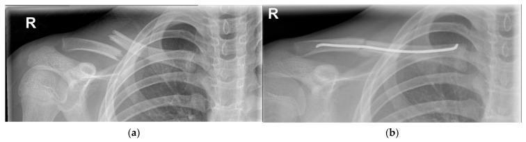
This comparative diagnostic image consists of two anteroposterior radiographs of the right shoulder and clavicle. Image (a) depicts a complete midshaft (Group I/Allman Type 1) clavicle fracture with extreme displacement and significant shortening. The distal fragment is positioned superiorly and posteriorly relative to the proximal fragment, with no evidence of primary bone callus formation. Image (b) shows the same clavicle following surgical intervention for internal fixation. A radiopaque intramedullary wire is visible, spanning the fracture site through the medullary canal to restore anatomical alignment and stability. The wire extends from the medial aspect through the lateral portion of the clavicle. These images illustrate the radiological presentation of a severe clavicular fracture and its subsequent management via intramedullary nailing. The clinical significance highlights surgical indications for highly displaced pediatric or adult clavicle fractures where conservative management may be insufficient for functional recovery.
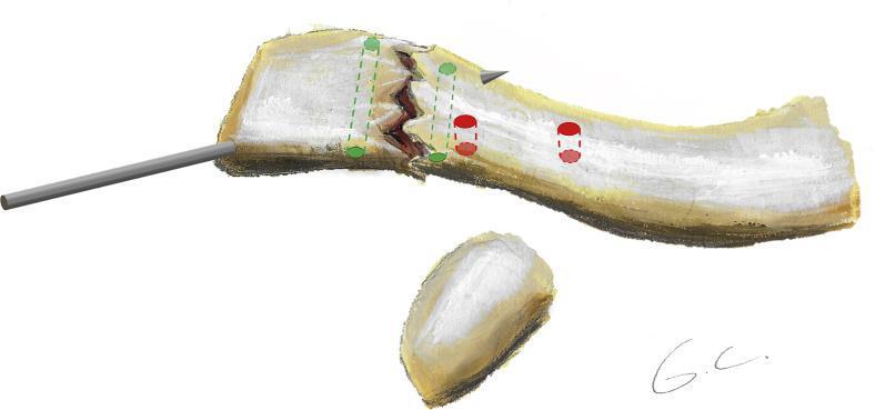
An anatomical diagram illustrating the surgical reduction and fixation planning for a distal clavicle fracture, likely a Neer Type II injury. The illustration depicts the distal third of the clavicle with a transverse fracture line. A metallic Kirschner wire (K-wire) is shown inserted laterally through the distal fragment into the proximal bone to maintain temporary reduction. The diagram highlights two distinct sets of surgical drill tunnels for definitive fixation: 1) Two green tunnels (2 mm diameter) oriented horizontally/anteroposteriorly across the fracture site, intended for figure-of-8 and figure-of-0 fracture cerclage using suture tape. 2) Two red tunnels (3.2 mm diameter) oriented vertically on the medial side of the fracture, intended for coracoclavicular cerclage using high-strength sutures. The placement of the medial tunnels reflects the anatomical insertion points of the coracoclavicular ligaments. This surgical strategy combines direct fracture site stabilization with stabilization of the coracoclavicular interval, addressing both bone and ligamentous components of the injury.
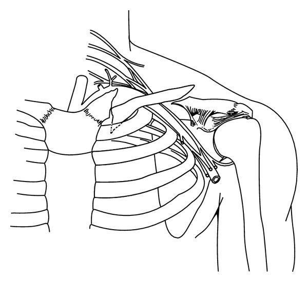
This anatomical illustration depicts a double fracture pattern of the left clavicle in an adult male. The drawing highlights two distinct fracture sites: an established medial nonunion and an acute lateral fracture. The medial fracture, located in the proximal third of the clavicle shaft, is characterized by a prominent, inferiorly directed bony spike. This spike is shown in close anatomical proximity to the lung apex and the first rib, illustrating the potential risk for intrathoracic complications such as pneumothorax. The lateral fracture is situated in the distal third of the clavicle shaft, presenting as a relatively clean, acute break without the significant remodeling seen in the medial nonunion. The illustration includes skeletal landmarks such as the sternum, rib cage, and humeral head, along with neurovascular structures passing beneath the clavicle. This diagram serves as an educational tool for identifying complex clavicular fracture patterns and understanding their spatial relationship to critical thoracic and vascular anatomy.
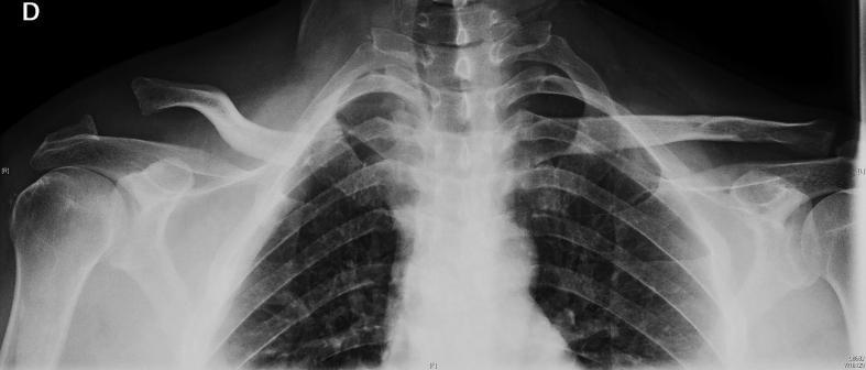
Anteroposterior (AP) chest and shoulder radiograph demonstrating a comparative view of the bilateral acromioclavicular (AC) joints. On the patient's right side, there is a high-grade acromioclavicular joint dislocation (Rockwood type IV) characterized by significant superior and posterior displacement of the distal clavicle relative to the acromion, with an increased coracoclavicular distance. Additionally, there is evidence of an Allman type III fracture of the proximal clavicle and asymmetry at the sternoclavicular region, suggesting a 'floating clavicle' injury pattern (bipolar clavicle injury). The contralateral (left) shoulder exhibits normal anatomical alignment of the acromioclavicular and sternoclavicular joints, serving as a baseline for comparison. This diagnostic image is intended for educational use in orthopedic trauma, illustrating complex clavicular fractures and ligamentous disruptions.
Allman Classification of Clavicle Fractures
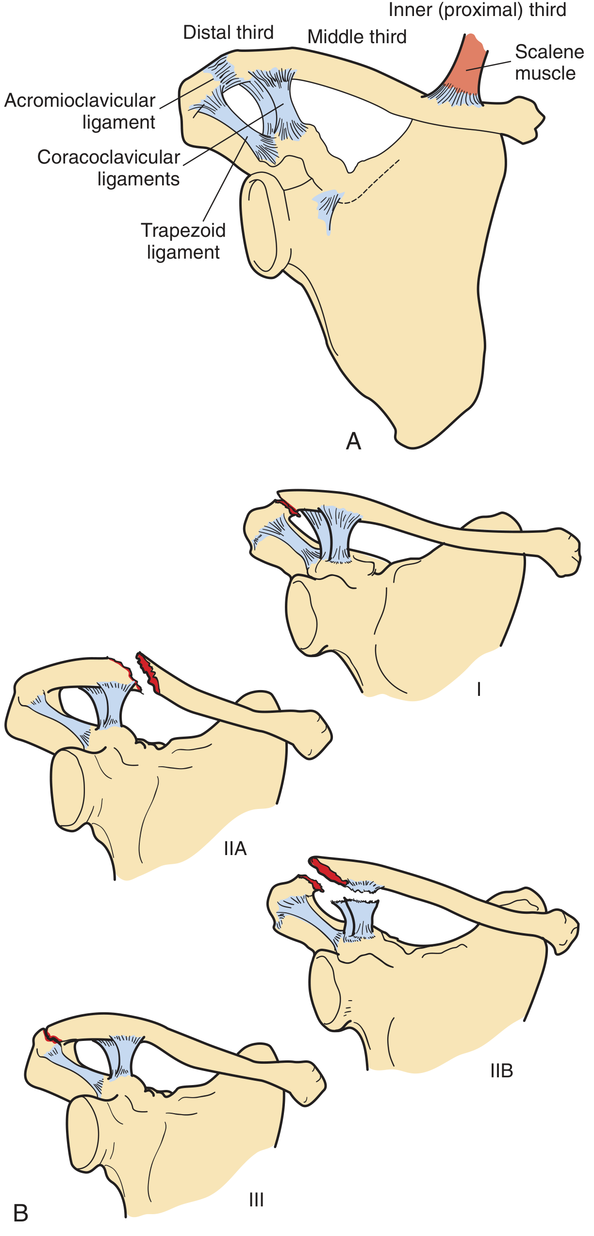
Group I - Middle Third Fractures
- Incidence: Most common, ~80% of all clavicle fractures
- Location: The middle third (diaphysis/shaft), between the coracoclavicular ligaments medially and the costoclavicular ligament laterally
- Mechanism: Direct blow or indirect force transmitted through an outstretched hand
- Deforming forces:
- The proximal fragment is displaced superiorly and posteriorly by the pull of the sternocleidomastoid (SCM) muscle
- The distal fragment is displaced inferiorly and anteriorly by the weight of the arm and the pectoralis major
- Clinical features: Pain, swelling, tenderness, visible or palpable deformity at the midshaft; the shoulder appears drooped
- Why common here: The middle third lacks the reinforcing ligamentous attachments of the lateral and medial thirds, making it mechanically the weakest zone
- Complications: Neurovascular injury (brachial plexus, subclavian vessels), nonunion (rare, <1%), pneumothorax
- Treatment: Mostly conservative - figure-of-eight brace or arm sling. Surgery reserved for skin tenting, significant shortening (>2 cm), neurovascular compromise, open fractures, or floating shoulder
Group II - Distal (Lateral) Third Fractures
- Incidence: ~15% of clavicle fractures
- Location: Lateral to the coracoclavicular (CC) ligaments, near the acromioclavicular (AC) joint
- Neer's subclassification (the most important extension of Allman's scheme):
| Subtype | Description | Ligament Status | Stability |
|---|---|---|---|
| Type I | Fracture between or lateral to the CC ligaments (between conoid and trapezoid, or lateral to trapezoid) | Both CC ligaments intact, AC ligament intact | Stable, minimal displacement |
| Type IIA | Fracture medial to the CC ligaments (conoid and trapezoid both attached to distal fragment) | Both CC ligaments intact - both on distal fragment | Unstable - proximal fragment displaced superiorly |
| Type IIB | Fracture between the two CC ligaments | Conoid ligament torn; trapezoid ligament intact on distal fragment | Unstable |
| Type III | Fracture involving the articular surface of the AC joint | No disruption of the CC ligaments | Stable, but risk of post-traumatic AC joint arthritis |
| Type IV (pediatric) | Periosteal sleeve fracture - clavicle displaces superiorly out of an intact periosteal tube | Ligaments attached to periosteal sleeve | Pseudo-dislocation appearance |
| Type V | Comminuted fracture with CC ligaments attached only to an inferior comminuted fragment (not to main proximal or distal fragments) | CC ligaments not attached to either main fragment | Very unstable |
- Clinical significance: Type IIA and IIB are unstable because the proximal fragment is pulled up by the SCM while the distal fragment remains anchored by the CC ligaments (attached to coracoid). This results in the characteristic superior displacement of the proximal fragment.
- Treatment: Types I, III: conservative. Types IIA, IIB, V: often surgical fixation due to instability and high nonunion rate (~30%)
Group III - Medial (Proximal) Third Fractures
- Incidence: Rarest, ~5% of clavicle fractures
- Location: Medial third, near the sternoclavicular (SC) joint
- Mechanism: Usually high-energy trauma (MVA, direct blow)
- Diagnosis challenge: Plain radiographs are often inadequate because of overlapping thoracic structures. CT scan is the investigation of choice to define the fracture pattern and rule out posterior displacement
- Important mimics: In patients under 25 years, the medial clavicular epiphysis has not yet fused (fuses ~25 years) - these injuries are often Salter-Harris fractures of the medial epiphysis rather than true SC dislocations
- Key concern: Posterior displacement of the medial fragment can impinge on mediastinal structures (trachea, esophagus, great vessels, brachial plexus) - a potentially life-threatening situation
- Treatment: Most are managed conservatively. Posteriorly displaced fractures compressing mediastinal structures require urgent reduction (closed or open)
Limitations of the Allman Classification
- Does not account for fracture displacement, comminution, or shortening - all of which influence treatment and prognosis
- Led to refinements such as the Nordqvist classification (added displacement/comminution subtypes) and the Edinburgh/Robinson classification (a comprehensive prognostic scheme based on 1,000+ patients that adds intra-articular extension, displacement, and comminution; however, it reverses the traditional numbering - medial = Type I, midshaft = Type II, distal = Type III - which causes confusion since "Type II" for distal fractures is deeply entrenched in the literature)
- Rockwood and Green's Fractures in Adults, 10th ed. (2025), Classification of Clavicle Fractures, p. 1199-1201
- Pfenninger and Fowler's Procedures for Primary Care, 3rd ed., Clavicle Fractures, p. 1317
Clavicle fracture
What aspect of clavicle fracture would you like covered?
clavicle fracture plate fixation ORIF surgical treatment
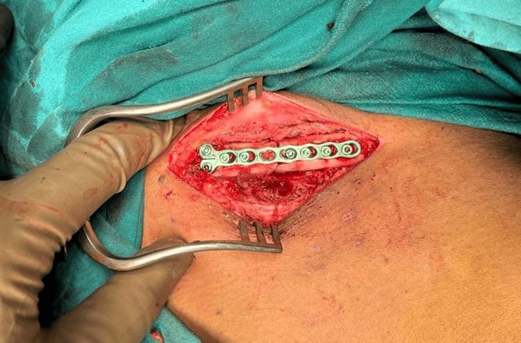
An intraoperative clinical photograph showing the open reduction and internal fixation (ORIF) of a medial end clavicle fracture. The surgical site is exposed through an anterior approach, with the skin and underlying soft tissues retracted using a self-retaining retractor to reveal the clavicle. A 2.4-mm mini fragment system locking compression plate (LCP) is fixed to the anterior surface of the bone across the fracture line. The metallic plate features seven holes, with multiple locking screws visible, providing stable fixation and alignment of the cortical bone. The surrounding surgical field is bordered by green sterile drapes, and the exposed muscle and connective tissues show normal surgical hyperemia. This image demonstrates orthopedic surgical techniques for stabilizing clavicular fractures using low-profile plating systems.
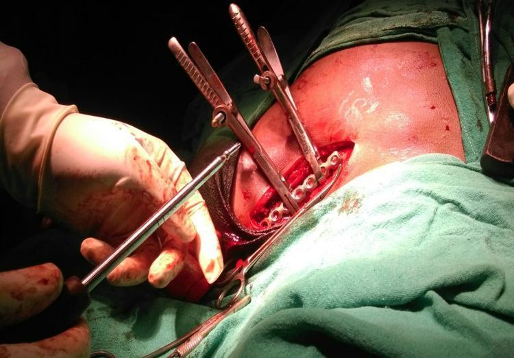
An intraoperative clinical photograph demonstrating an Open Reduction Internal Fixation (ORIF) of a midshaft clavicle fracture. The image shows a sterile surgical field with an incision exposing the clavicle's anterosuperior surface. A contoured anatomical locking compression plate is positioned across the fracture site. Two pointed reduction clamps are applied to maintain cortical alignment and bone-to-plate contact. A gloved hand is visible using a screwdriver or drill bit to secure fixation screws through the plate's circular and elongated dynamic compression holes. The surgical area is bounded by green sterile drapes and shows minor physiological hemorrhage typical of orthopedic procedures. This visual illustrates the surgical technique for managing displaced clavicular fractures, highlighting the relationship between the anatomical plate, temporary reduction clamps, and permanent screw fixation for interfragmentary compression and stability.
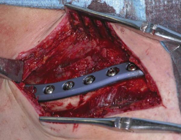
A clinical photograph capturing an intraoperative view of an Open Reduction and Internal Fixation (ORIF) procedure for a displaced fracture of the middle third of the clavicle. The image shows a surgically exposed anatomical region with visible soft tissue dissection and presence of blood. A contoured, metallic, light-blue orthopedic compression plate is positioned across the fracture site to stabilize the bone fragments. The plate is secured with multiple metallic cortical screws, which are fully seated within the plate's pre-drilled holes. Self-retaining retractors and other surgical instruments are positioned at the margins of the incision to maintain visibility of the operative field. This visual illustrates standard orthopedic trauma management and the use of internal fixation hardware to promote primary bone healing in clavicular fractures.
Management of Clavicle Fractures
GROUP I - Midshaft (Middle Third) Fractures (~80%)
Nonoperative Treatment (Standard of Care for Most)
| Device | Key Features |
|---|---|
| Broad arm sling | Preferred in most guidelines; more comfortable, less skin complications |
| Figure-of-eight brace | Keeps shoulders retracted; leaves elbow and hand free; associated with more skin irritation and pressure sores |
- Neither device provides reliable anatomical reduction; the goal is symptom relief, not reduction
- Sling worn for 4-6 weeks (adults), less in children
- Gentle pendulum exercises started early
- Active range of motion as pain permits
- Radiographic union confirmed before return to contact/high-demand activities (typically 8-12 weeks)
- Nonunion rate with conservative treatment: ~11% (higher in displaced fractures)
Indications for Operative Treatment
- Open fracture
- Neurovascular compromise (subclavian vessel or brachial plexus injury)
- Skin tenting / impending skin breakdown
- Displacement >1 shaft width / shortening >1.5-2 cm
- Comminution with rotational deformity / scapular winging
- Floating shoulder (ipsilateral clavicle + scapula neck fracture)
- Multiple ipsilateral upper rib fractures
- Polytrauma requiring early upper limb weight-bearing
- Elite athletes or patients with high functional demands
- Female sex + unfavorable fracture pattern (increased nonunion risk)
Operative Options
1. Open Reduction and Internal Fixation (ORIF) with Plate - Gold Standard
- Approach: Anterior or superior approach; incision along skin tension lines
- Plate position: Superior (most common) or anterior-inferior (potentially safer re: subclavian vessels)
- Implants: Pre-contoured anatomic locking compression plates; combination of locking and non-locking screws; minimum 3 bicortical screws each side of fracture
- Lag screw for large butterfly fragments before plating
- Outcomes: Union rate 94-100%; nonunion rate ~1.3-2.2%; earlier return to function vs conservative
- Outpatient procedure
- Sling for comfort; pendulum exercises from day 1
- Wound check at 10-14 days
- Resisted exercises and strengthening from 6 weeks (if radiograph acceptable)
- No contact sports for 12 weeks
- Routine implant removal NOT recommended; if required, wait minimum 1 year post-union to reduce refracture risk
2. Intramedullary (IM) Nailing/Pinning
- Advantages: Smaller incision, less soft tissue disruption, better cosmesis, easier implant removal
- Disadvantages: Poor rotational and axial control with unlocked devices; NOT suitable for comminuted fractures; high hardware prominence/removal rates with smooth pins
- Newer locked IM devices improve on traditional unlocked TENs (titanium elastic nails)
- RCTs comparing unlocked TENs vs plate fixation show comparable union rates but higher hardware irritation and removal rates with IM nailing
- Patient selection: Non-comminuted fractures with adequate IM canal diameter
GROUP II - Distal (Lateral) Third Fractures (~15%)
| Neer Type | Stability | Preferred Treatment |
|---|---|---|
| Type I (lateral to or between CC ligaments, ligaments intact) | Stable | Conservative: sling x 4-6 weeks |
| Type IIA (both CC ligaments on distal fragment, proximal displaced up) | Unstable | Operative fixation |
| Type IIB (conoid ruptured, trapezoid intact) | Unstable | Operative fixation |
| Type III (intra-articular, AC joint) | Stable | Conservative; watch for late AC arthrosis |
| Type IV (pediatric physeal) | Variable | Usually conservative (periosteum intact, remodels) |
| Type V (comminuted, CC ligaments on neither main fragment) | Very unstable | Operative fixation |
Conservative Treatment for Distal Fractures
- Same protocol as midshaft: sling x 2 weeks, pendulum exercises, expand ROM at 6 weeks
- Despite high nonunion rates (~20-33% for displaced types), most patients remain asymptomatic - only ~20-47% of those with nonunion require secondary surgery
- DASH scores and satisfaction remain high even with fibrous union
Operative Options for Distal Fractures
- Pre-contoured locking plate fixation - most popular currently; provides direct fracture fixation; challenge is achieving adequate purchase in the small distal fragment
- Hook plate fixation - plate hooks under acromion, maintains reduction; effective but must be removed at 3-6 months (risks subacromial impingement)
- Coracoclavicular (CC) fixation - suture button device, suture anchor, or CC screw; stabilizes the clavicle to the coracoid but does not directly fix the fracture
- Tension band wiring - older technique, now less favored
- Transacromial K-wire fixation - simple but requires removal; risk of wire migration
- Arthroscopically assisted techniques - emerging; minimally invasive
GROUP III - Medial (Proximal) Third Fractures (~5%)
- Most are managed conservatively with a sling
- CT scan mandatory before treatment to define fracture anatomy and posterior displacement
- Posteriorly displaced fractures - risk impingement on trachea, esophagus, subclavian vessels, or brachial plexus; require urgent reduction
- Closed reduction first (under general anesthesia, with cardiothoracic/vascular surgery on standby)
- Open reduction if closed fails or if impingement persists
- Medial epiphyseal fractures in patients <25 years (unfused epiphysis) - usually conservative; reduction often unstable but remodels
- SC joint involvement: anterior displacement - conservative; posterior - urgent reduction
Summary Table: Treatment by Group
| Group | Location | Incidence | First-line | Surgery When? |
|---|---|---|---|---|
| I | Midshaft | ~80% | Sling / figure-of-eight | Displaced, shortened, open, NV injury, high demand |
| II (Neer I, III) | Distal, stable | Sling | Rarely | |
| II (Neer IIA, IIB, V) | Distal, unstable | ~15% total | Operative | Standard for unstable types |
| III | Medial | ~5% | Sling | Posterior displacement with mediastinal compromise |
Complications
| Complication | Notes |
|---|---|
| Nonunion | Midshaft: ~1-3% operative, ~11% conservative (displaced); Distal: ~20-33% conservative |
| Malunion | Shortening/clavicular deformity; can cause pain, impaired shoulder mechanics |
| Post-traumatic AC arthritis | Particularly after Neer Type III fractures |
| Refracture | Risk increased with premature implant removal; wait >1 year post-union |
| Supraclavicular nerve injury | Numbness below incision; usually improves; rare painful neuroma |
| Subclavian vessel / brachial plexus injury | Rare iatrogenic; more risk with medial fractures |
| Pneumothorax | Associated with high-energy midshaft fractures; always auscultate the chest |
| Infection / hardware prominence | Particularly with IM devices |
- Rockwood and Green's Fractures in Adults, 10th ed. (2025), Treatment Options for Midshaft and Lateral Clavicle Fractures, pp. 1206-1221
- Miller's Review of Orthopaedics, 9th ed., Clavicle Fracture, p. 932
- Pfenninger and Fowler's Procedures for Primary Care, 3rd ed., Clavicle Fractures, p. 1317