
Varicose vein treatment of 27 yr old boy
- Conservative: compression stockings, elevation, lifestyle changes
- Minimally invasive: endovenous laser ablation (EVLA), radiofrequency ablation (RFA), sclerotherapy
- Surgical: saphenofemoral ligation + stripping (for large truncal veins)
- In a young patient, underlying causes (deep vein reflux, congenital factors) should be ruled out first
"varicose veins"[MeSH Terms] AND treatment
varicose veins treatment endovenous laser ablation sclerotherapy diagram
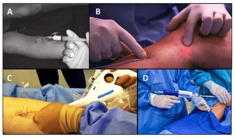
This composite of four clinical photographs illustrates various endovenous treatment modalities for saphenous vein insufficiency and varicose veins. Image A shows ultrasound-guided foam sclerotherapy, featuring a syringe connected to a catheter for injecting chemical sclerosants. Image B demonstrates Endovenous Laser Ablation (EVLA), where a thin laser fiber is inserted into the vein; a red transillumination light on the skin surface indicates the laser tip position. Image C depicts mechanochemical ablation (MOCA), showing a specialized motorized handle device that combines mechanical endothelial damage with simultaneous sclerosant infusion. Image D displays cyanoacrylate embolization (medical glue injection), featuring a gun-like delivery system used to deploy an adhesive agent into the vein lumen via a catheter. All procedures are minimally invasive, performed through small percutaneous access points on the lower extremity. The images serve to compare thermal and non-thermal, non-tumescent (NTNT) techniques in modern phlebology, highlighting the diverse instrumentation used for venous occlusion.
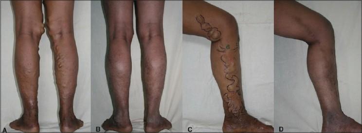
This composite clinical photograph (labeled A-D) illustrates the comparative outcomes of endovenous laser ablation for the treatment of varicose veins. Panels A and C display the preoperative status, showing significant vascular abnormalities of the lower extremities. Key features include prominent, dilated, and tortuous superficial veins (varicosities) primarily distributed along the posterior and medial aspects of the calves and thighs. These veins appear as raised, snake-like protrusions beneath the skin. In panel C, surgical markings delineate the planned treatment course along the Great Saphenous Vein distribution. Associated skin changes such as hyperpigmentation and darkening near the ankles are visible, suggesting chronic venous stasis and hemosiderin deposition. Panels B and D provide postoperative views following treatment with a 980 nm Diode laser. They demonstrate a marked reduction in venous distension, with the leg contours appearing significantly smoother and the pathological bulging resolving. The series highlights the efficacy of endovenous laser intervention in treating symptomatic venous insufficiency and its associated clinical manifestations.
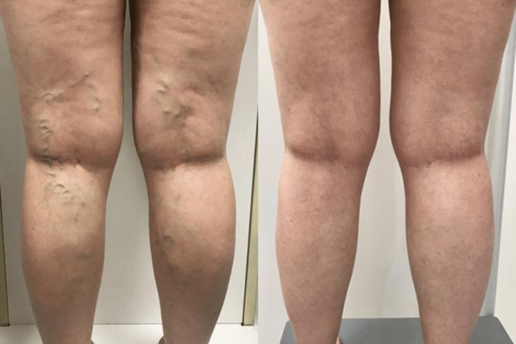
This clinical photograph presents a side-by-side comparison of a patient's lower limbs from a posterior view, illustrating the efficacy of endovenous laser ablation (EVLA) for chronic venous insufficiency. The 'before' image (left) demonstrates extensive, bilateral varicose veins characterized by dilated, tortuous, and protuberant superficial veins primarily localized to the popliteal fossa, posterior thighs, and calves. These varicosities exhibit a bluish hue and create a palpable, irregular skin surface. The 'after' image (right) shows the results three months post-treatment, revealing a dramatic resolution of the visible varicosities. The previously bulging vessels are no longer prominent, and the skin contour appears smoother and more uniform. This comparison serves as an educational tool for identifying clinical manifestations of varicose veins and evaluating therapeutic outcomes in vascular surgery or dermatology. Key concepts include venous reflux, saphenous vein incompetence, and minimally invasive thermal ablation techniques.
PMID: 37652254
Varicose Vein Treatment in a 27-Year-Old Male
First Step: Evaluation
- Duplex ultrasound (DUS) - mandatory to assess saphenofemoral junction (SFJ) competence, great saphenous vein (GSV) reflux, deep vein reflux, and perforator incompetence
- CEAP classification to stage disease severity (C0-C6)
- Exclude secondary causes: previous DVT, May-Thurner syndrome, Klippel-Trenaunay syndrome (relevant given young age)
- Check for family history - varicose veins have a strong genetic component
Treatment Options
1. Conservative (First Line - All Patients)
- Graduated compression stockings: 20-30 mmHg for simple varicose veins; 30-40 mmHg if edema or skin changes are present
- Elevate legs periodically, avoid prolonged standing
- Regular exercise - strengthens the calf muscle pump
- Weight management if applicable
Compression garments improve symptoms but are not curative and do not prevent progression. - Harrison's Principles of Internal Medicine 22E
2. Endovenous Thermal Ablation (Preferred Minimally Invasive - 1st choice for truncal reflux)
a) Endovenous Laser Ablation (EVLA)
- A catheter is introduced percutaneously at the knee level under ultrasound guidance and advanced to just below the SFJ
- Laser energy is delivered as the catheter is withdrawn - heat injures the endothelium, causing thrombosis and fibrosis → vein occlusion
- 1-year occlusion rates >90%; 5-year rates remain high
- Adverse effects: pain, paresthesias, bruising, hematoma, hyperpigmentation
b) Radiofrequency Ablation (RFA)
- Same principle as EVLA but uses radiofrequency energy
- Slightly lower 5-year occlusion rates than EVLA, but comparable outcomes
- A 2024 meta-analysis (Jiang et al., J Vasc Surg Venous Lymphat Disord, PMID 38316290) comparing EVLA vs RFA found both are effective with minor differences in post-procedure pain (RFA slightly less painful)
Harrison's 22E - "Average 1- and 5-year occlusion rates exceed 90% following endovenous laser therapy"
3. Nonthermal, Nontumescent (NTNT) Ablation (Emerging options)
a) Cyanoacrylate Embolization (VenaSeal / Medical Glue)
- A tissue adhesive is injected via catheter into the GSV, causing fibrosis and occlusion
- 1-year occlusion rates ~90%
- No tumescent anesthesia required; minimal post-procedure compression needed
- Adverse effect: superficial thrombophlebitis, foreign body reaction
b) Mechanochemical Ablation (MOCA - ClariVein)
- Rotating wire damages the endothelium mechanically + simultaneous liquid sclerosant infusion
- ~90% 1-year occlusion
- Adverse effects: ecchymoses, hematoma, hyperpigmentation
4. Sclerotherapy (For smaller veins, tributaries, telangiectasias)
- Chemical (e.g., sodium tetradecyl sulfate, polidocanol) injected into the vein to cause fibrosis
- Foam sclerotherapy (using Tessari technique) is preferred for larger reticular and varicose veins - increases contact area
- Liquid sclerotherapy for spider veins and telangiectasias
- Often combined with EVLA/RFA after truncal ablation to treat residual varicosities
- Complications: hyperpigmentation, telangiectatic matting, skin ulceration (rare), DVT (rare with foam)
Goldman-Cecil Medicine - "Laser and surgical treatments appear superior to liquid or foam sclerotherapy" for varicose vein treatment broadly
5. Ambulatory Phlebectomy (Stab Avulsion)
- Small (2-3 mm) stab incisions are made and varicose tributaries are hooked out with a phlebectomy hook under local anesthesia
- Excellent cosmetic results; performed as an outpatient
- Often combined with EVLA/RFA to remove bulging tributaries not addressed by ablation alone
- Fitzpatrick's Dermatology notes ambulatory phlebectomy is commonly combined with endovenous ablation for junction treatment
6. Surgical Options (Now less commonly used)
- High ligation + stripping of the GSV (Trendelenburg procedure)
- Saphenofemoral junction ligation (SFJ ligation)
- Multiple stab avulsions for tributaries
2023 SVS/AVF/AVLS Guidelines Summary (PMID 37652254)
- Duplex ultrasound evaluation before any intervention
- Endovenous thermal ablation (EVLA or RFA) as preferred over open surgery for truncal reflux
- Compression therapy is recommended before and after procedures
- Sclerotherapy remains the standard for spider veins
- Foam sclerotherapy as an option for truncal and tributary veins when thermal ablation is not suitable
Treatment Algorithm for a 27-Year-Old
| Finding | Recommended Treatment |
|---|---|
| Isolated telangiectasias / spider veins | Sclerotherapy or laser |
| Reticular veins only | Foam sclerotherapy |
| GSV reflux + varicose tributaries | EVLA/RFA + ambulatory phlebectomy ± foam sclerotherapy |
| SFJ incompetence + large GSV | EVLA/RFA (preferred) or ligation + stripping |
| Failed endovenous / recurrent | Surgical re-exploration or repeat sclerotherapy |
| Congenital / complex anatomy | MDT assessment; avoid aggressive ablation |
Visual Reference
Key Points for a Young Patient (27 yrs)
- Rule out secondary causes - young age raises suspicion for congenital venous anomaly, previous DVT, or pelvic pathology
- EVLA or RFA is the preferred first-line intervention for truncal GSV reflux - excellent long-term outcomes with low recurrence
- Surgery is rarely the first choice now unless anatomy is unsuitable for thermal ablation
- Follow-up compression post-procedure reduces recurrence
- He is at low surgical risk and an ideal candidate for minimally invasive procedures