

Is This pathology a cyst Patient has a history of trauma on 21 teeth Xray shows this
periapical radiolucent lesion radicular cyst radiograph
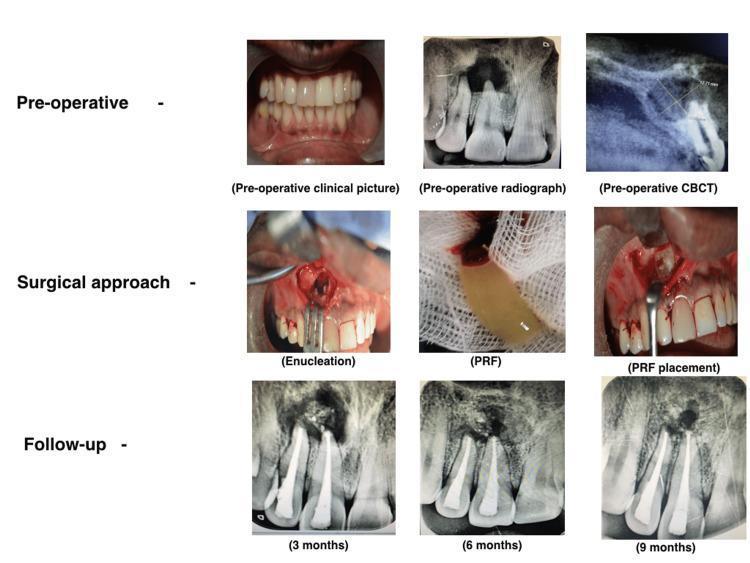
A multi-panel medical compilation illustrating the management of a periapical lesion (radicular cyst) associated with the maxillary right central incisor (tooth 11). The content is organized into three rows: Pre-operative, Surgical approach, and Follow-up. The pre-operative row includes a clinical photograph showing tooth discoloration, a periapical radiograph with a large radiolucency, and a sagittal CBCT view measuring the lesion's labio-palatal extent. The surgical approach row shows the intraoperative enucleation of the cyst, the preparation of yellowish Platelet-Rich Fibrin (PRF), and its subsequent placement into the surgical cavity. The follow-up row consists of three serial periapical radiographs at 3, 6, and 9 months, demonstrating progressive healing. These radiographs show the tooth with a radio-opaque retrograde filling material (MTA) and a gradual decrease in the periapical radiolucent area, replaced by increasing radiopacity indicative of successful bone regeneration and ossification.
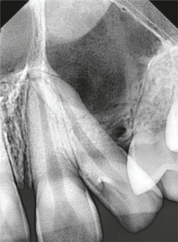
This intraoral periapical (IOPA) radiograph depicts the maxillary anterior and premolar region, specifically involving the left lateral incisor and first premolar. The primary finding is a large, ill-defined radiolucent lesion located between the roots of these teeth. The pressure from this lesion has caused significant distal and mesial deflection of the adjacent roots, increasing the inter-radicular space. Superior to this primary radiolucency, a dense, partial radiopacity is visible, suggesting the presence of an unerupted impacted tooth, possibly the maxillary canine. Additionally, a smaller, well-defined circular radiolucency is noted at the alveolar crest between the lateral incisor and first premolar. The overall radiographic presentation is highly suggestive of a pericoronal or cystic lesion, such as a dentigerous cyst, associated with an impacted tooth. The image illustrates key diagnostic features in oral and maxillofacial pathology, including root divergence and the characteristic radiolucency associated with odontogenic cysts in a pediatric or adolescent patient.
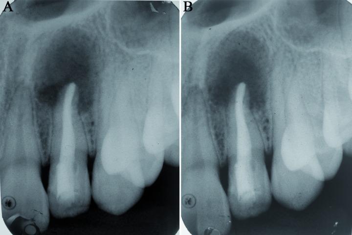
Two side-by-side intraoral periapical (IOPA) radiographs (A and B) demonstrate the post-surgical healing of a large periapical lesion in the maxillary anterior region. The central focus is a maxillary lateral incisor that has undergone endodontic treatment, evidenced by a dense, uniform radiopaque root canal filling extending to the apex. Radiograph A, representing a 6-week follow-up, shows a significant, well-defined radiolucent area surrounding the root apex, indicative of a persistent bony defect following the enucleation of a radicular cyst. Radiograph B, at a 12-week follow-up, illustrates progressive bone regeneration. The previously dark radiolucent area exhibits increased radiographic density, with a visible trabecular bone pattern filling the defect from the periphery toward the center. This comparison highlights the typical timeline and radiographic appearance of successful bone healing and apical tissue repair after surgical intervention and root canal therapy. The surrounding alveolar bone and adjacent teeth remain stable throughout the observation period.
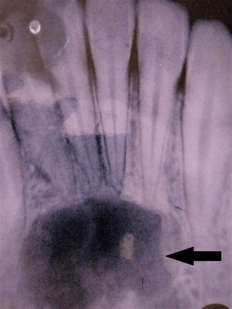
This intraoral periapical (IOPA) radiograph displays the mandibular anterior region, revealing a large, unilocular radiolucency located at the apices of the incisors and canine teeth. The lesion is characterized by a well-defined, corticated border (indicated by the black arrow), which distinguishes it from the surrounding normal trabecular bone. Centrally, the internal structure is predominantly homogenous and radiolucent, suggesting a cystic or fluid-filled cavity. Crucially, the radiograph shows that the roots of the associated teeth remain intact, with no evidence of external root resorption or significant displacement, despite being within the lesion's proximity. The finding is clinically significant for the differential diagnosis of odontogenic cysts, such as a radicular cyst (if teeth are non-vital) or a lateral periodontal cyst, and non-odontogenic lesions like a traumatic bone cyst. This image is an essential teaching tool for oral radiology, highlighting key features of benign periapical pathology including margin definition and effects on adjacent dental structures.
Radiographic Analysis: Periapical Lesion in Tooth 21
What the X-ray Shows
- Well-demarcated borders with a relatively smooth outline
- Extensive bone destruction in the periapical and coronal region
- Blunted root apex - consistent with apical root resorption
- Adjacent tooth showing external root resorption at the interface with the lesion
- The lesion lacks a thick sclerotic cortical rim around its entire periphery
Is It a Cyst? - Differential Diagnosis
1. Radicular Cyst (Most Likely) ✓
- Long-standing pulp inflammation secondary to advanced caries or local trauma (exactly your patient's history)
- Necrosis of pulp tissue traversing the root to exit at the apex into alveolar bone
- Granulation tissue development → epithelialization → cyst formation
| Feature | This X-ray | Radicular Cyst |
|---|---|---|
| Location | Periapical, at root apex | ✓ Classic location |
| Border | Well-demarcated, smooth | ✓ Typical |
| Shape | Unilocular radiolucency | ✓ Typical |
| Root resorption | Present (adjacent tooth) | ✓ Seen in larger cysts |
| Associated tooth | Root canal treated (implies prior non-vitality) | ✓ Required for diagnosis |
| History | Trauma to tooth 21 | ✓ Known etiology |
| Size | Large | ✓ Consistent |
2. Large Periapical Granuloma (Differential)
- Can look identical on X-ray, especially when large
- Distinguished only on histopathology - does NOT show true granulomatous inflammation despite the name
- Typically smaller than cysts but can overlap in size
- Does not have an epithelial lining (unlike a true cyst)
3. Odontogenic Keratocyst (OKC) - Must Exclude
- Can appear as a large, well-demarcated radiolucency
- More aggressive behavior, higher recurrence
- Usually has characteristic scalloped borders
- Requires histopathology to exclude definitively
4. Ameloblastoma - Less Likely
- Typically multilocular ("soap bubble" appearance)
- More commonly in the mandible
- Unilocular unicystic form can mimic this, but less common in anterior maxilla
Reference Image
Why Radicular Cyst is the Top Diagnosis Here
- Trauma history on tooth 21 - leads to pulp necrosis
- Root canal treated tooth - confirms prior non-vitality
- Large, well-defined unilocular radiolucency at the apex
- Root resorption of adjacent structures (pressure effect)
- Absence of multilocularity (argues against ameloblastoma)
Key Takeaway and Management
- For this size lesion: surgical enucleation + root-end surgery (apicoectomy) or extraction, depending on the restorability of the tooth
- Submit the entire specimen for histopathological examination to confirm diagnosis (and rule out OKC or rare neoplasm)
- If the tooth is to be retained: ensure the root canal treatment is adequate, then enucleate the cyst